Gallery
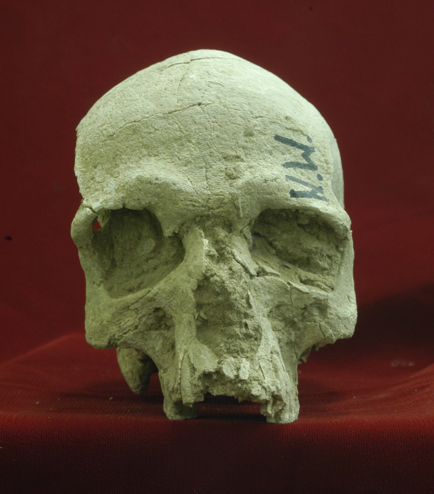
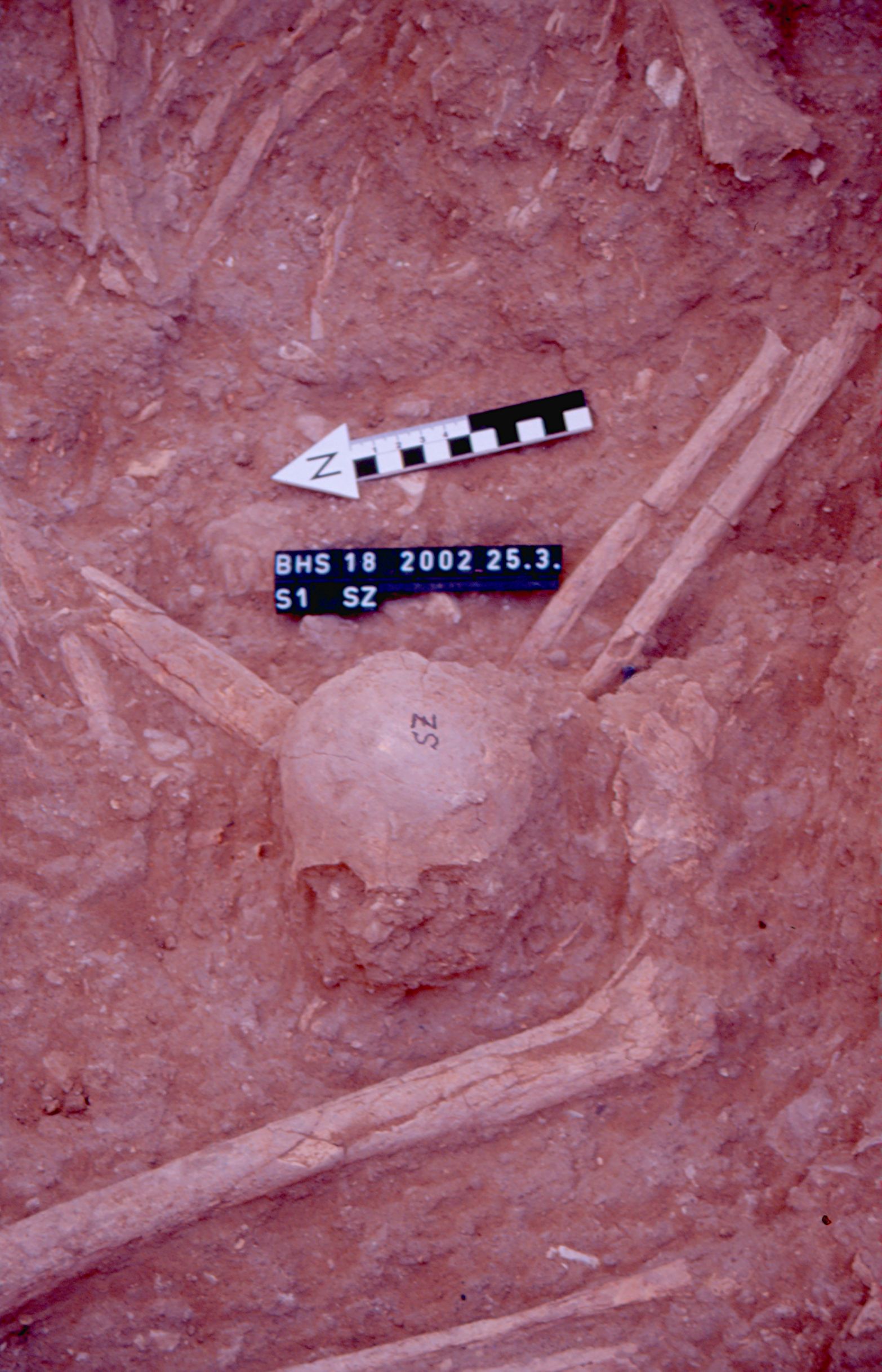
Inidvidual AB:
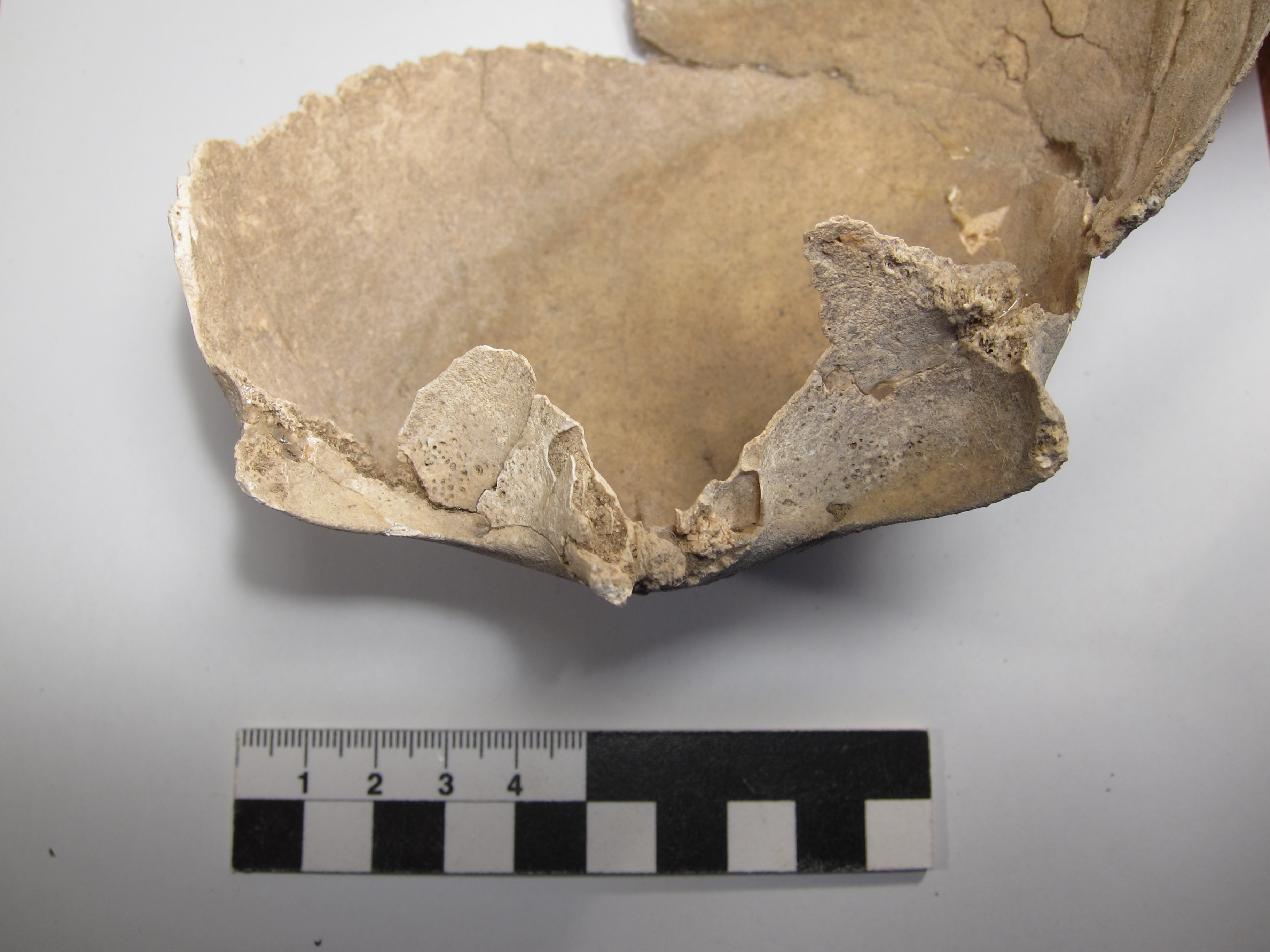
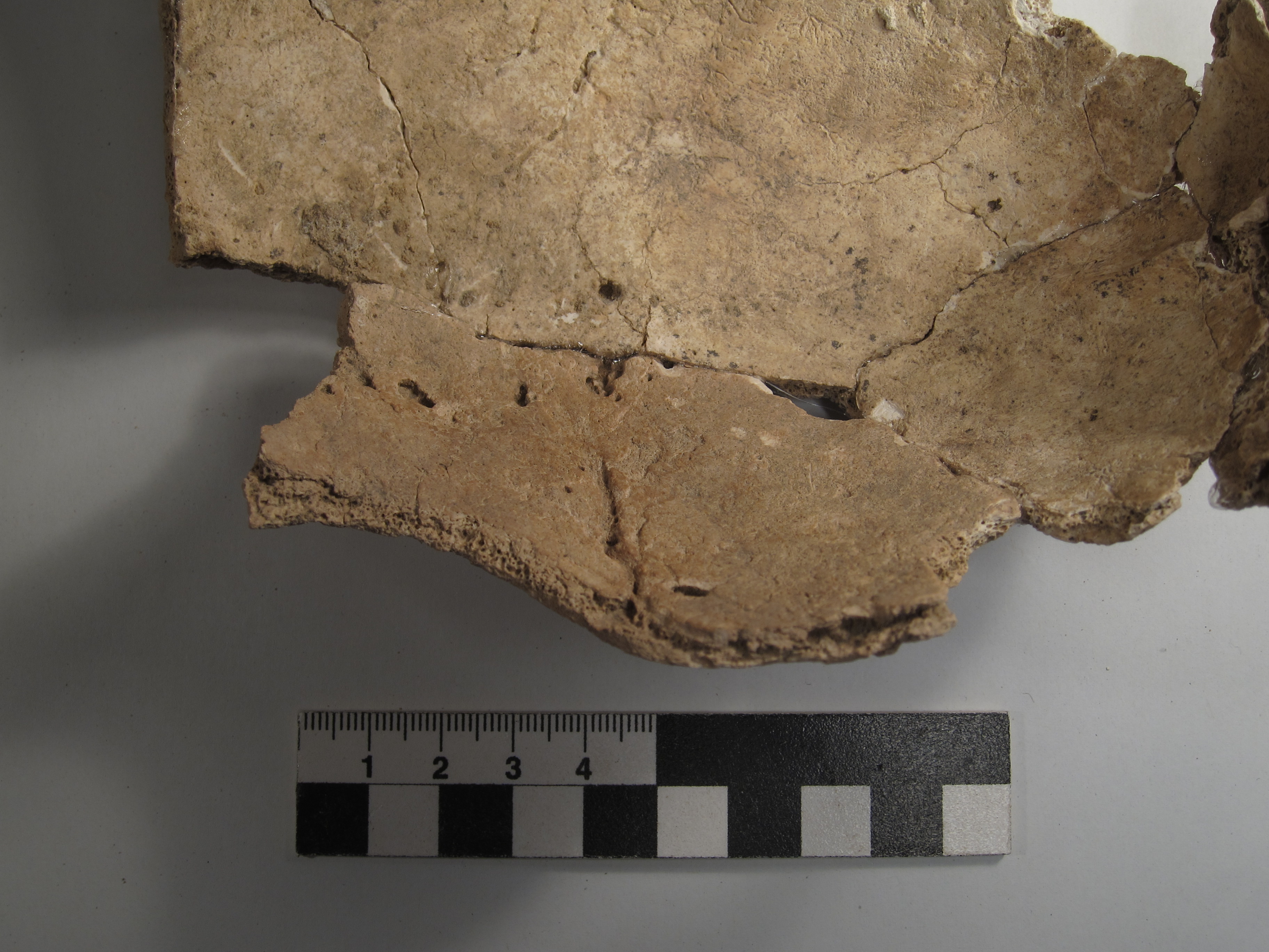
Cribra orbitalia
Full Resolution
Full Resolution
Inidvidual AH:
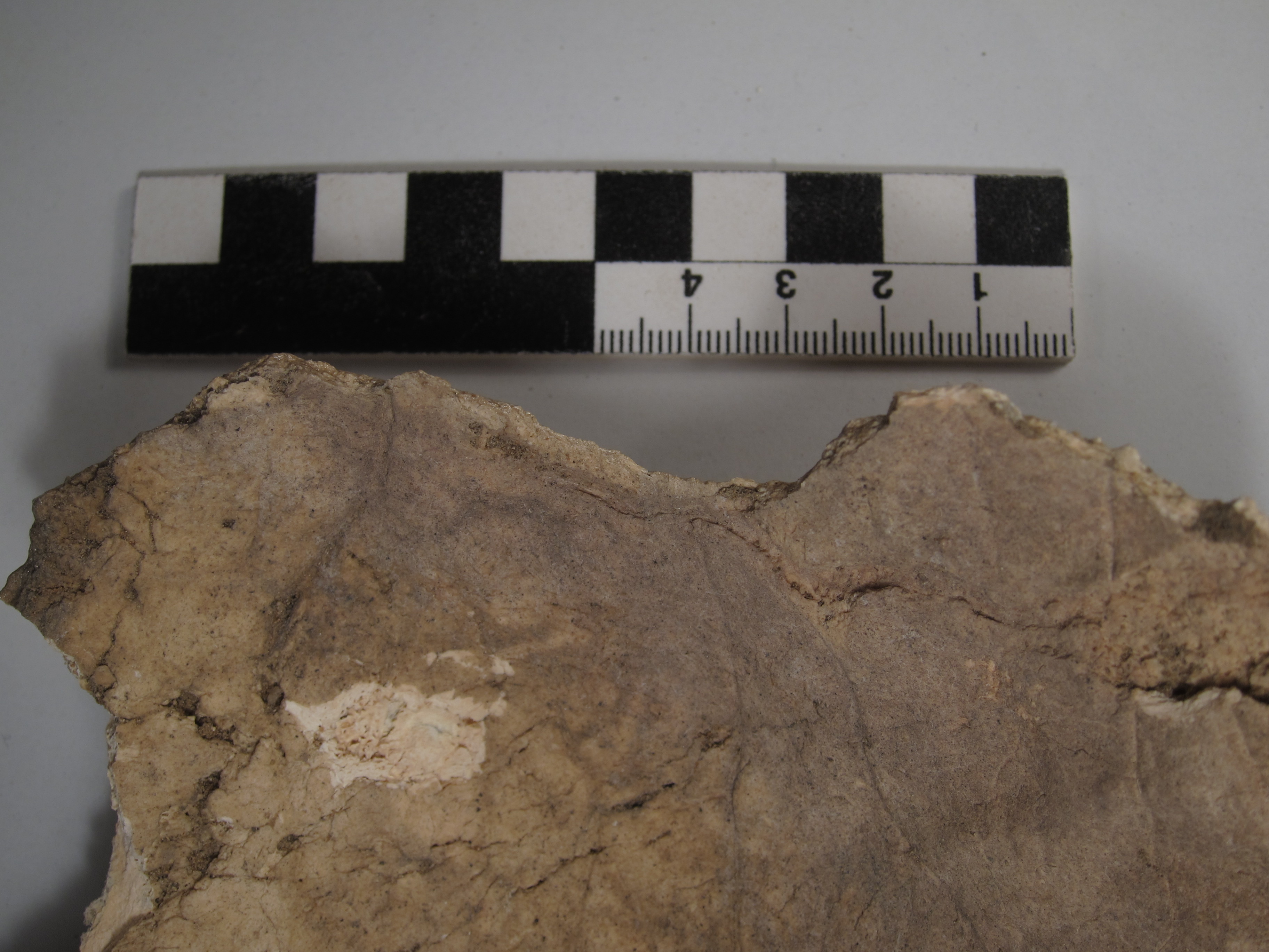
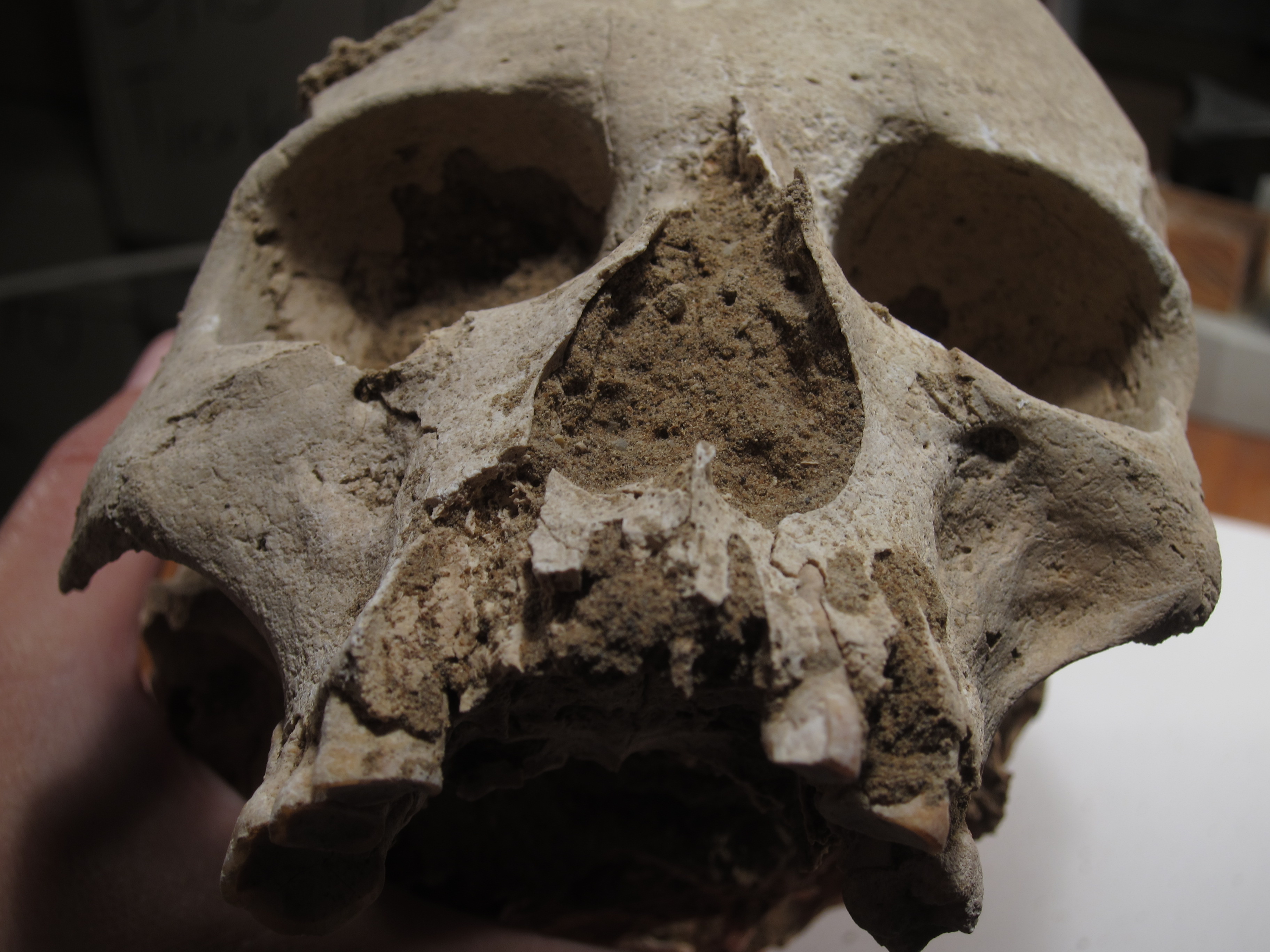
Frontal bone with a small oval depression fracture (18 x 12 mm) on the right frontal eminence; the surrounding bone displays signs of inflammation.
Full Resolution
Full Resolution
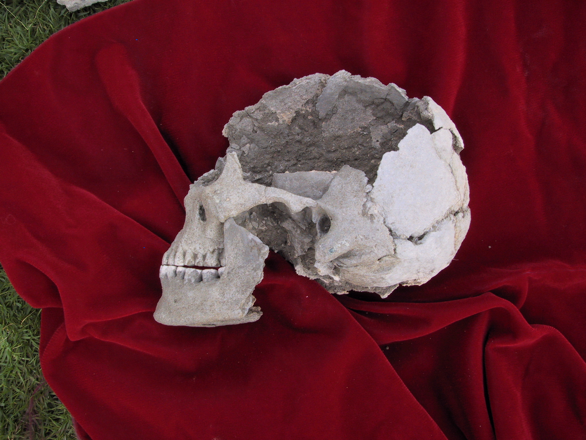
Inidvidual AJ:
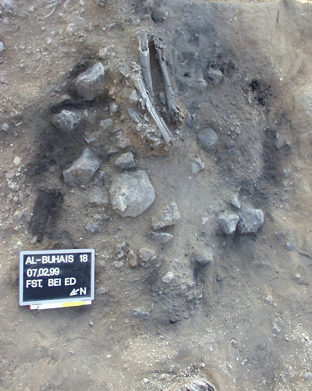
Skull AJ with fracture and Osteomyelitis
Full Resolution
Full Resolution

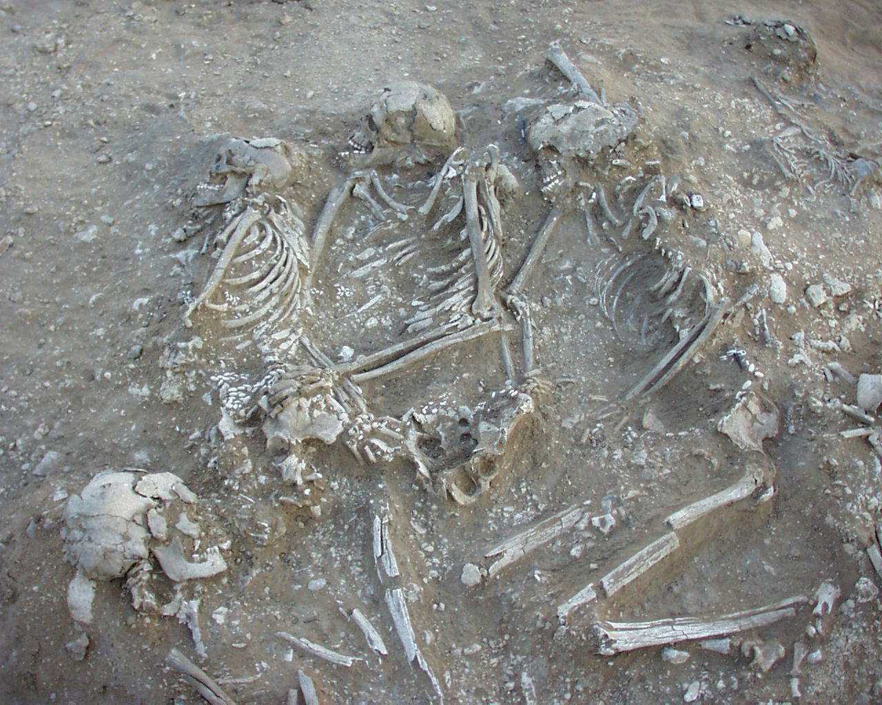
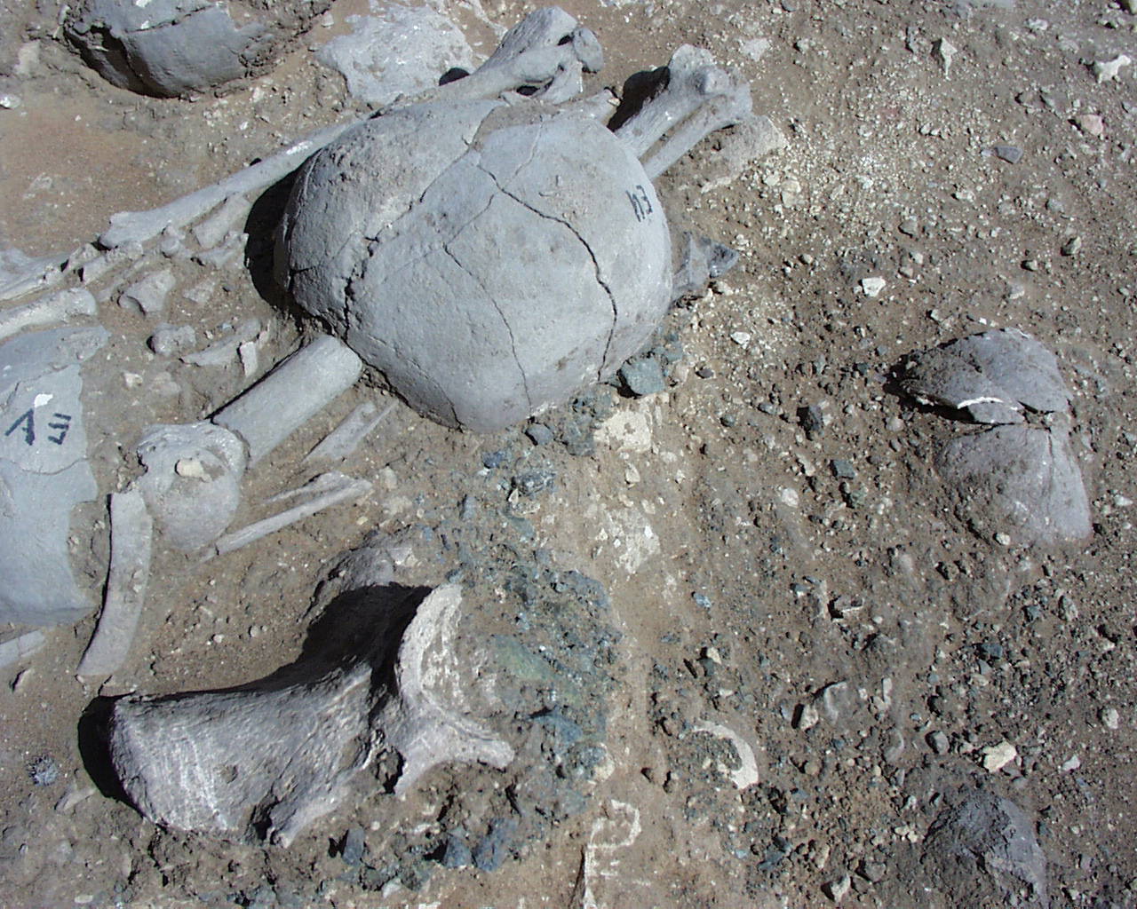
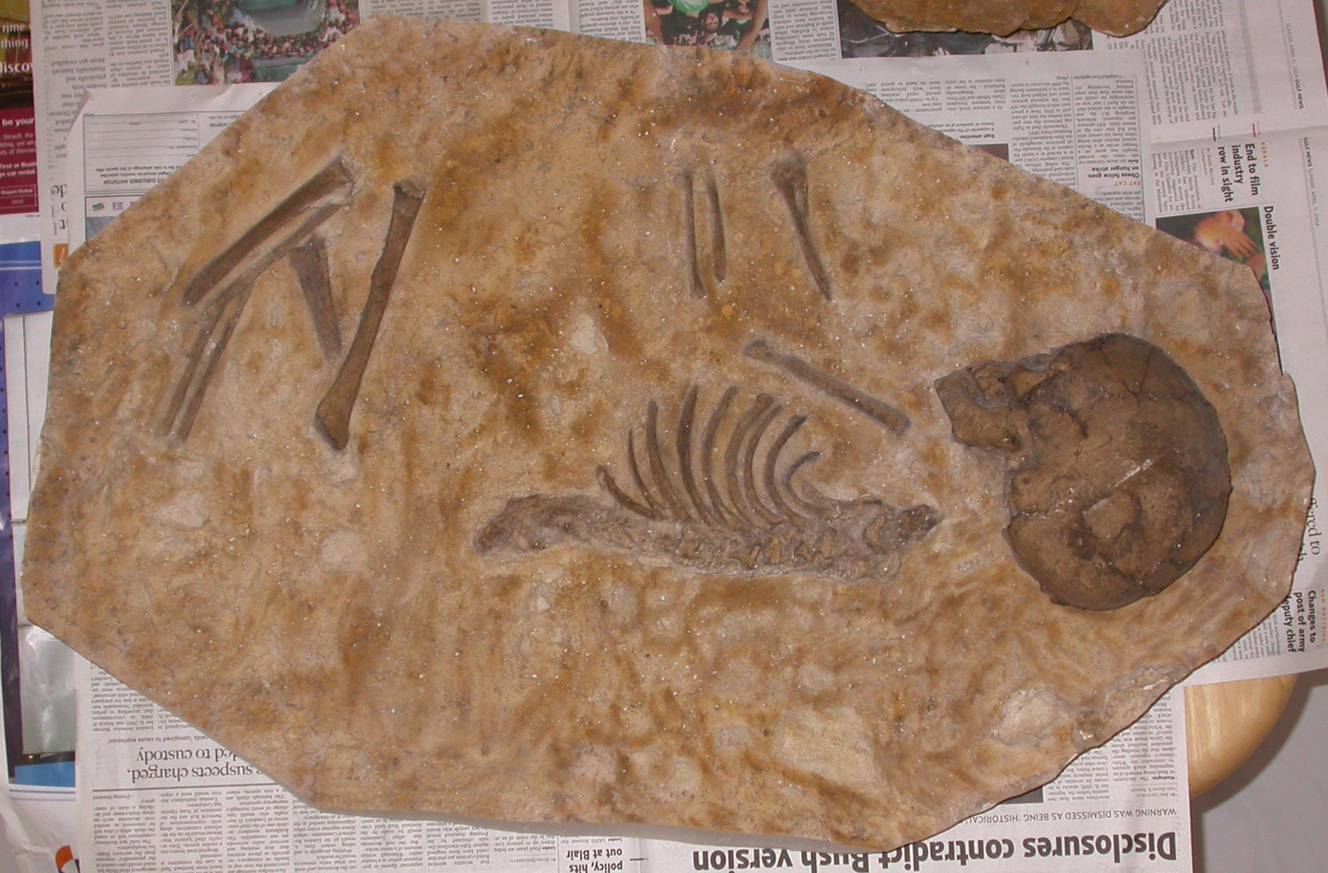
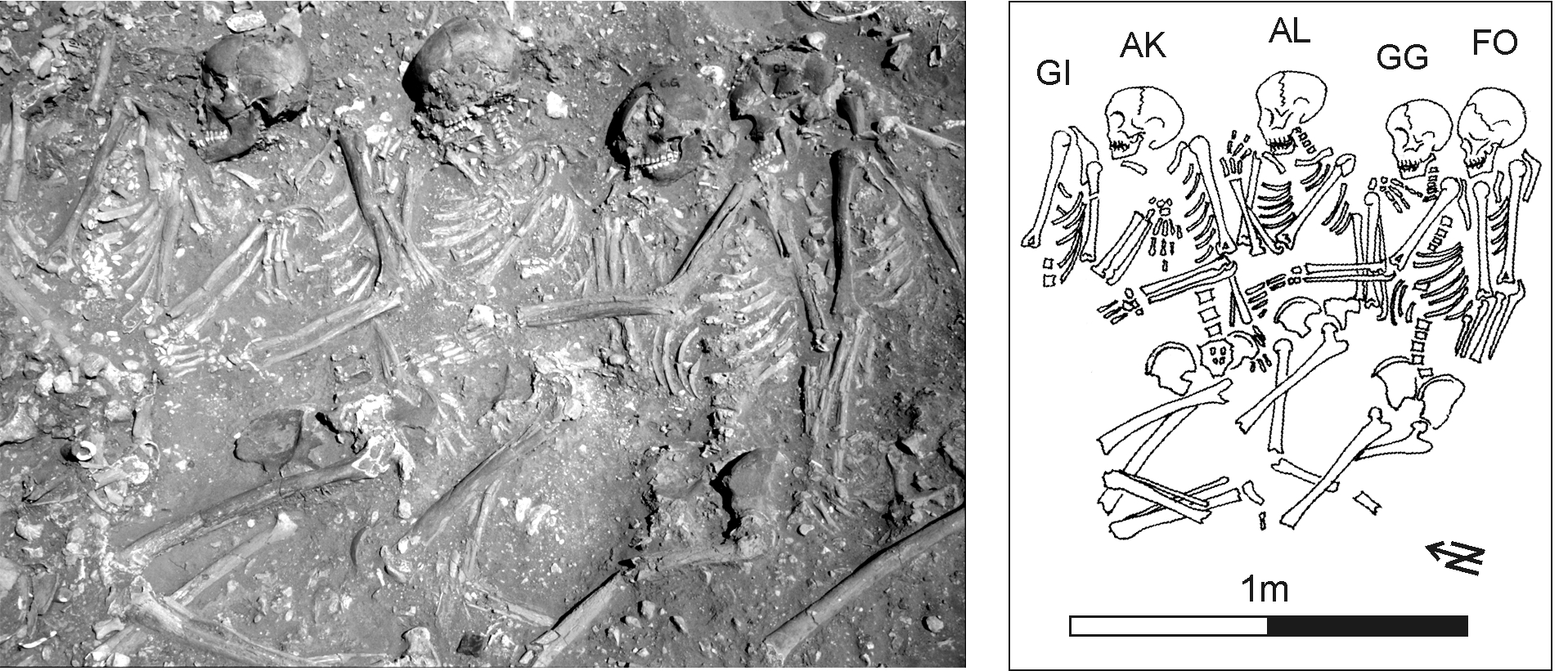
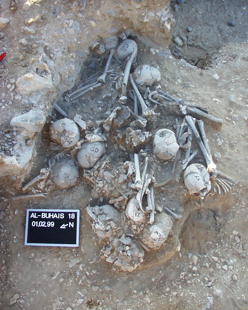
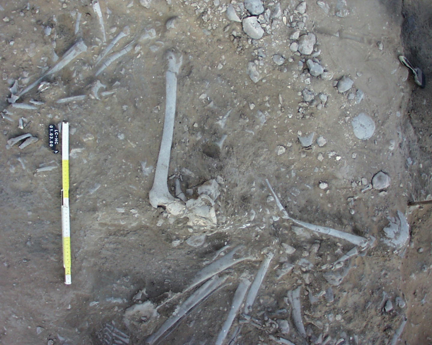
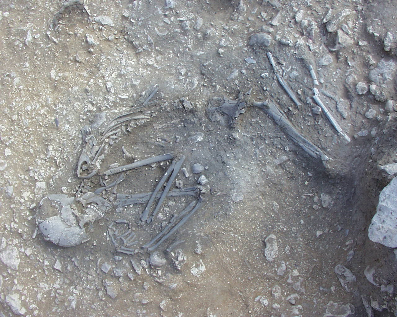
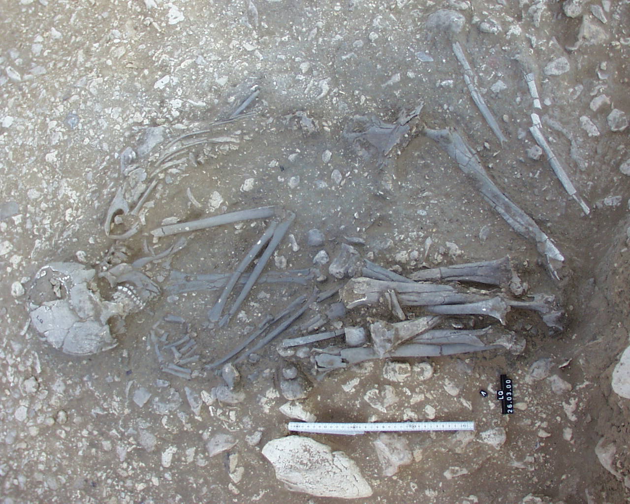
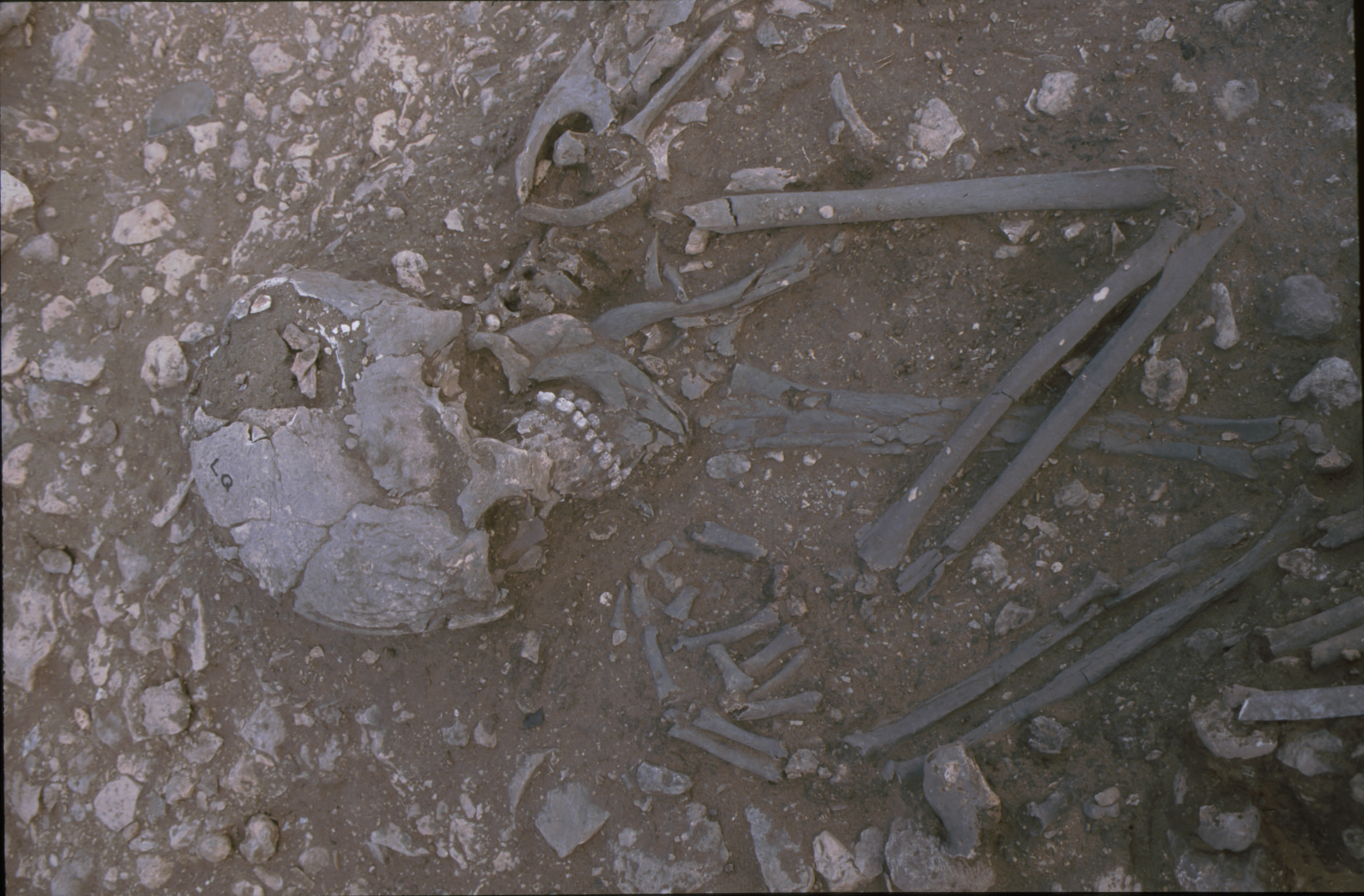
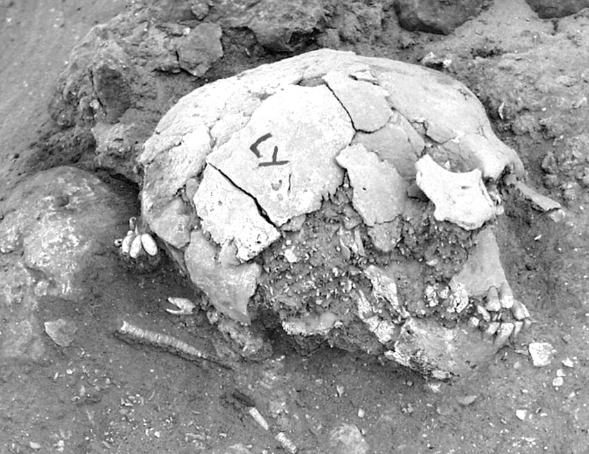
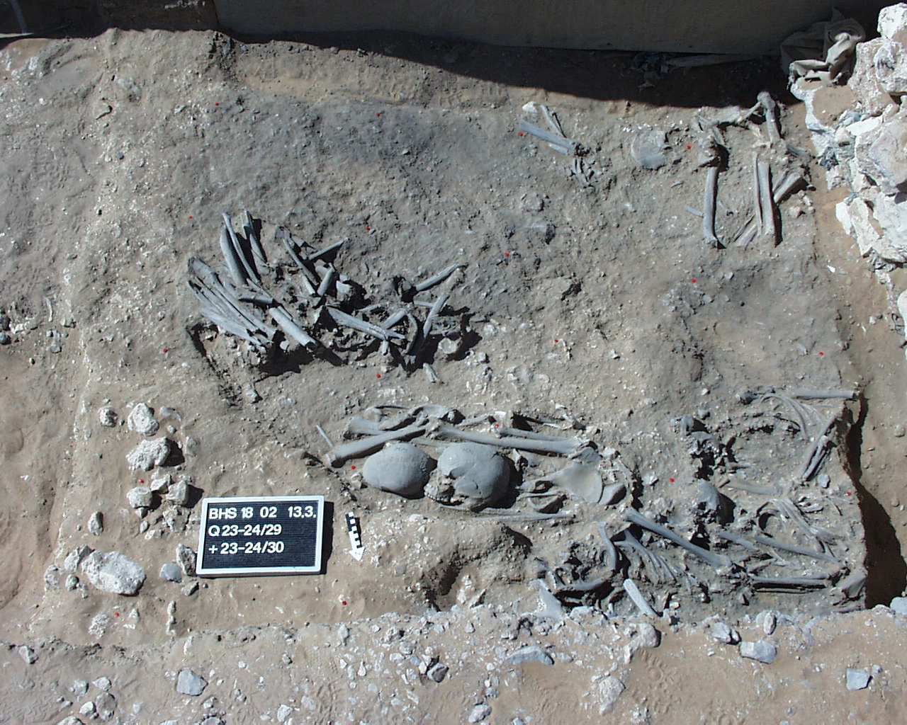
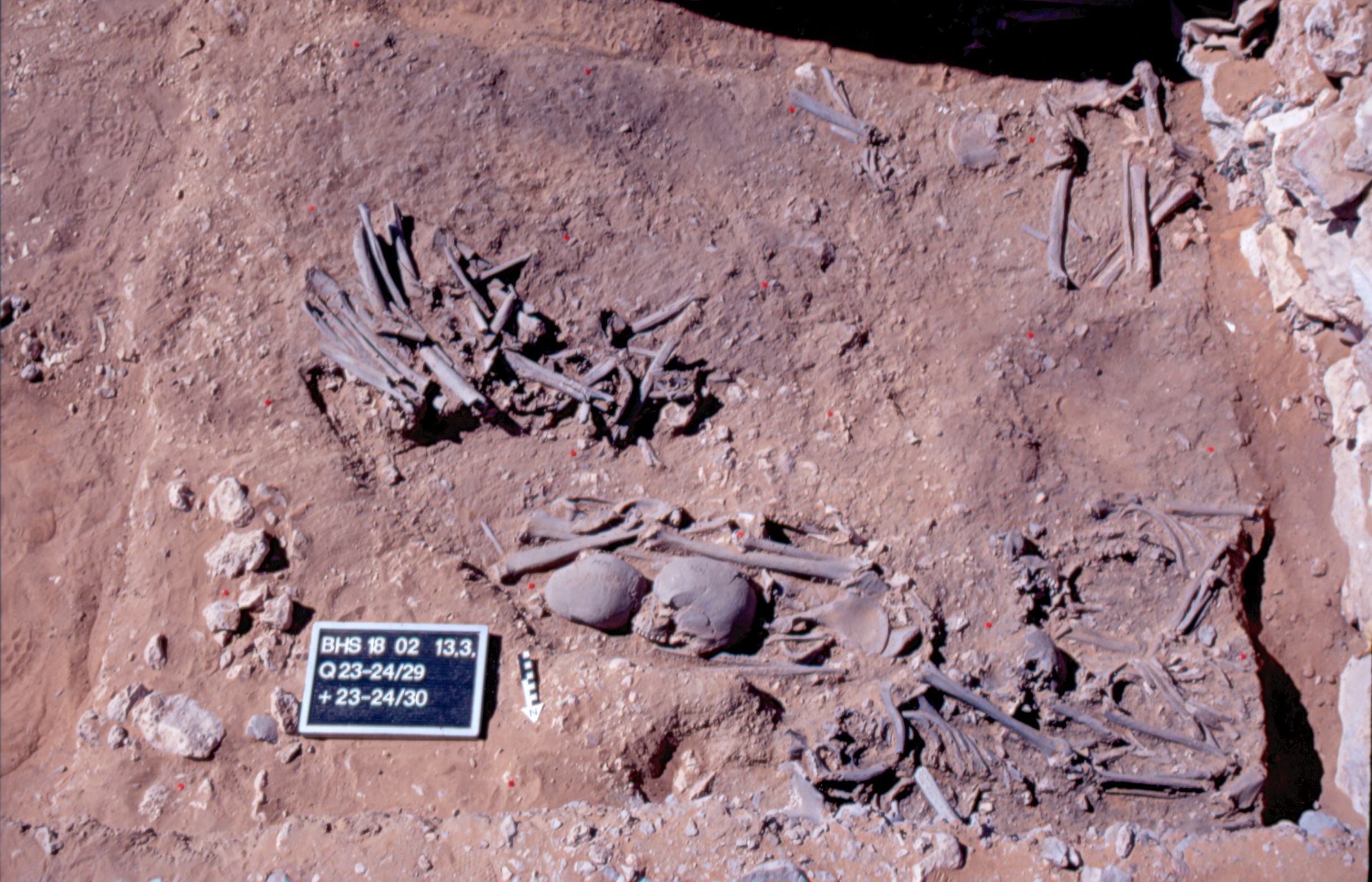
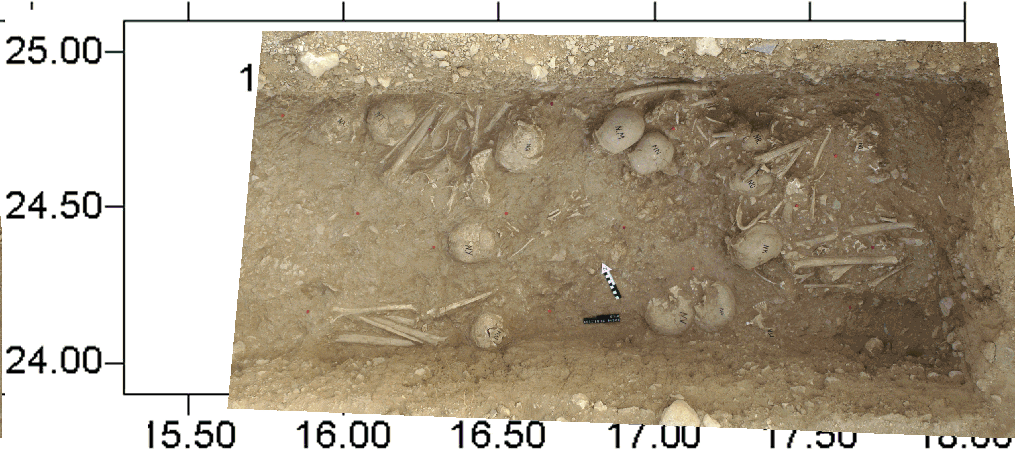
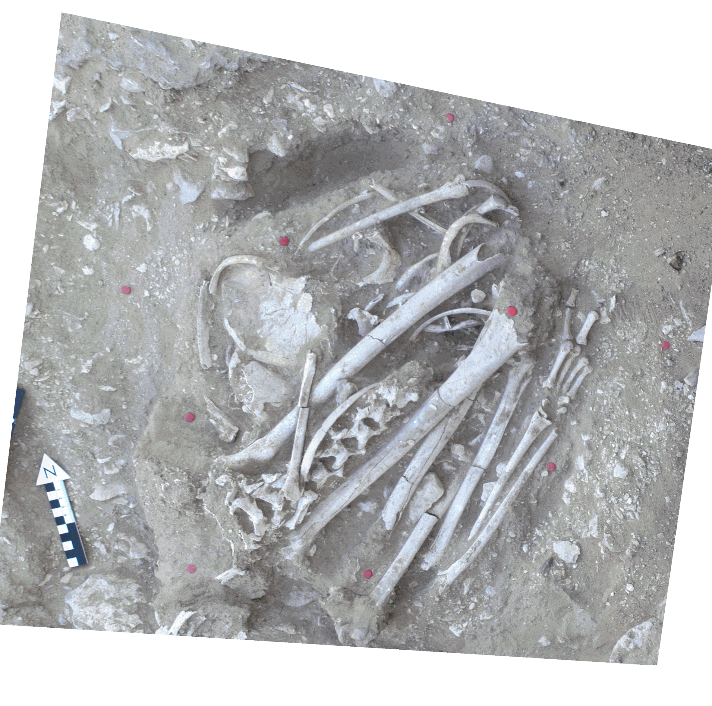
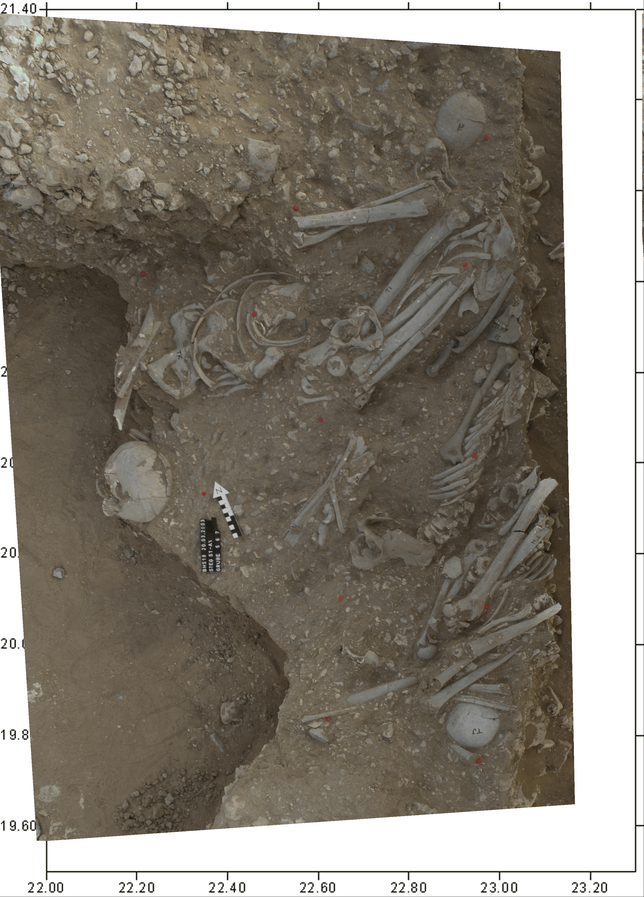
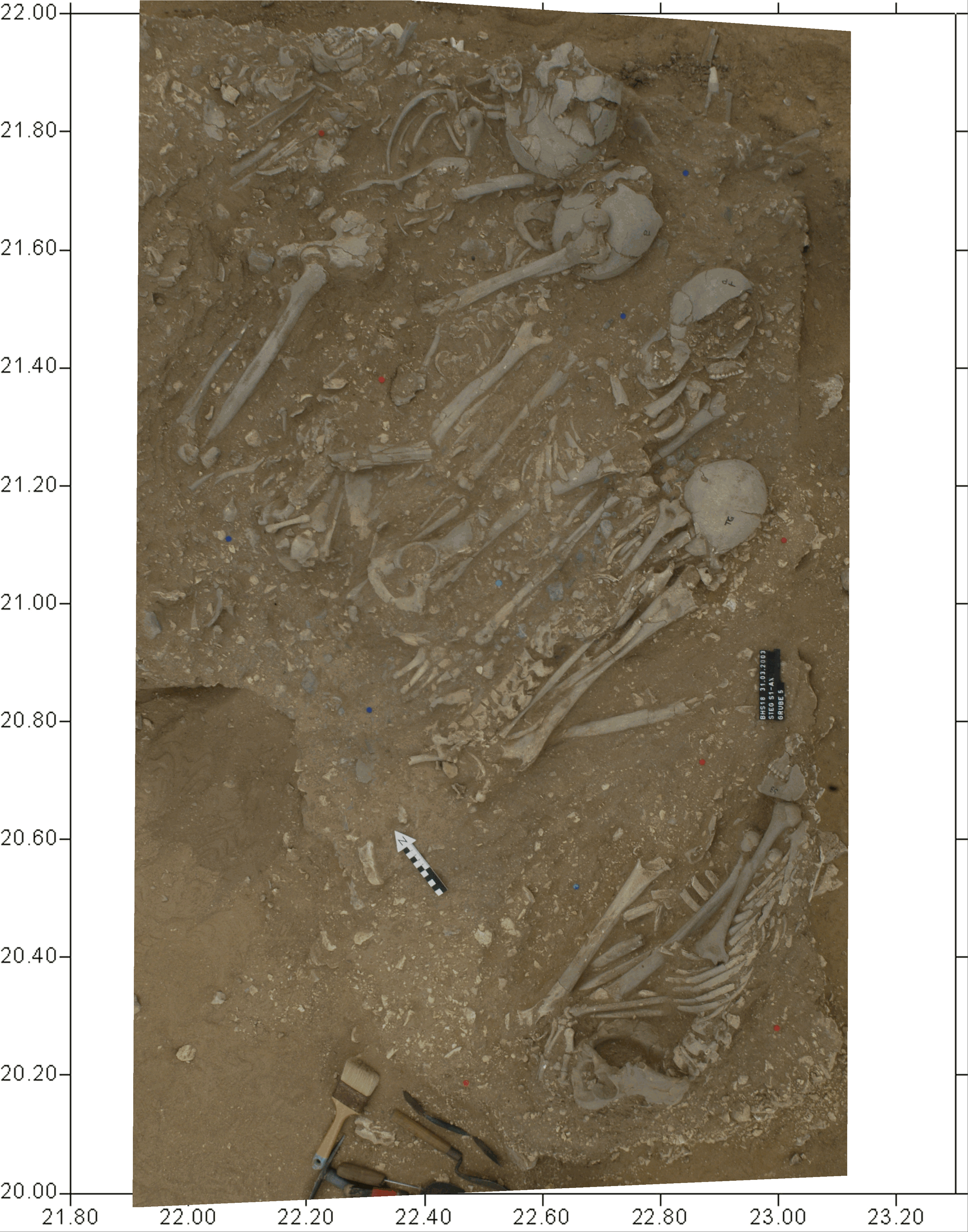
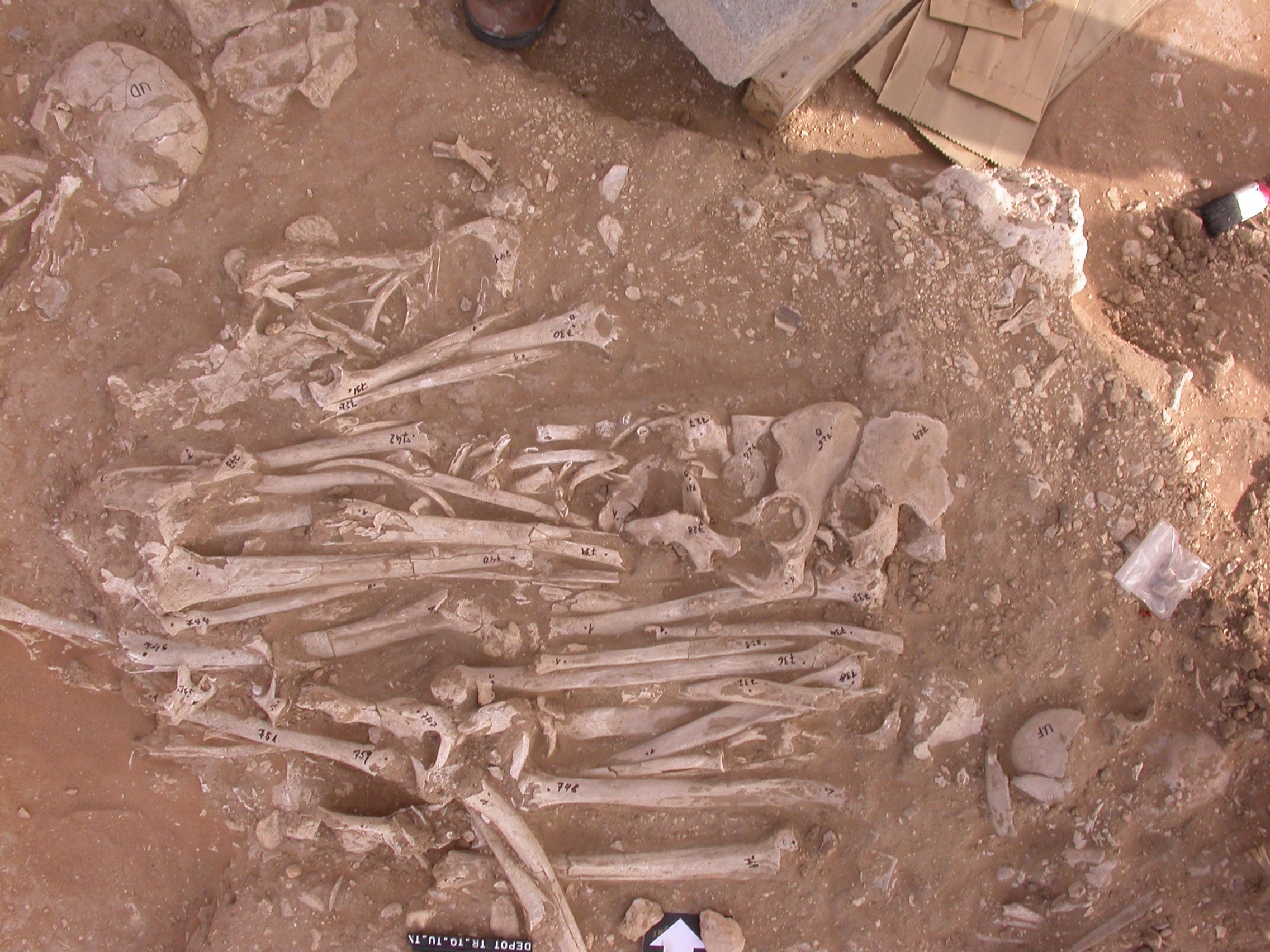
Inidvidual AK:
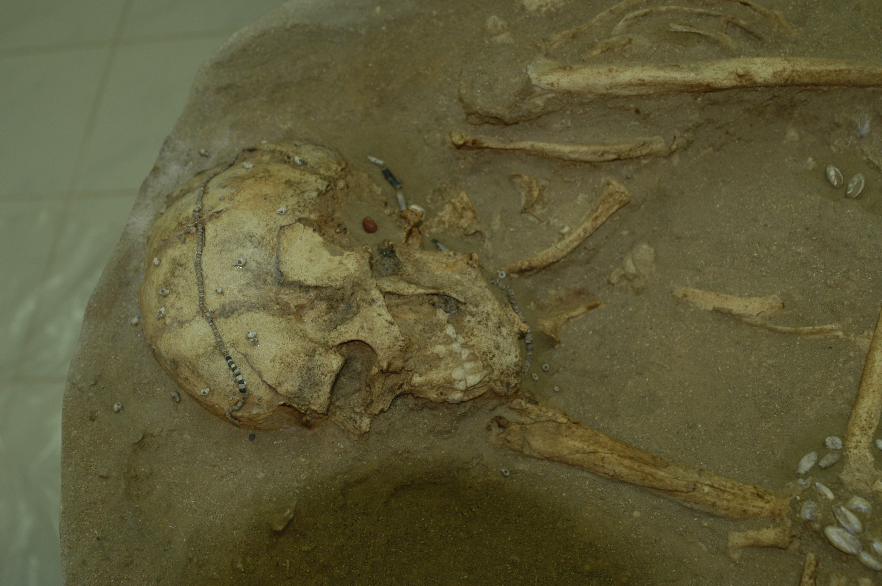
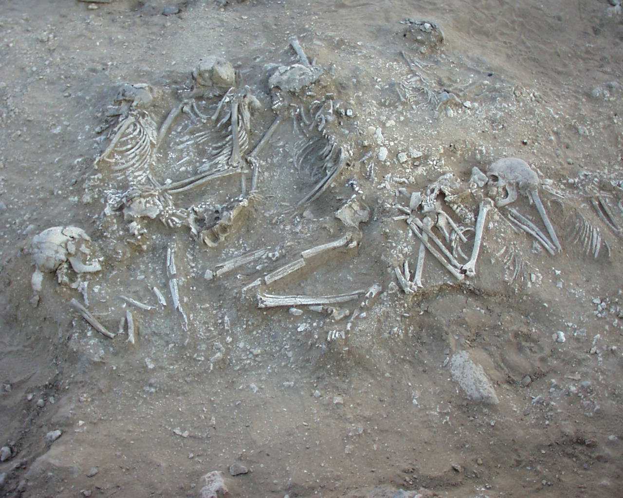
Multiple burial of 5 individuals, in situ and drawing
Full Resolution
Full Resolution

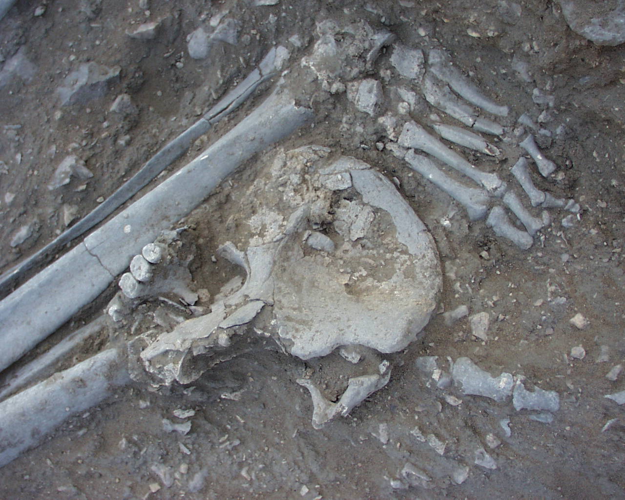
Inidvidual AL:
Multiple burial
Full Resolution
Full Resolution
Inidvidual AM:
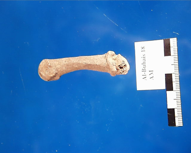
Individual AM with arthrotic right Metacarpus proximal
Full Resolution
Full Resolution
Inidvidual AM:
Mandible AM with intra-vitam tooth loss of tooth 45 and tooth attrition
Full Resolution
Full Resolution
Inidvidual AM:
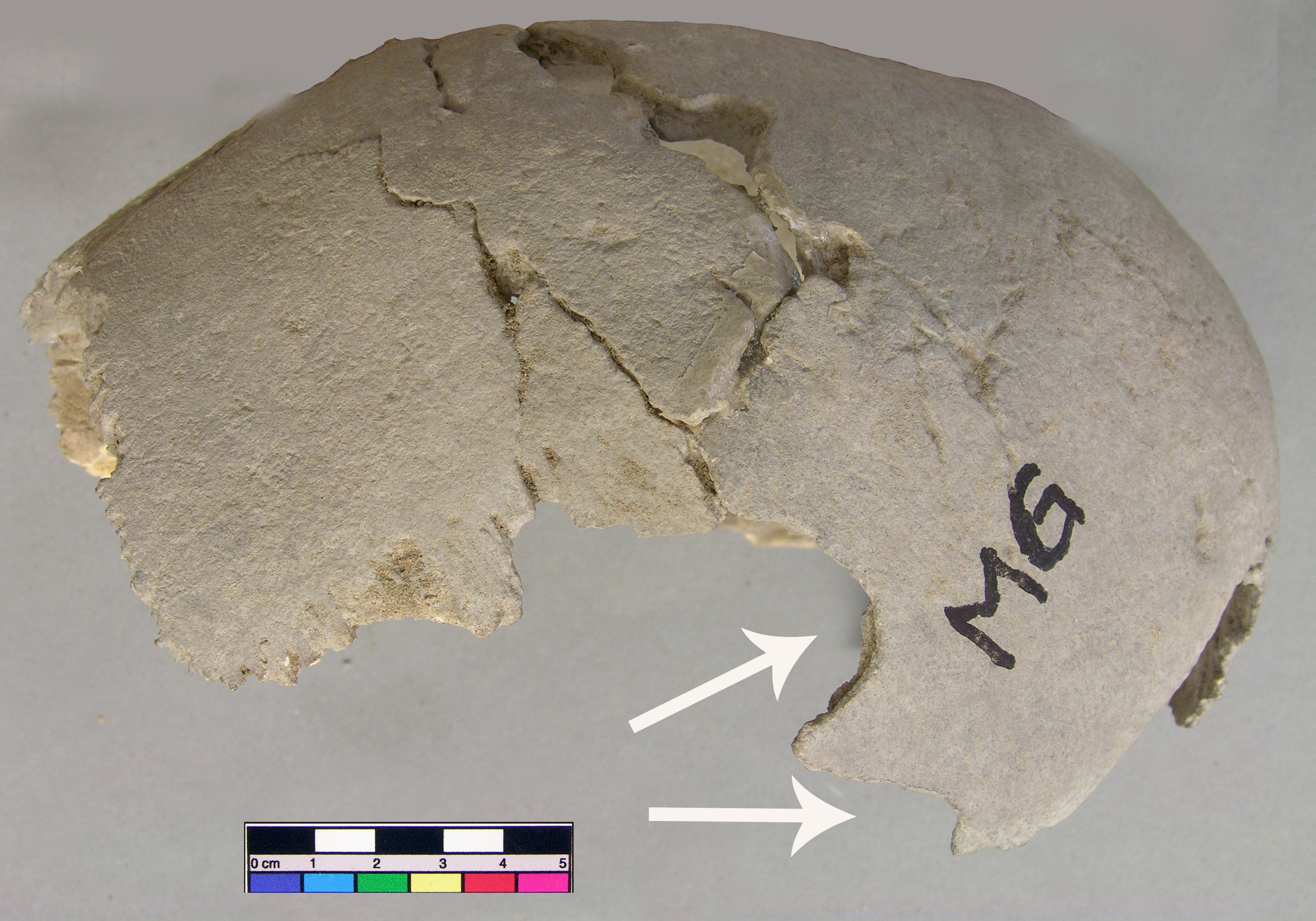
Unhealed fracture on the left parietal tuber caused by blunt trauma
Full Resolution
Full Resolution
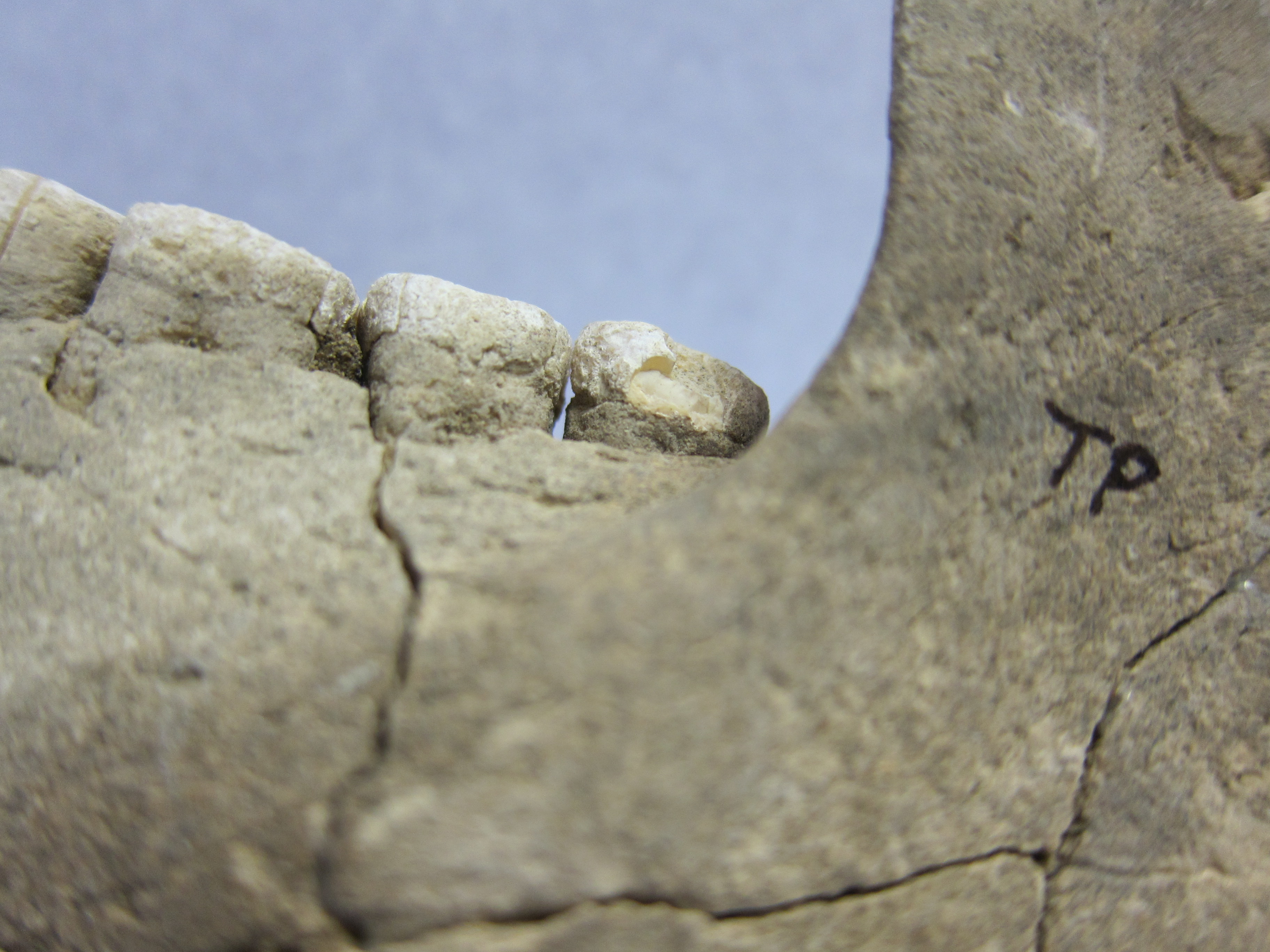
Inidvidual AV:
Arthrotic fovea dentis, individual AV
Full Resolution
Full Resolution
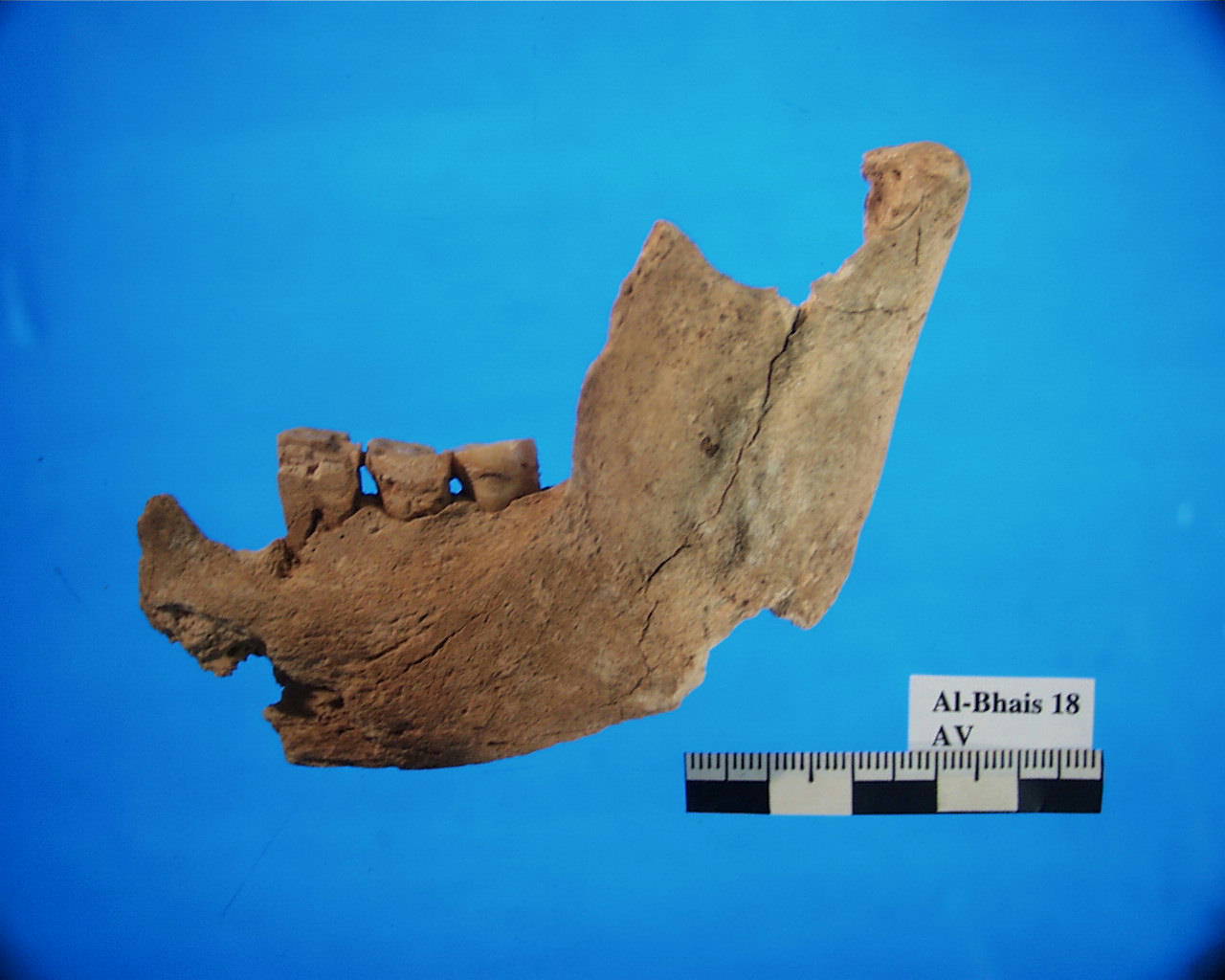
Inidvidual AV:
Mandible AV with cyst
Full Resolution
Full Resolution
Inidvidual AW:
left parietal bone with large unhealed perimortem fracture caused by blunt trauma. Due to the fragmentary preservation of the skull, its full extent cannot be examined.
Full Resolution
Full Resolution
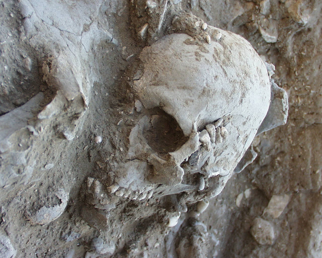
Inidvidual AY:
Skull AY with Porotic Hyperostosis
Full Resolution
Full Resolution
Inidvidual AY:
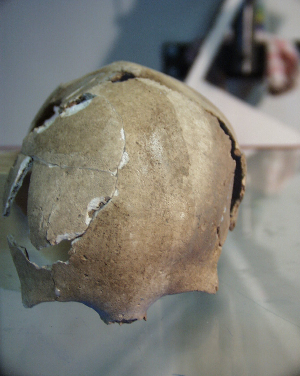
Scaphocephalic skull AY
Full Resolution
Full Resolution
Inidvidual AY:
Sample for REM
Full Resolution
Full Resolution
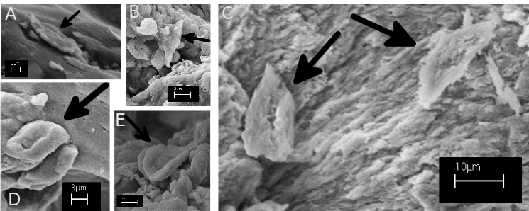
Inidvidual AY:
Unpublished SEM-images of possible sickle-cells detected in the marrow cavity of a rib of infant AY from Neolithic BHS18. A-C: possible examples of flat and elongated sickle cells. D: possible thicker and more oval shaped sickle-cell. E: possible target-cell-erythrocyte.
Full Resolution
Full Resolution
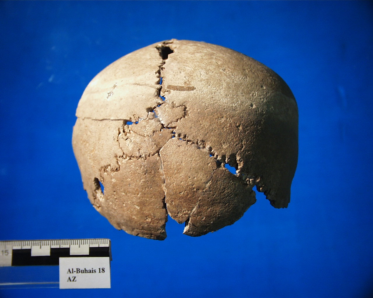
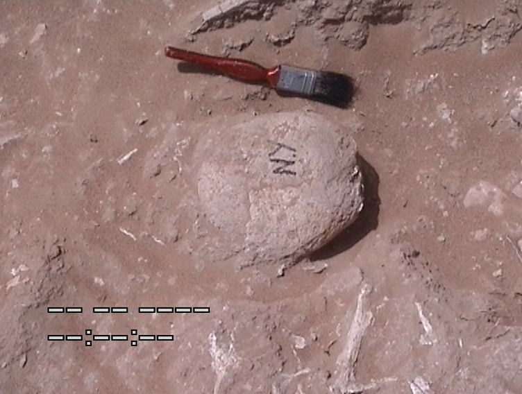
Inidvidual AZ:
Ossicula lambdoidea of individual AZ
Full Resolution
Full Resolution
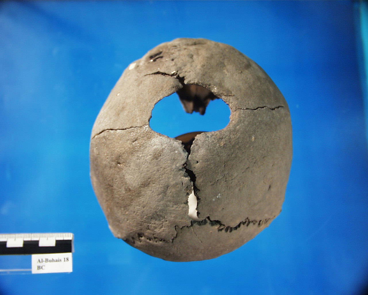
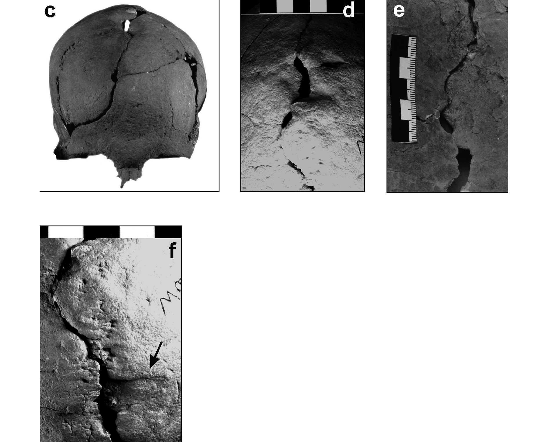
Inidvidual BC:
Skull BC with Trepanation, Trephination, Trepanning
Full Resolution
Full Resolution

Inidvidual BC:
Skull BC with Trepanation, Trephination, Trepanning
Full Resolution
Full Resolution
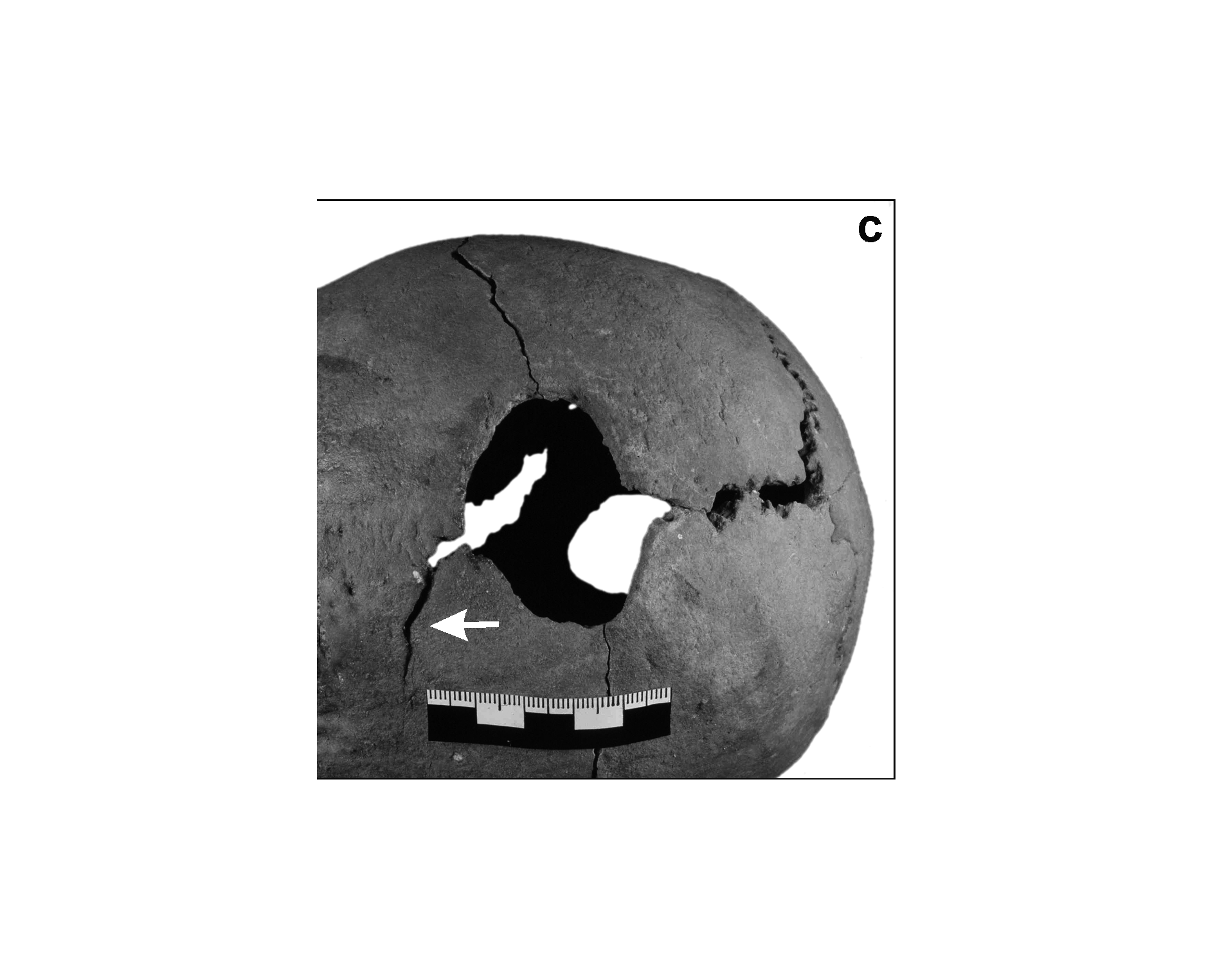
Inidvidual BC:
Trepanation
Full Resolution
Full Resolution
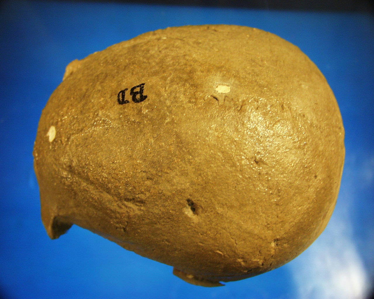
Inidvidual BD:
Skull BD with linear impression
Full Resolution
Full Resolution
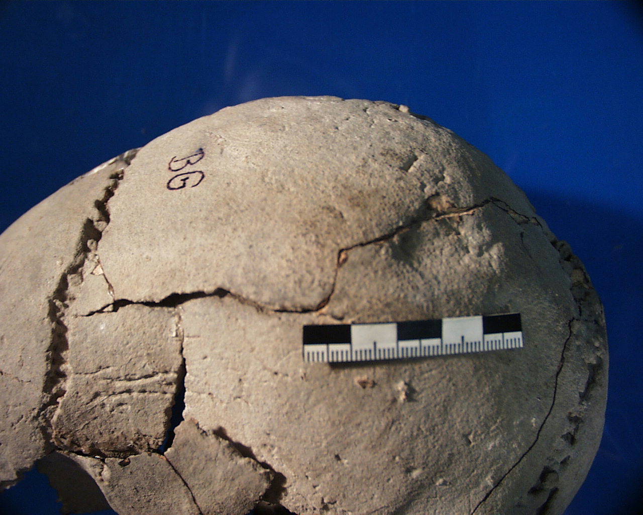
Inidvidual BG:
Skull BG with impression fracture
Full Resolution
Full Resolution
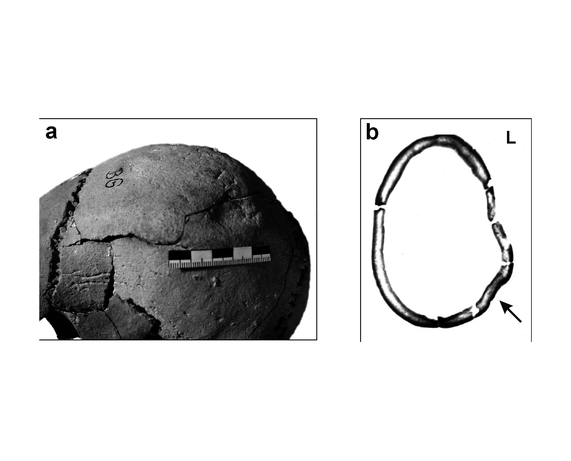
Inidvidual BG:
Skull with fracture, CT-scan
Full Resolution
Full Resolution
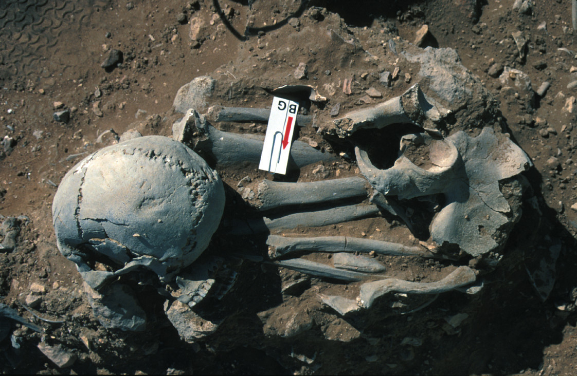
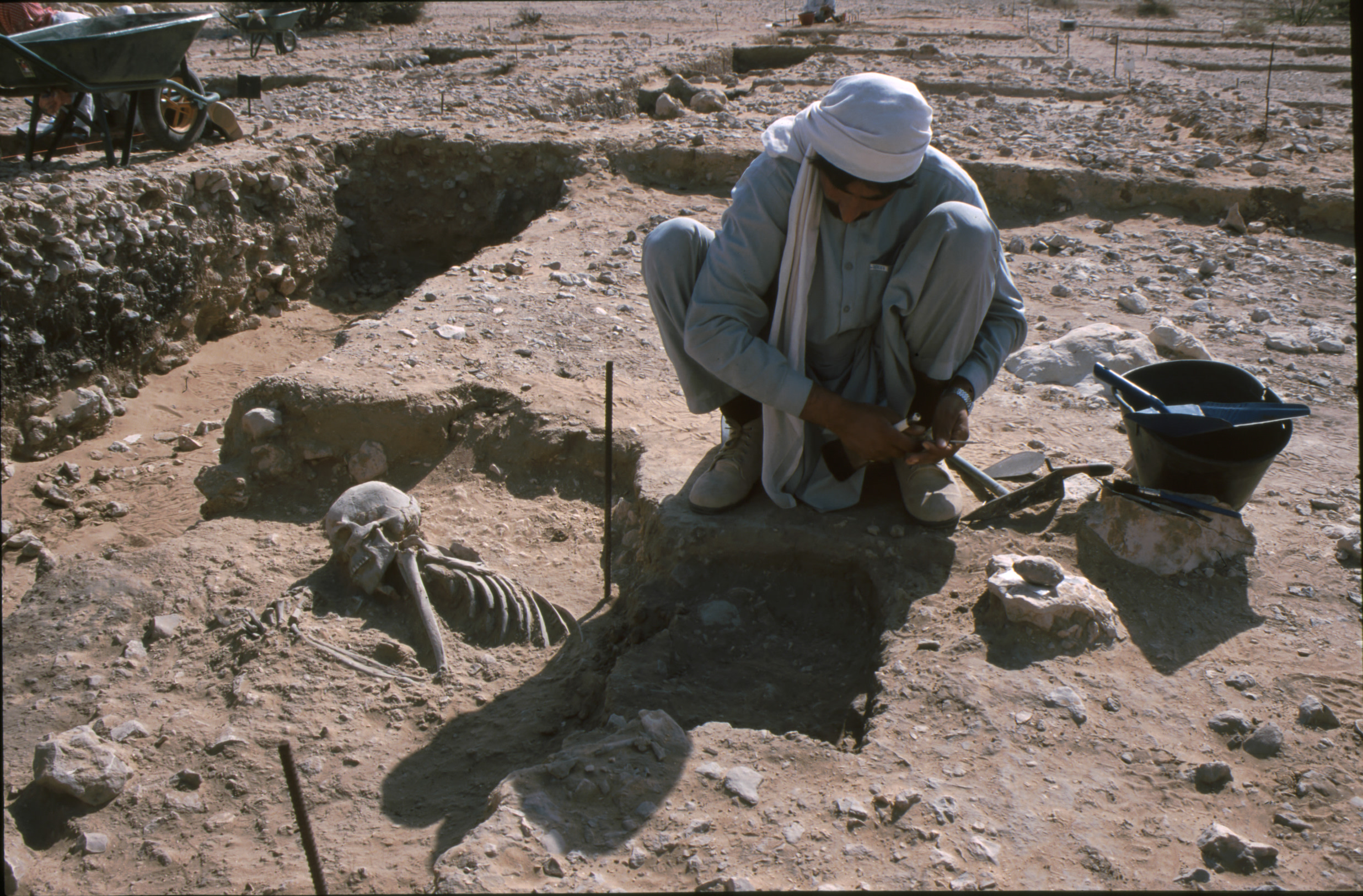
Inidvidual BG:
BG in situ
Full Resolution
Full Resolution
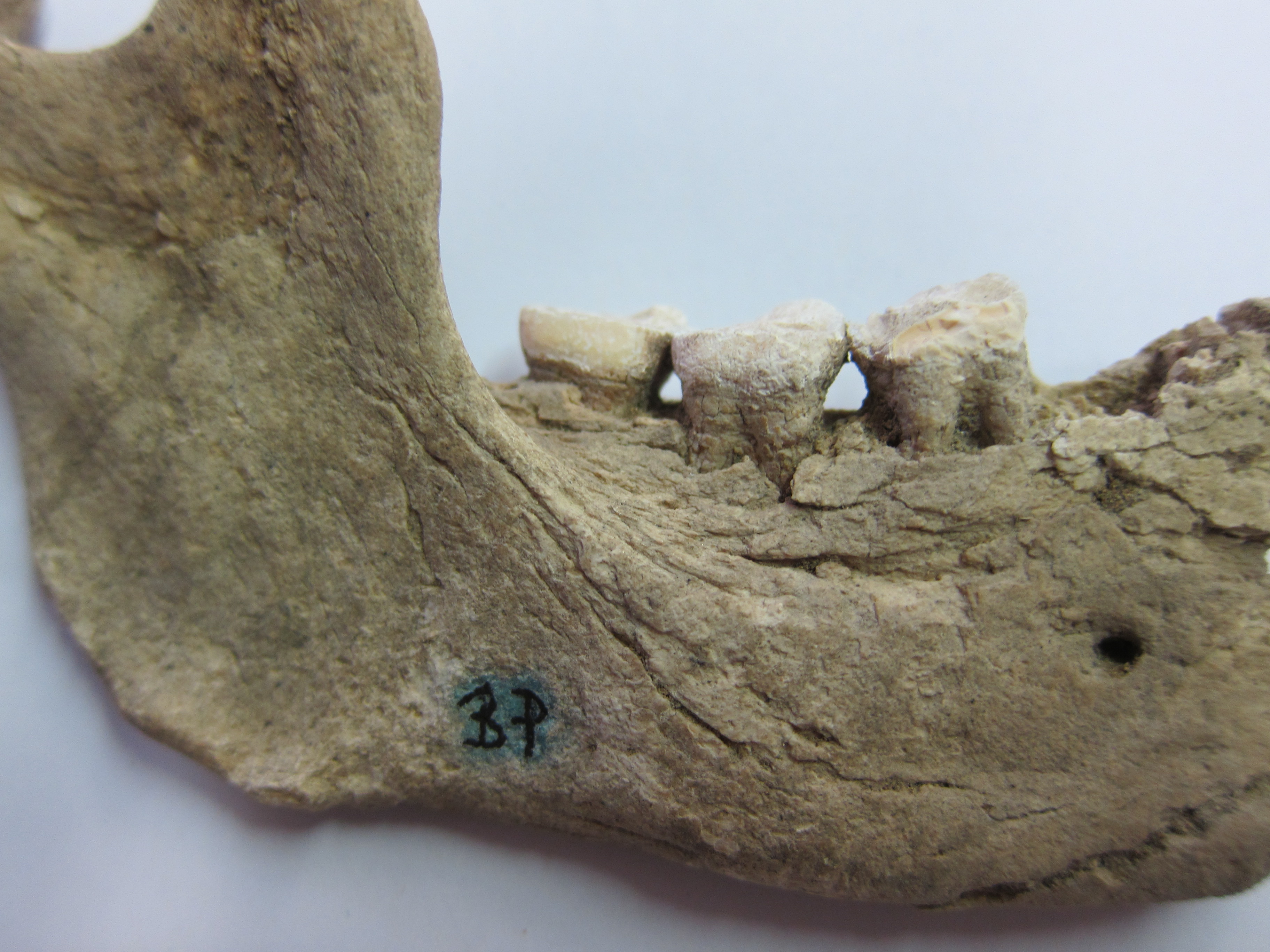
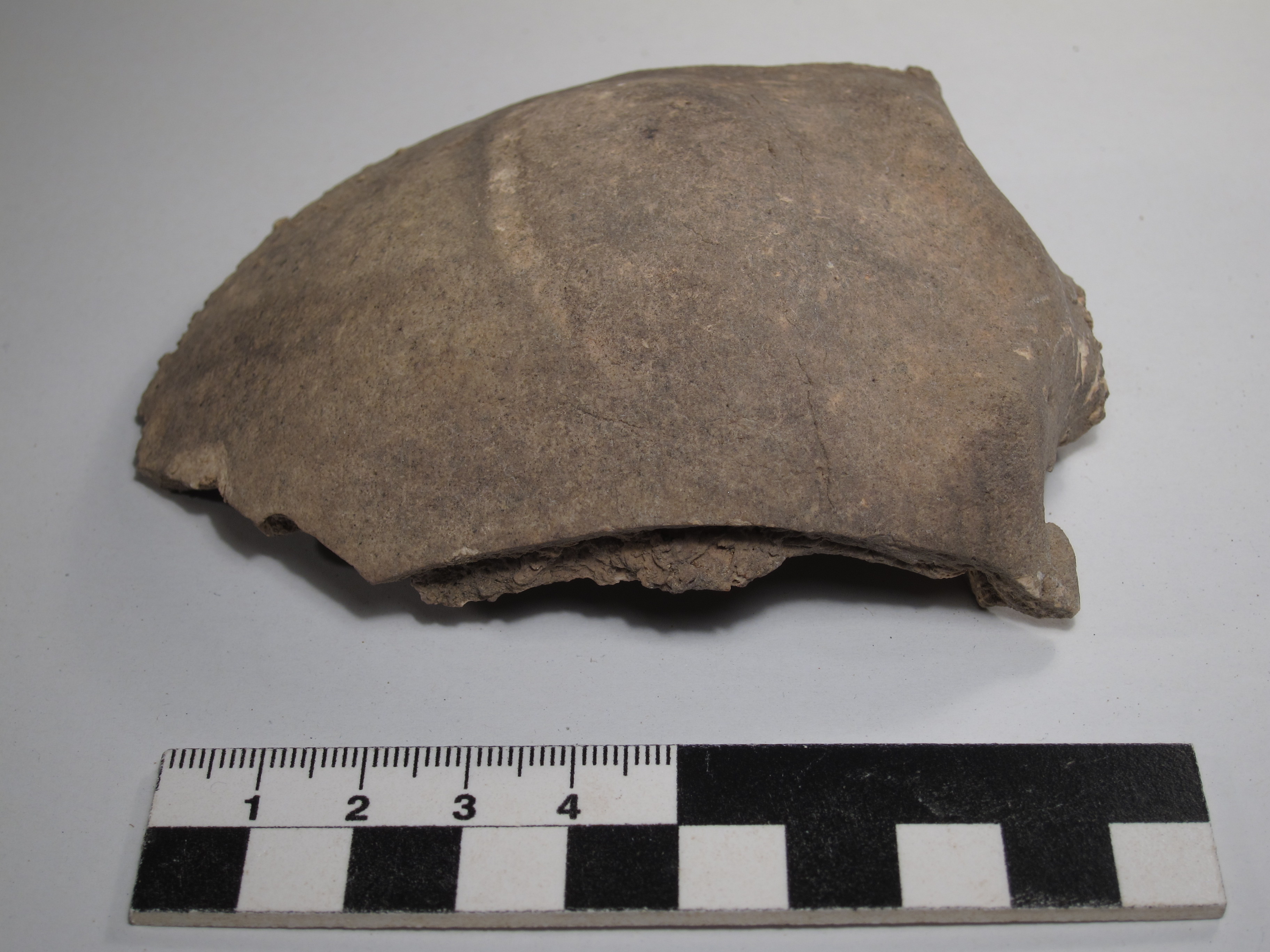
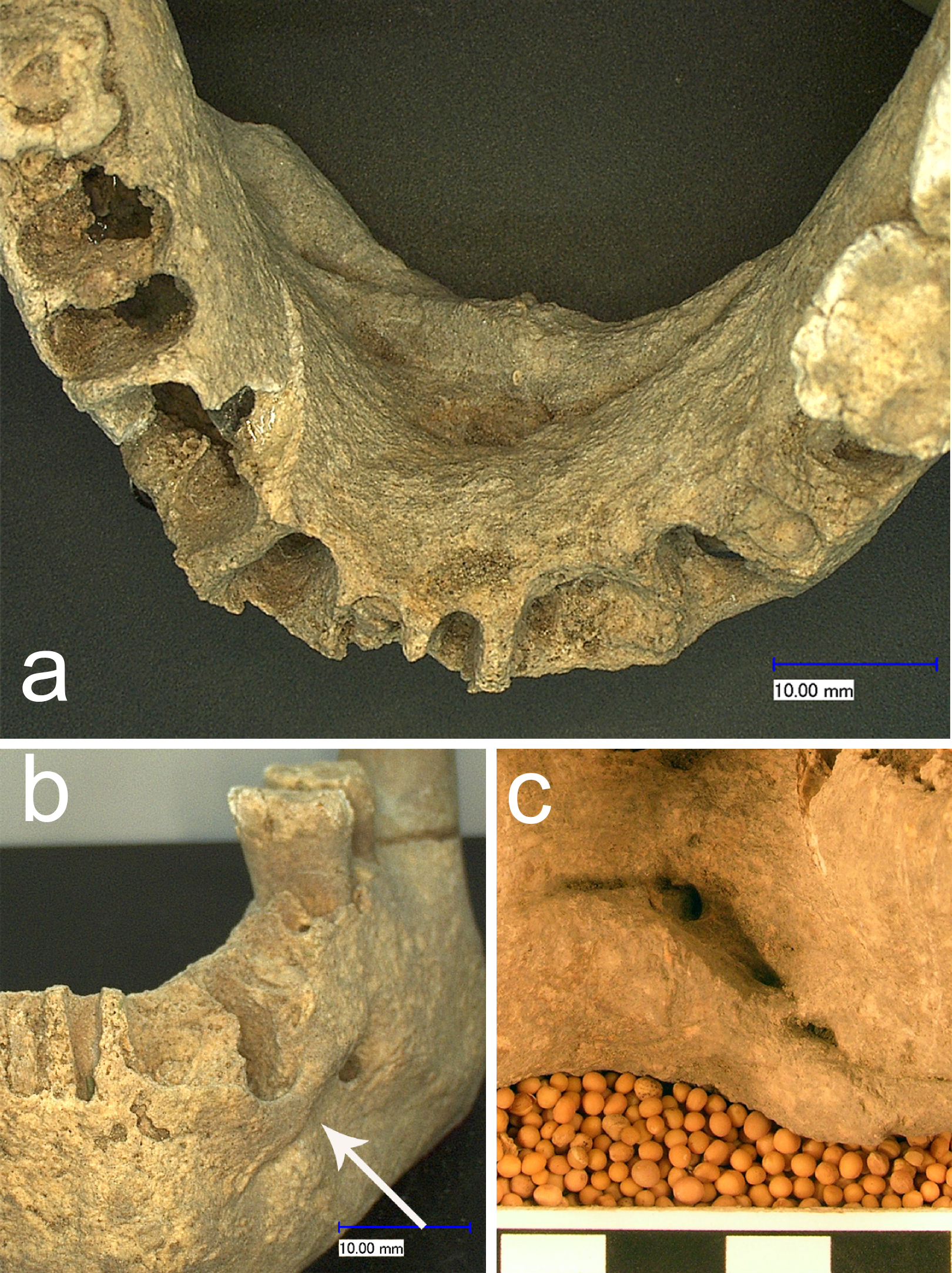
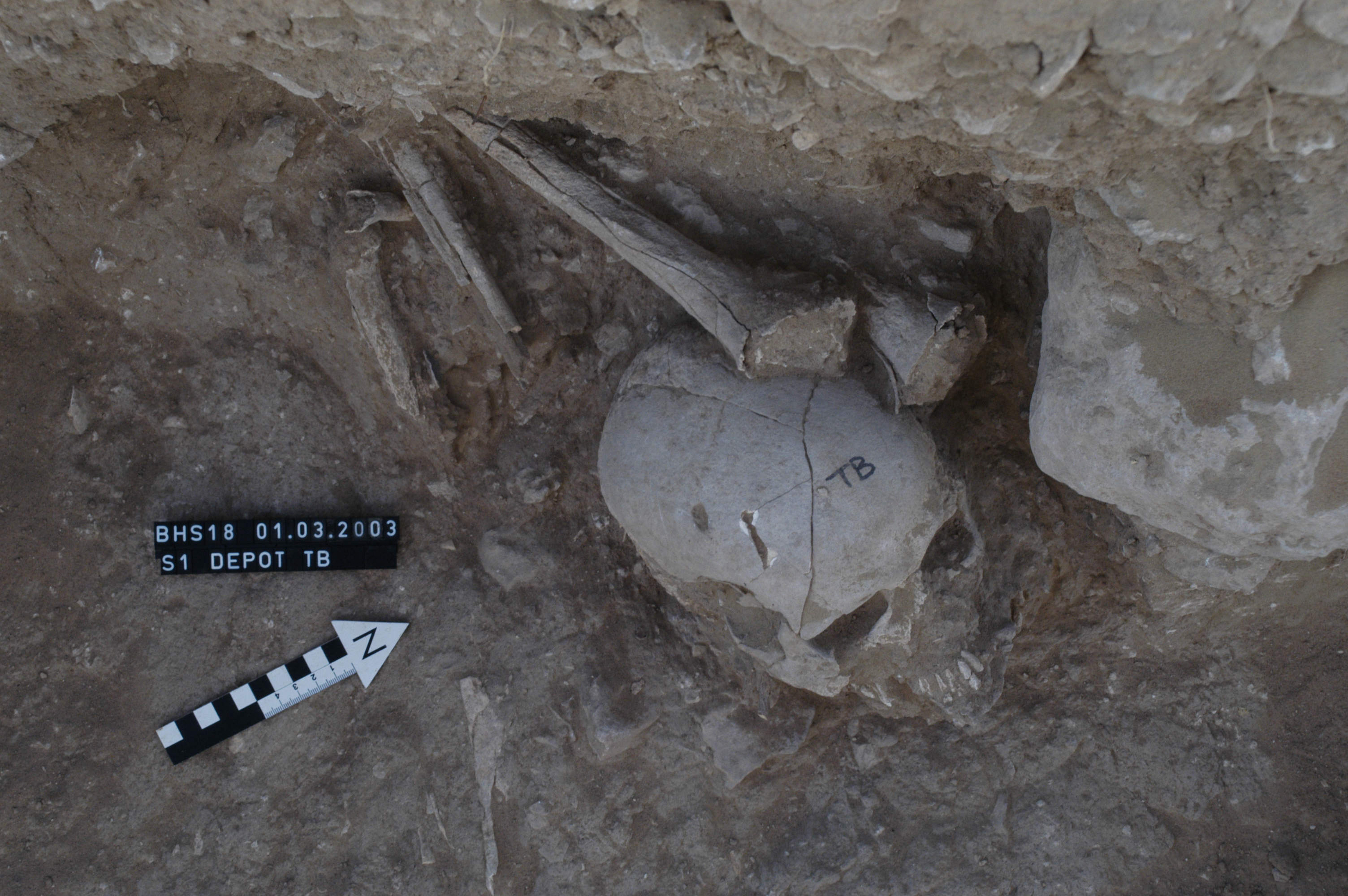
BP mandible
Full Resolution
Full Resolution
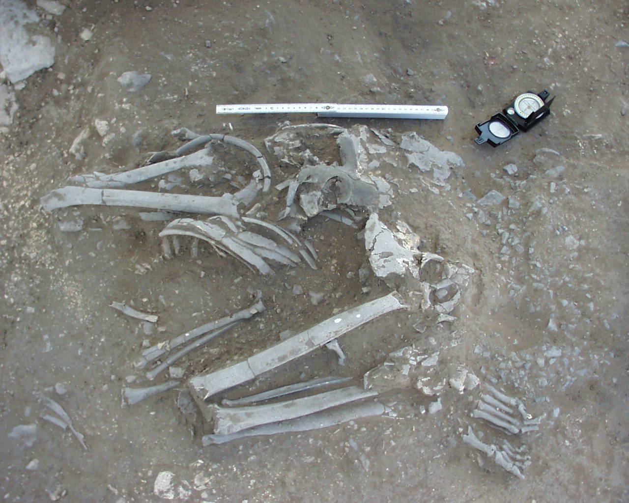
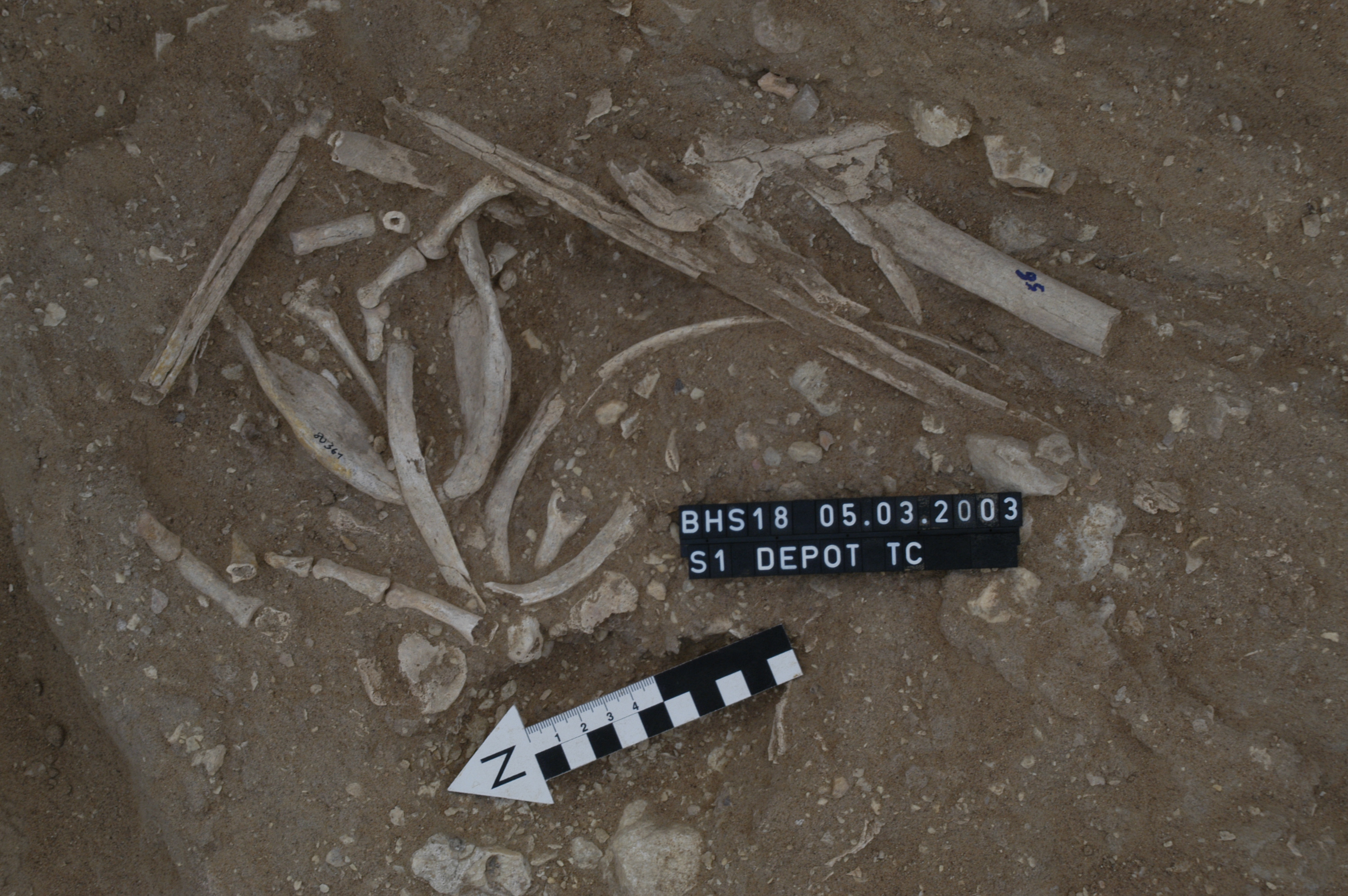
Inidvidual BS:
Burial in situ
Full Resolution
Full Resolution
Inidvidual BS:
BS in situ with fracture at the left parietal bone
Full Resolution
Full Resolution
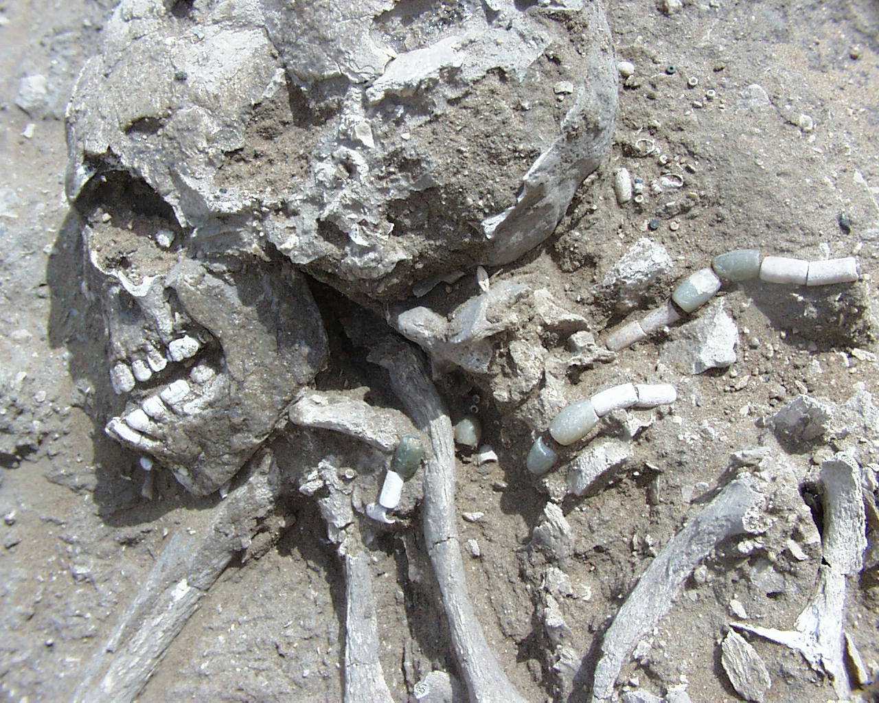
Inidvidual BX:
BX with pearl at the chin
Full Resolution
Full Resolution

Inidvidual BX:
BX, BY, HS and CL in situ
Full Resolution
Full Resolution

Inidvidual BX:
BX, BY, HS, CL, KG and KE in situ
Full Resolution
Full Resolution
Inidvidual BX:
Skull BX in situ with adornments
Full Resolution
Full Resolution
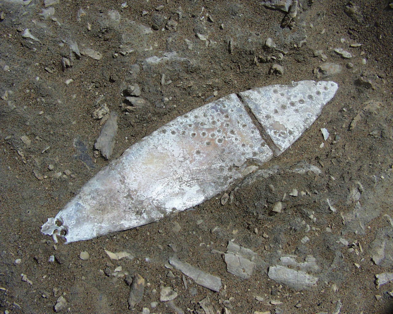
Inidvidual BX:
Pendant made of mother of pearl
Full Resolution
Full Resolution
Inidvidual BY:
BX, BY, HS and CL in situ
Full Resolution
Full Resolution

Inidvidual BY:
BX, BY, HS, CL, KG and KE in situ
Full Resolution
Full Resolution
Inidvidual BY:
Skull fragment BY
Full Resolution
Full Resolution
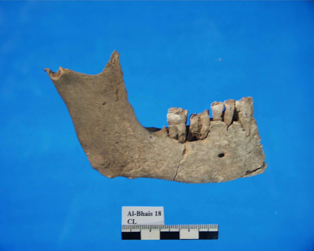
Inidvidual CL:
CL mandible with parodontitis
Full Resolution
Full Resolution
Inidvidual CL:
CL maxilla with Cyst
Full Resolution
Full Resolution

Inidvidual CL:
BX, BY, HS and CL in situ
Full Resolution
Full Resolution

Inidvidual CL:
BX, BY, HS, CL, KG and KE in situ
Full Resolution
Full Resolution
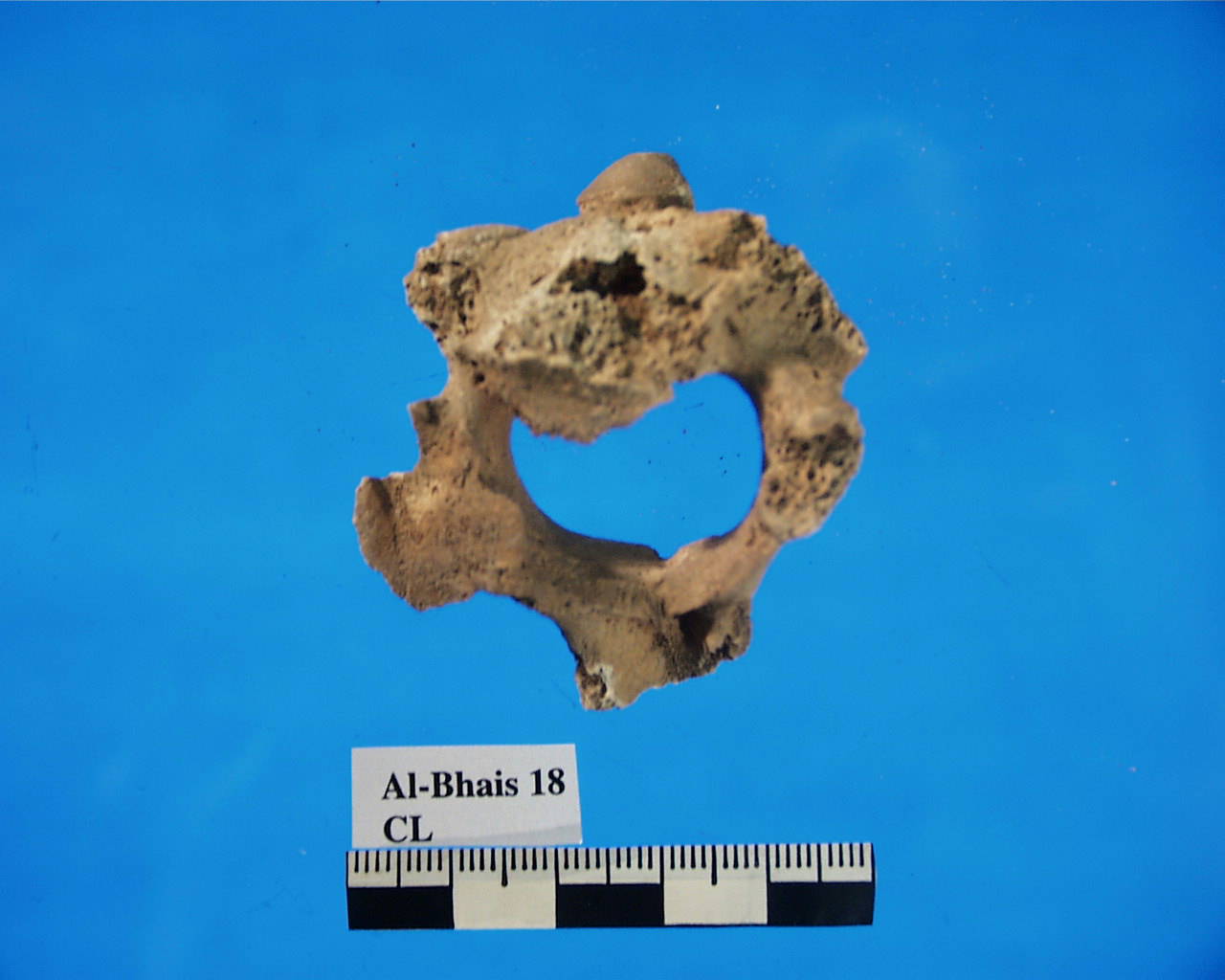
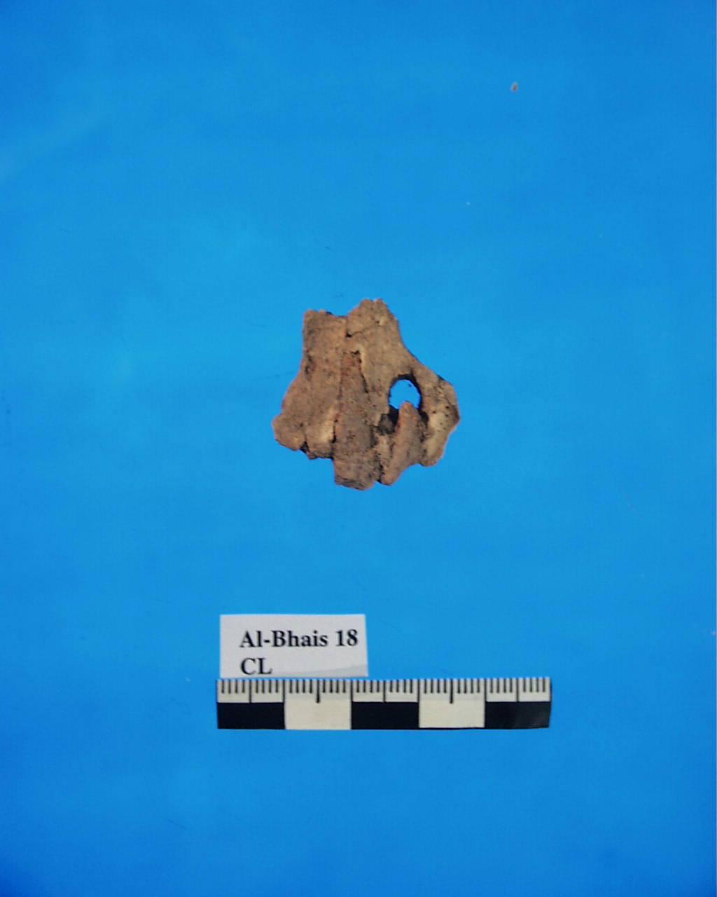
Inidvidual CL:
CL axis
Full Resolution
Full Resolution
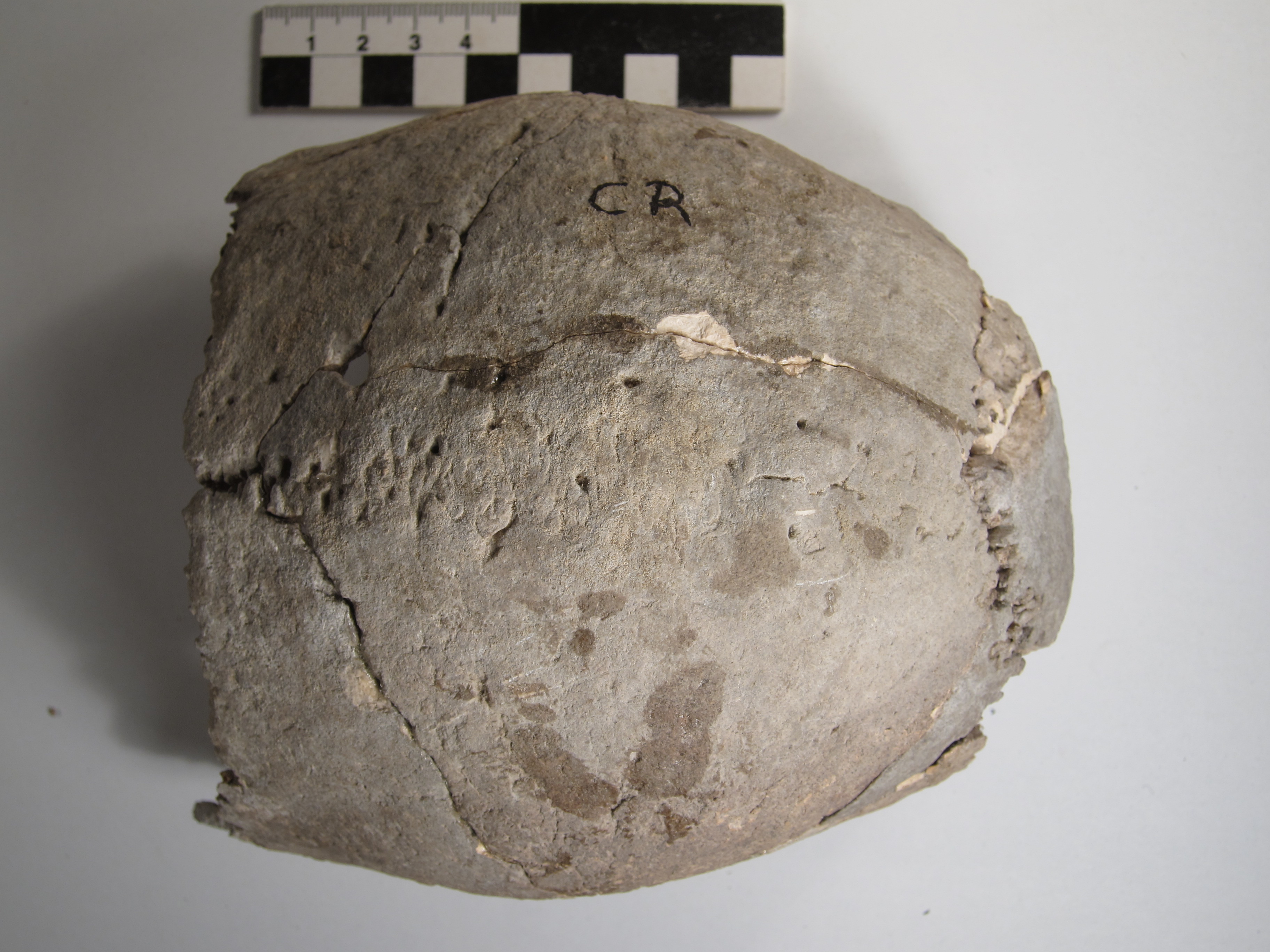
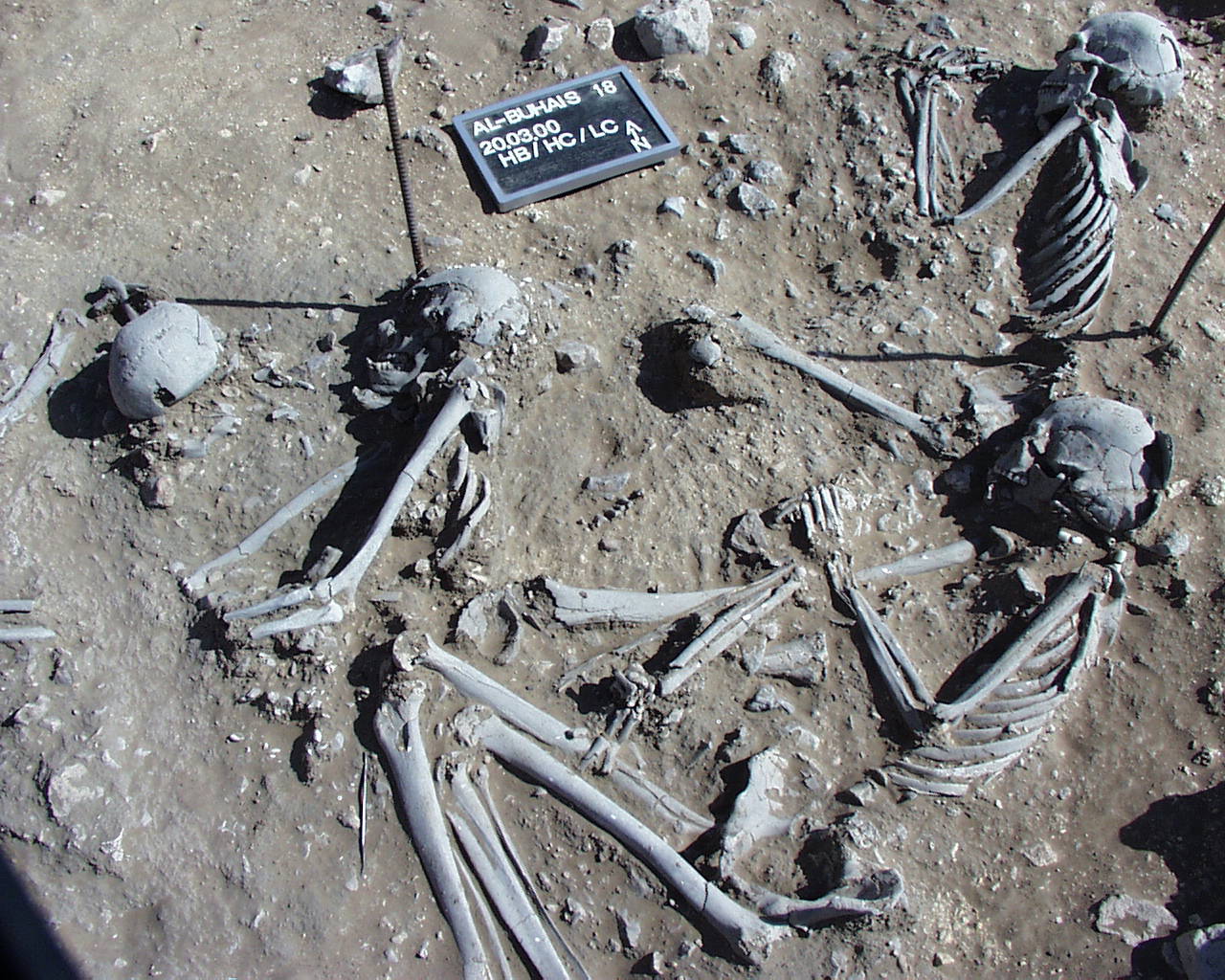
Inidvidual CR:
parietal with a healed depression fracture. Signs of inflammation at the sagittal suture, probably related to the injury.
Full Resolution
Full Resolution
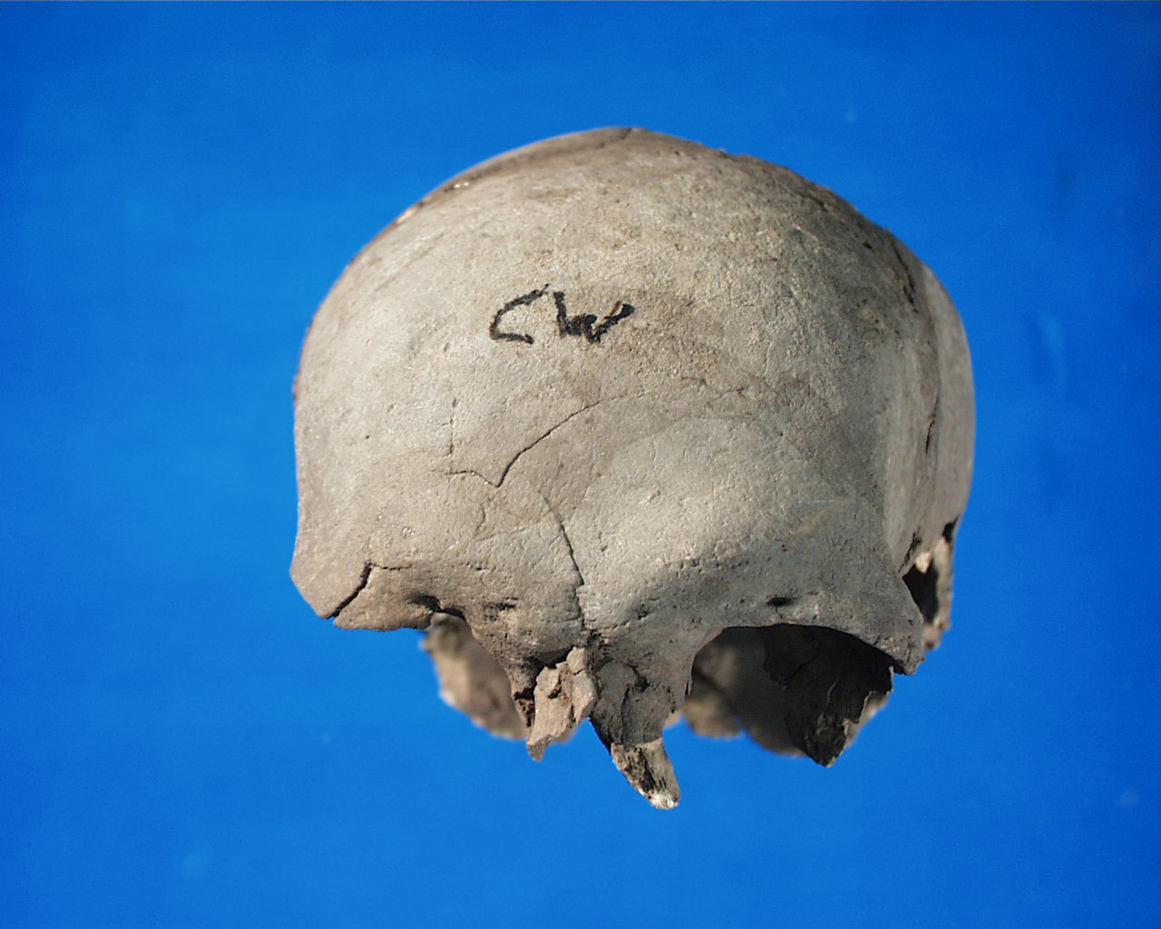
Inidvidual CW:
CW frontal
Full Resolution
Full Resolution
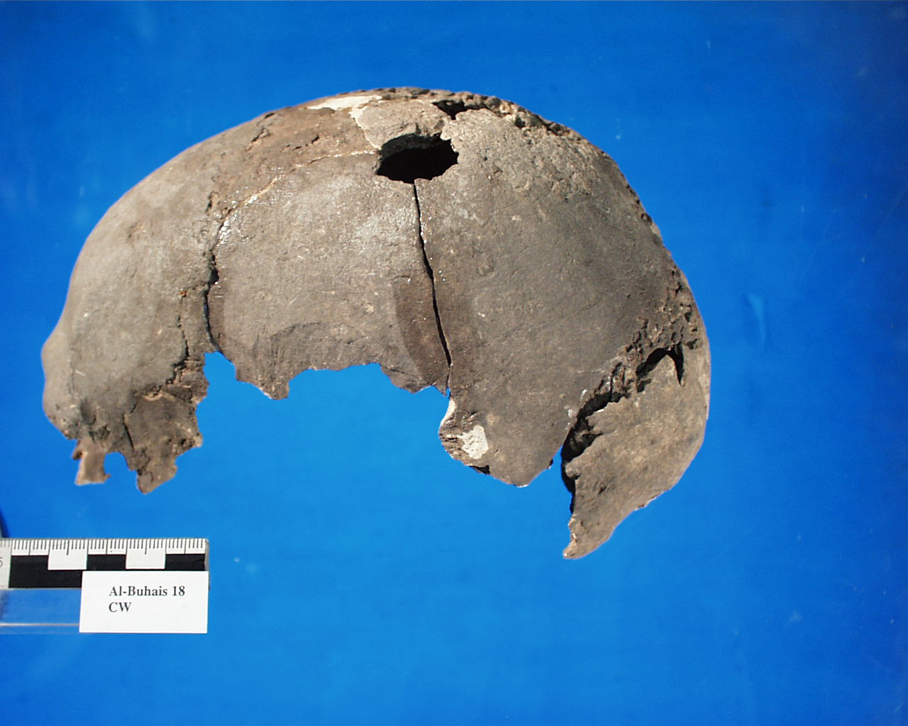
Inidvidual CW:
Skull CW with lethal fracture
Full Resolution
Full Resolution
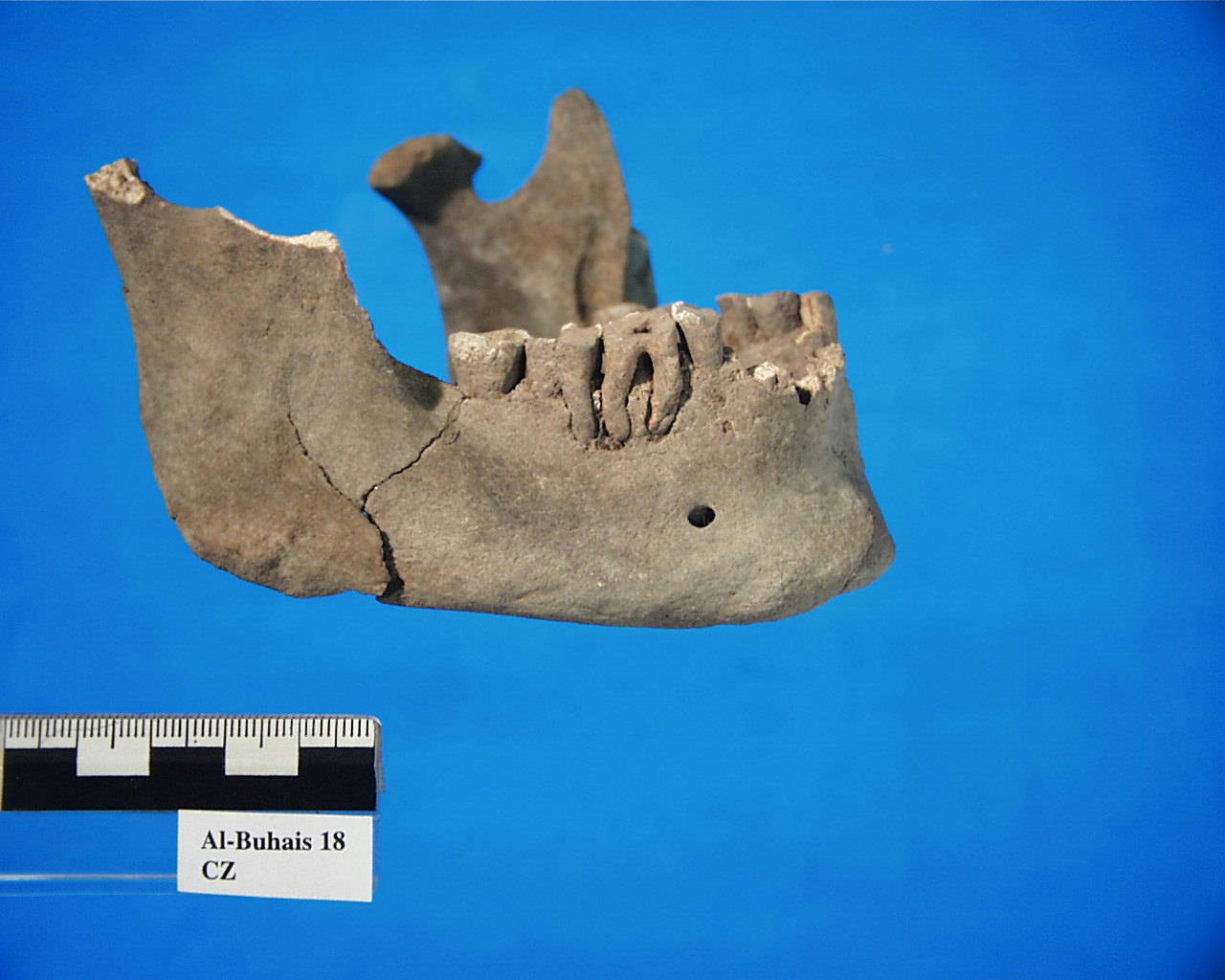
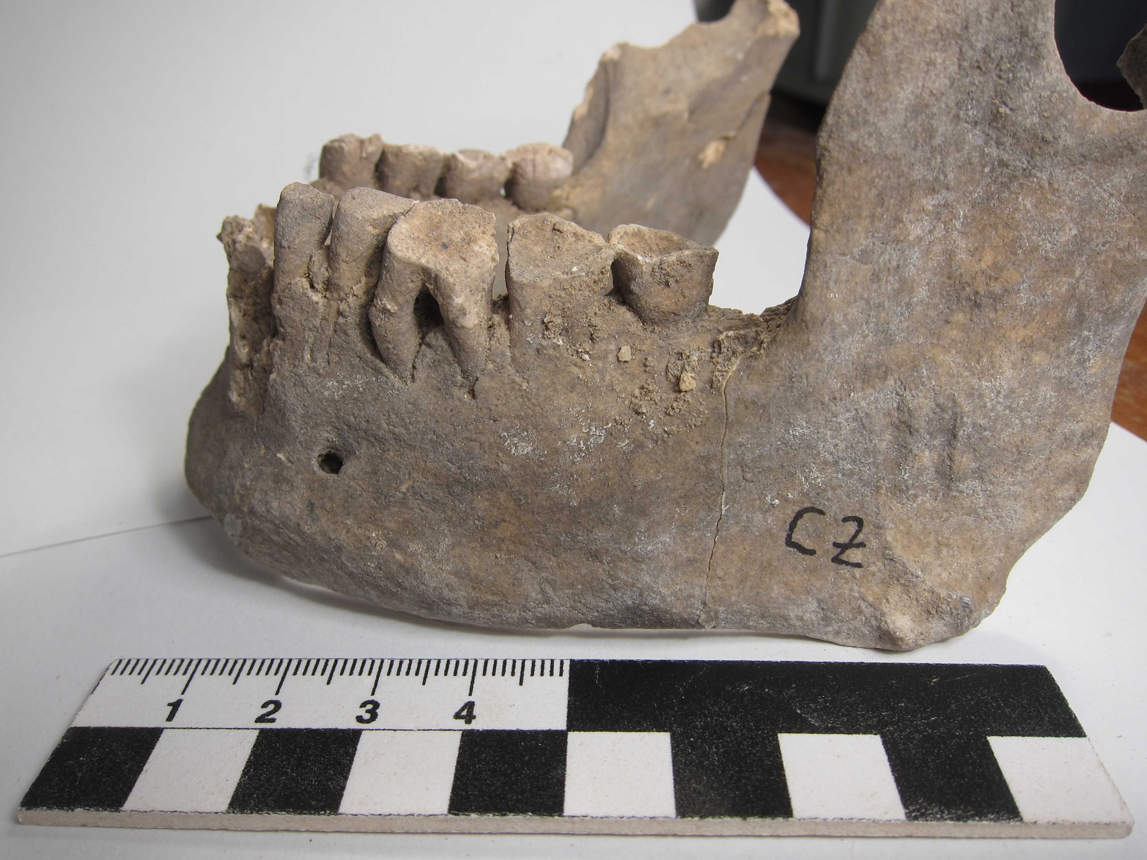
Inidvidual CZ:
Mandible CZ with Parodontitis
Full Resolution
Full Resolution
Inidvidual CZ:
Severe tooth attrition
Full Resolution
Full Resolution
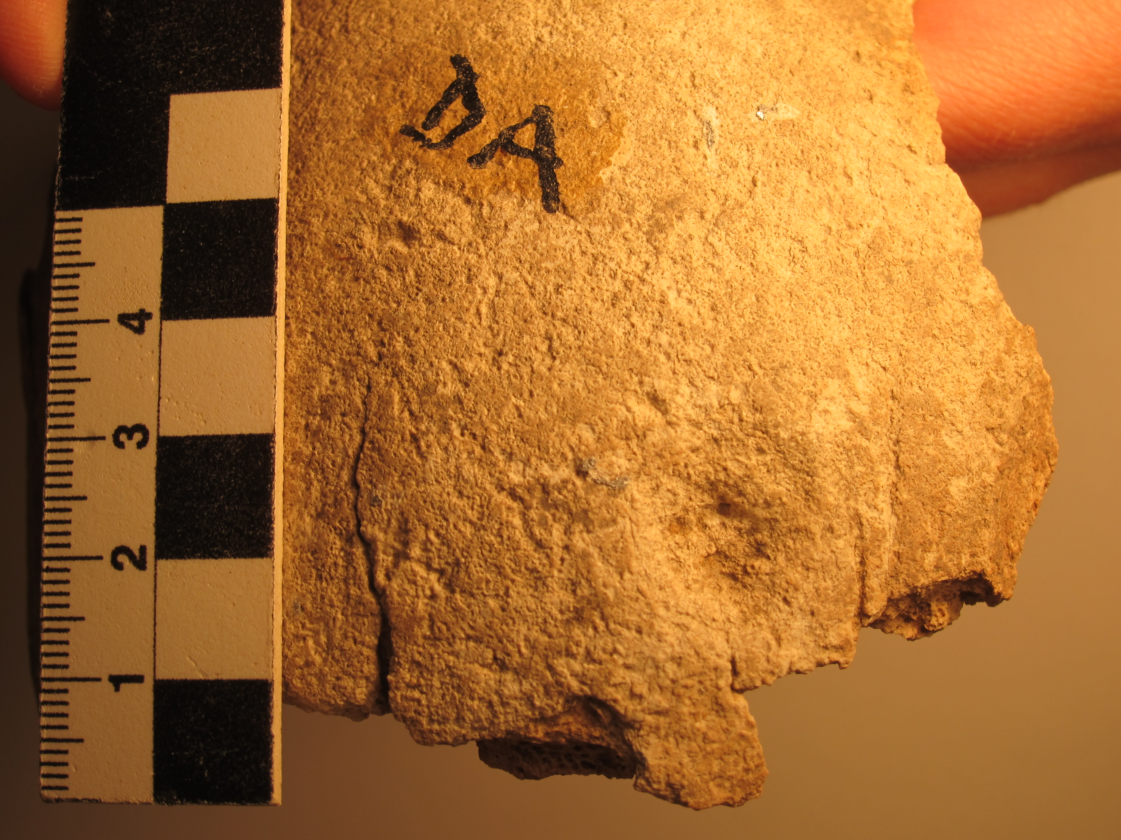
Inidvidual DA:
Healed fracture on frontal bone
Full Resolution
Full Resolution
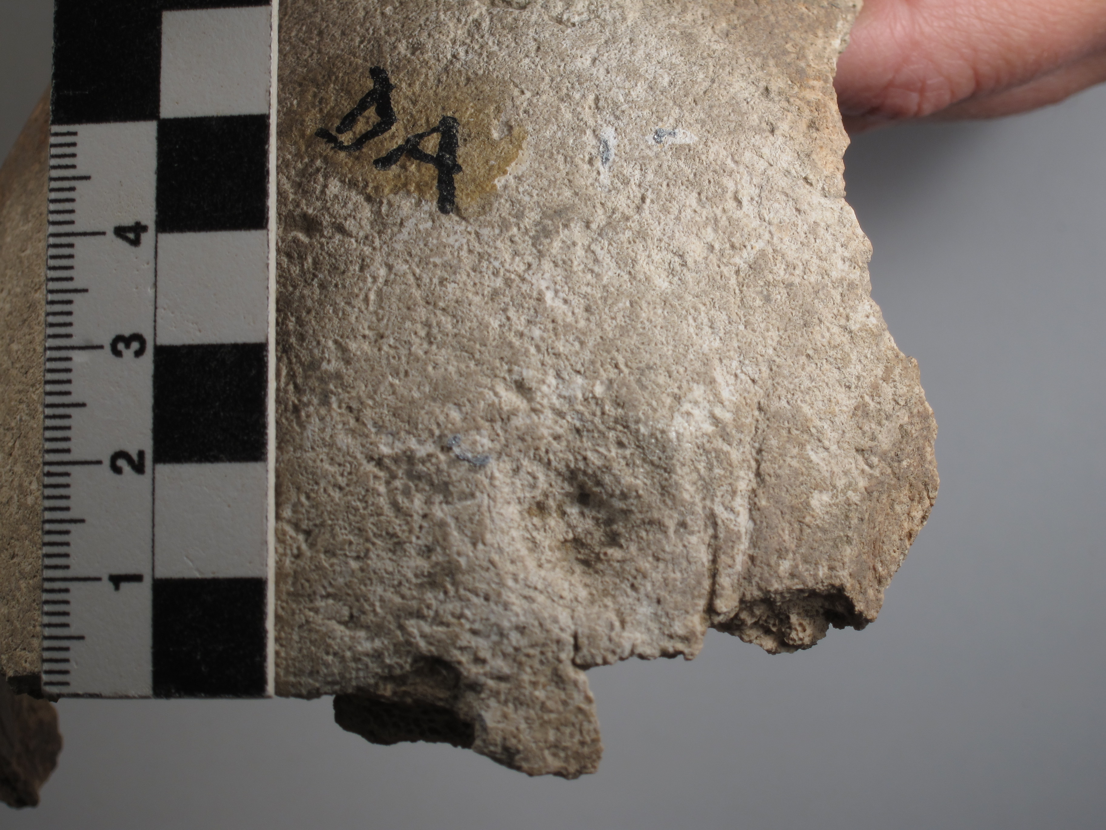
Inidvidual DA:
Healed fracture on frontal bone
Full Resolution
Full Resolution

Inidvidual DD:
Skull DD, lateral right
Full Resolution
Full Resolution
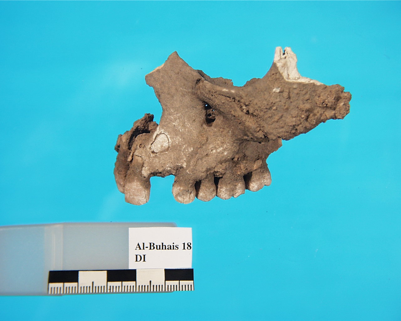
Inidvidual DI:
Retained deciduous tooth in the maxilla, individual DI
Full Resolution
Full Resolution
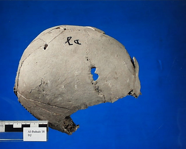
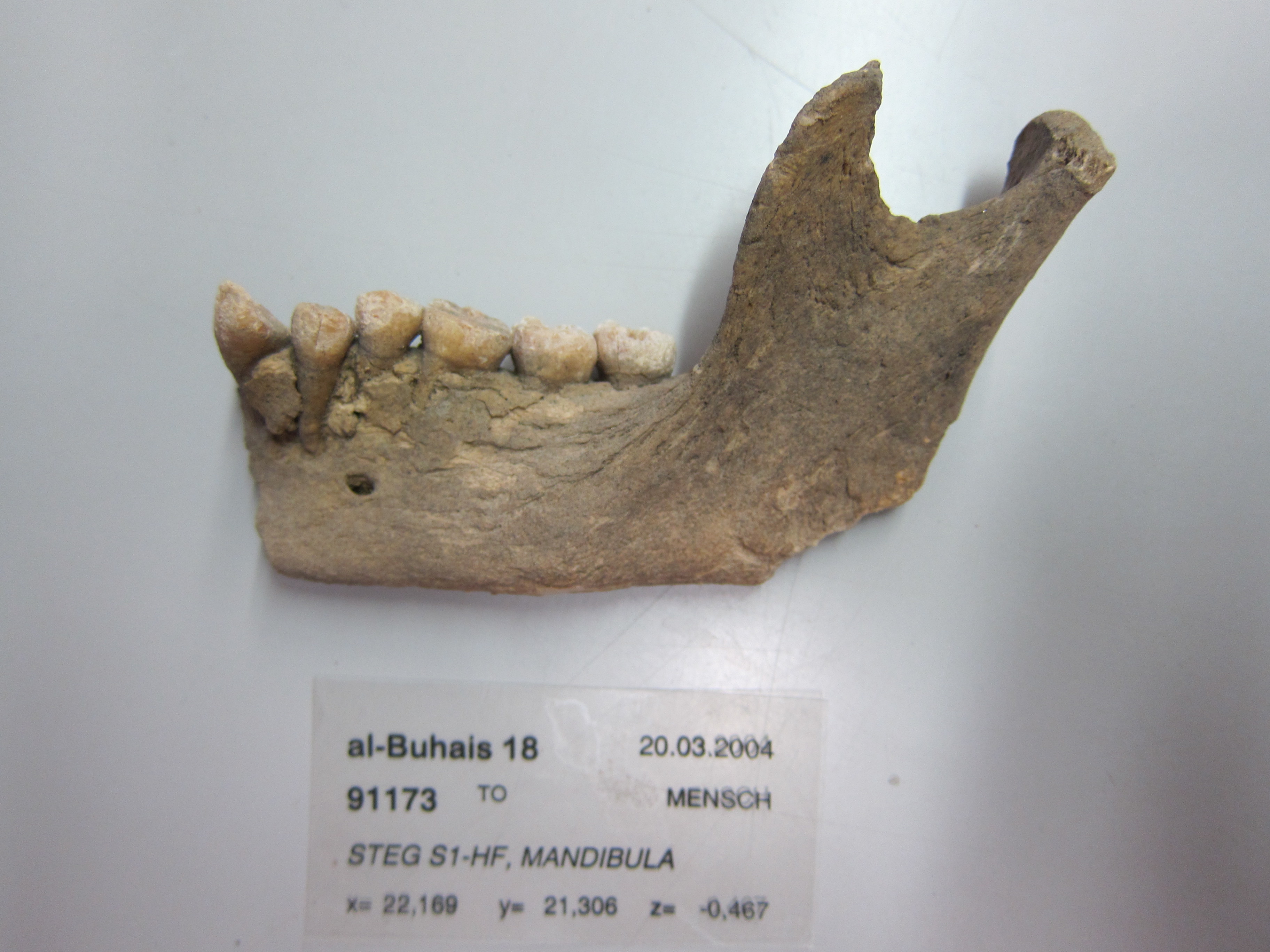
Inidvidual DJ:
DJ, sutura coronalis with hole
Full Resolution
Full Resolution
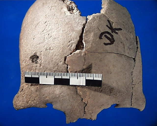
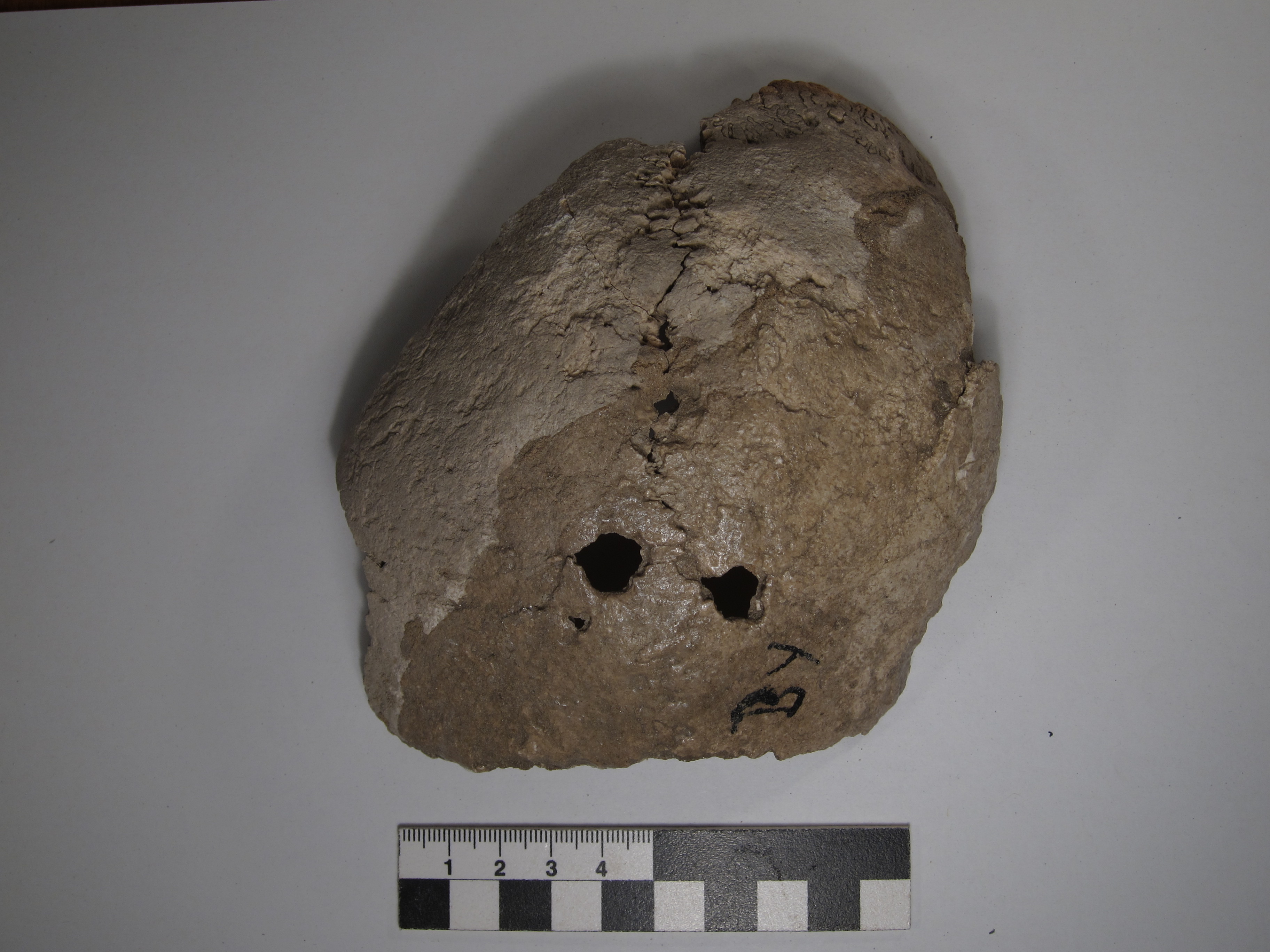
Inidvidual DK:
Skull DK with abscess or post-mortem defect in right frontal region
Full Resolution
Full Resolution
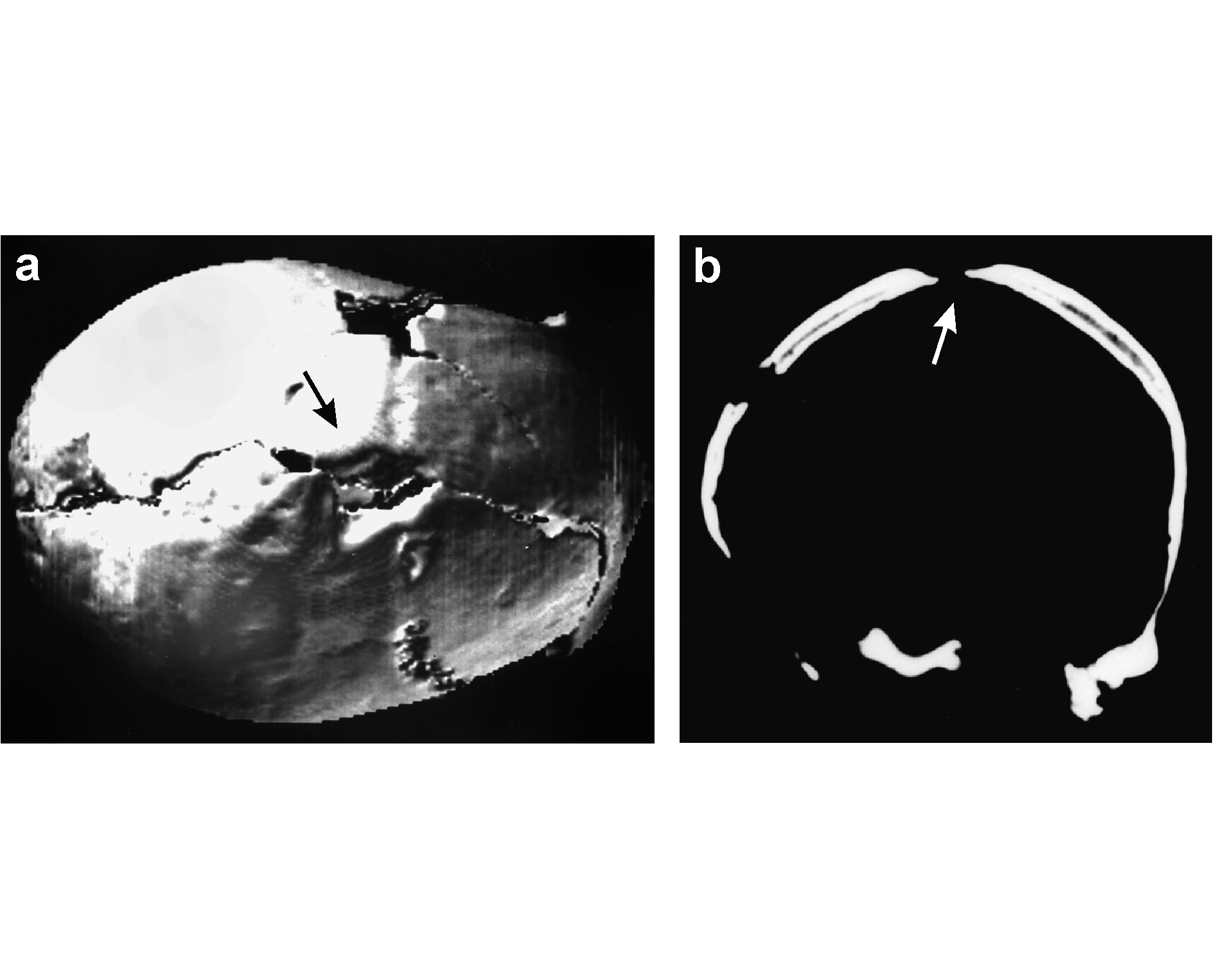
Inidvidual DW:
CT-scan of trephined area, trepanation
Full Resolution
Full Resolution
Inidvidual DW:
Cranium DW, 2 head injuries
Full Resolution
Full Resolution
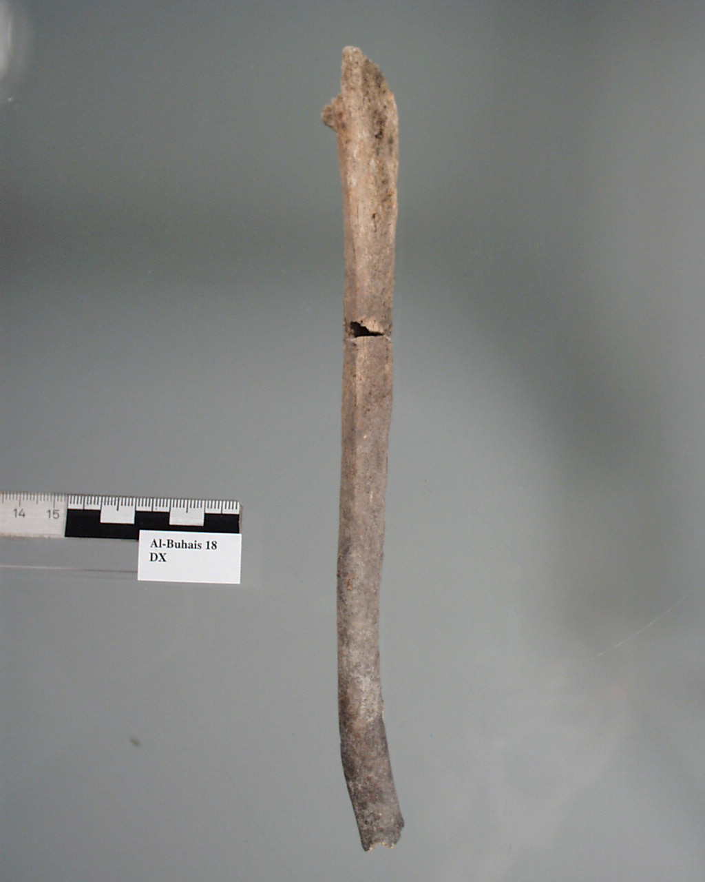
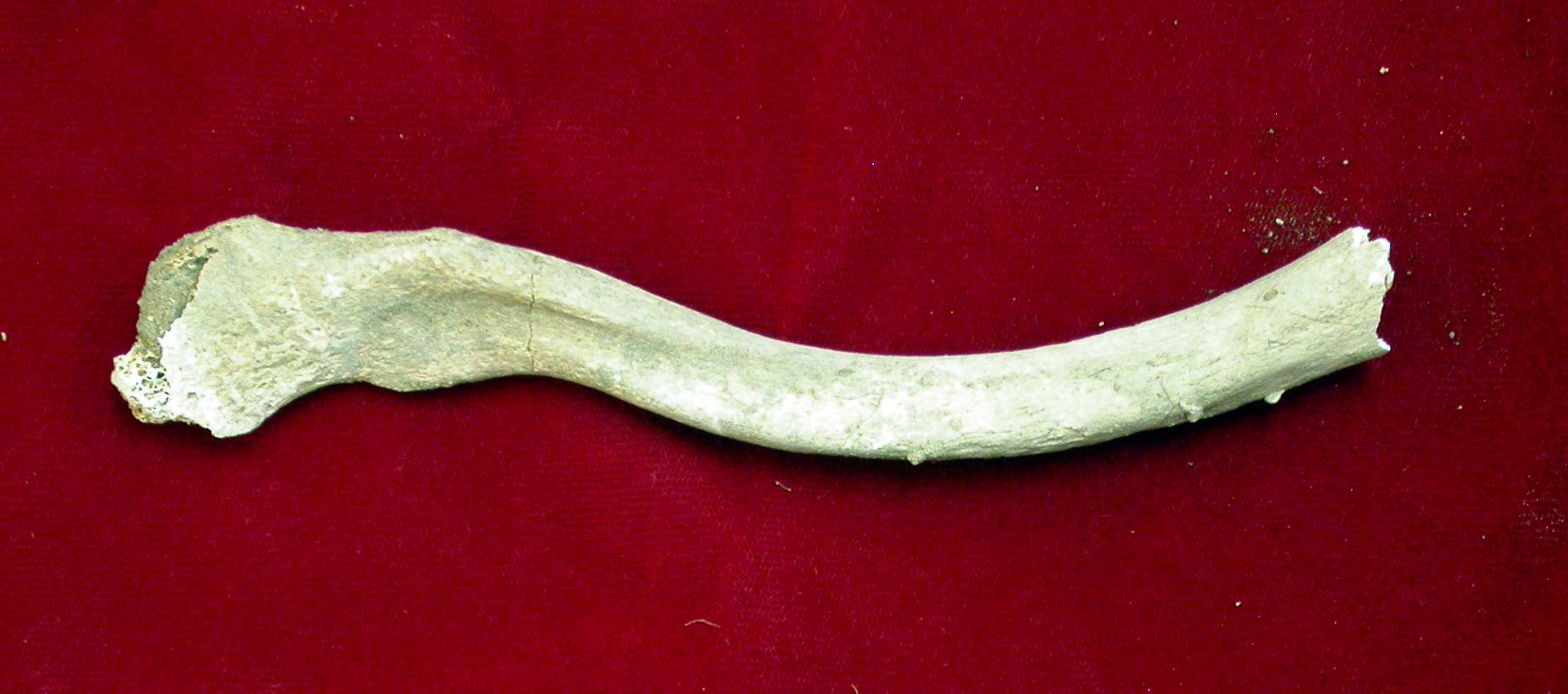
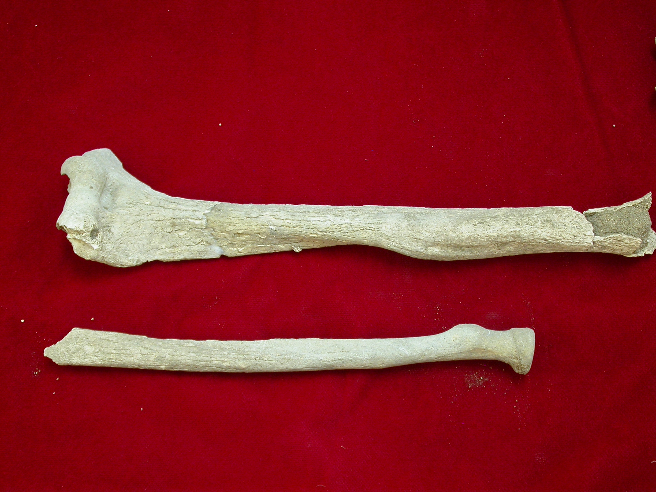
Inidvidual DX:
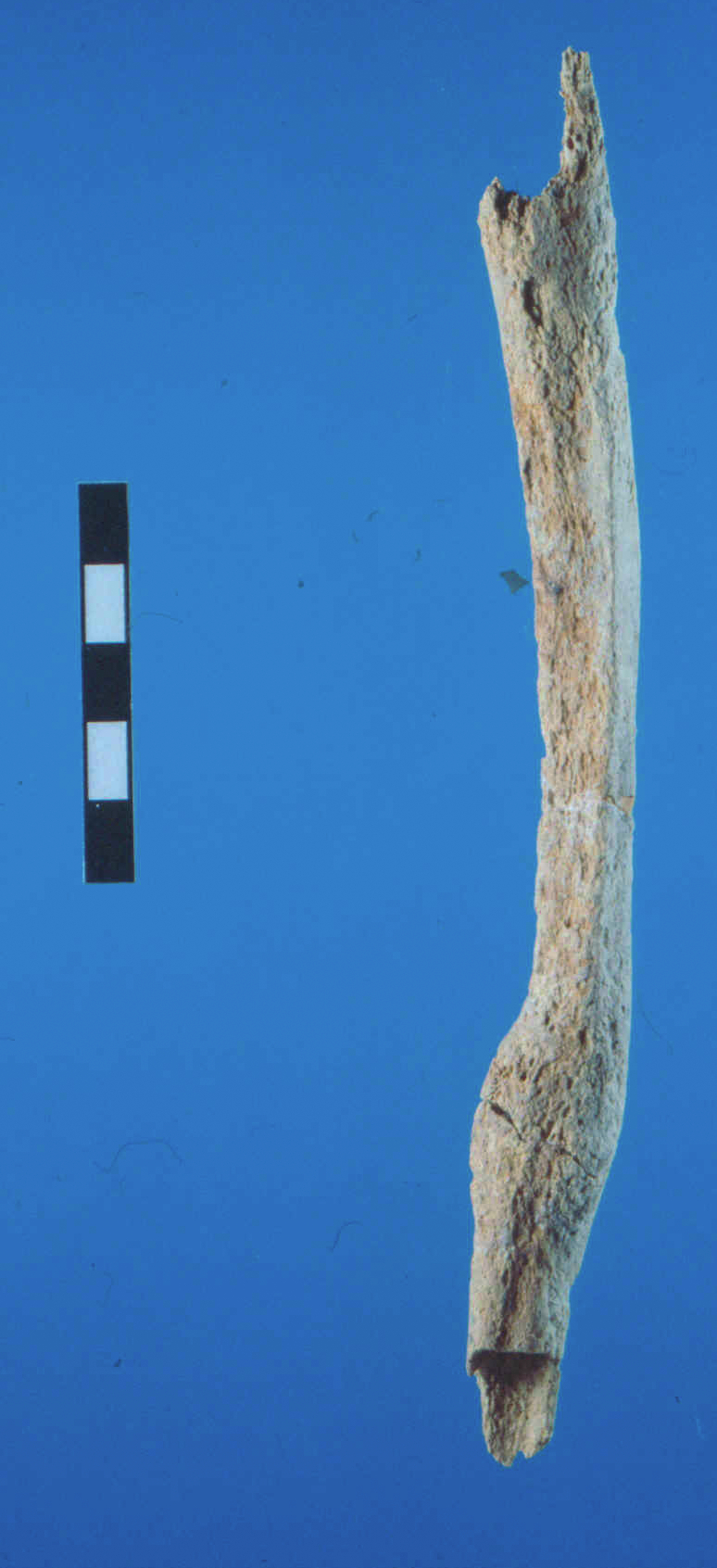
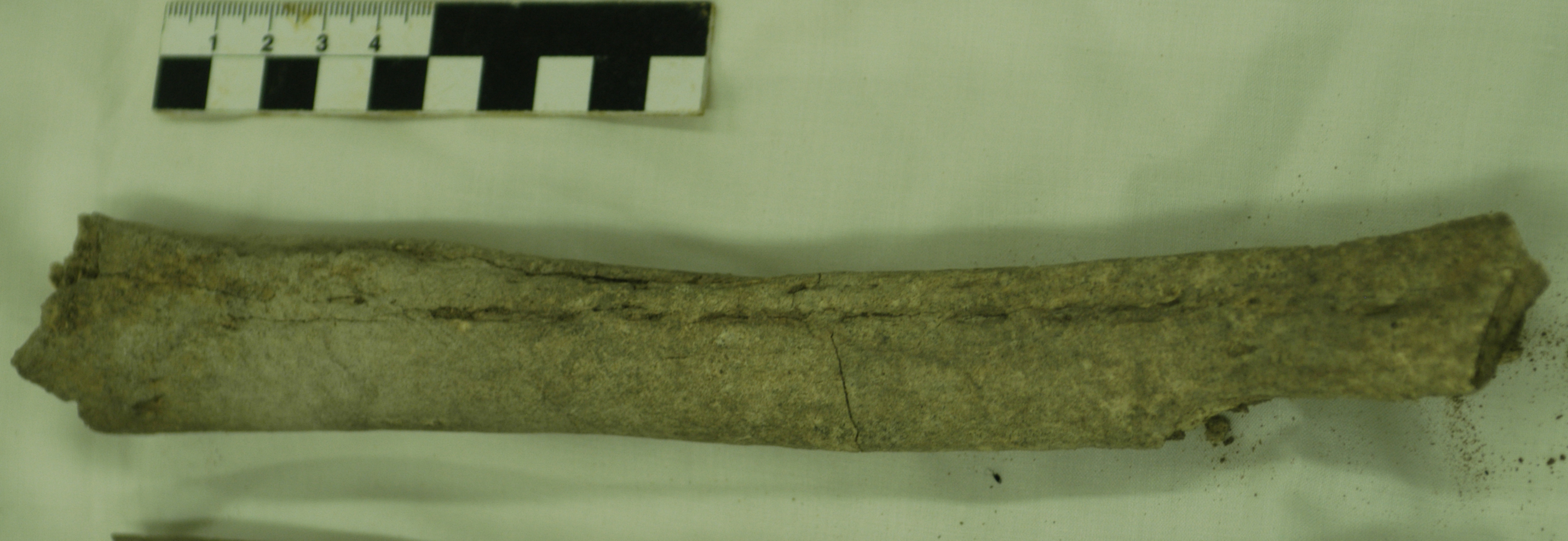
Right Ulna DX, slightly deformed
Full Resolution
Full Resolution
Inidvidual EA:
EA with unhealed peri-mortem fracture
Full Resolution
Full Resolution
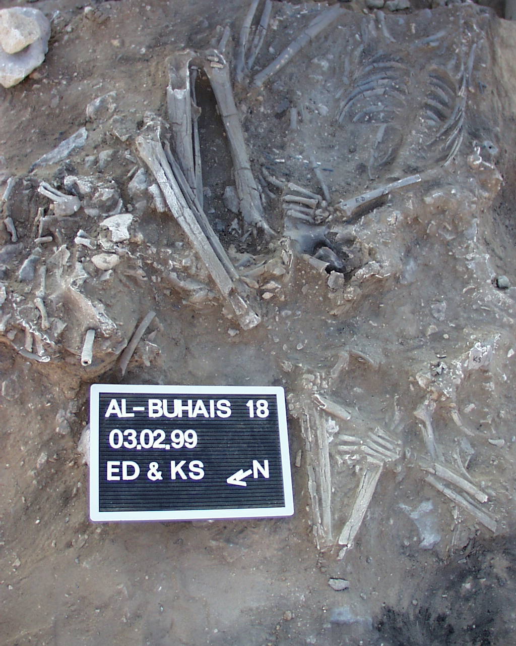
Inidvidual ED:
ED, black (burnt?) mandible
Full Resolution
Full Resolution

Inidvidual ED:
ED and KS in situ
Full Resolution
Full Resolution
Inidvidual ED:
Fireplace near ED
Full Resolution
Full Resolution
Inidvidual EE:
Cast EE made by Marc Haendel, Detail head
Full Resolution
Full Resolution
Inidvidual EE:
Cast EE made by Marc Haendel, processed picture
Full Resolution
Full Resolution
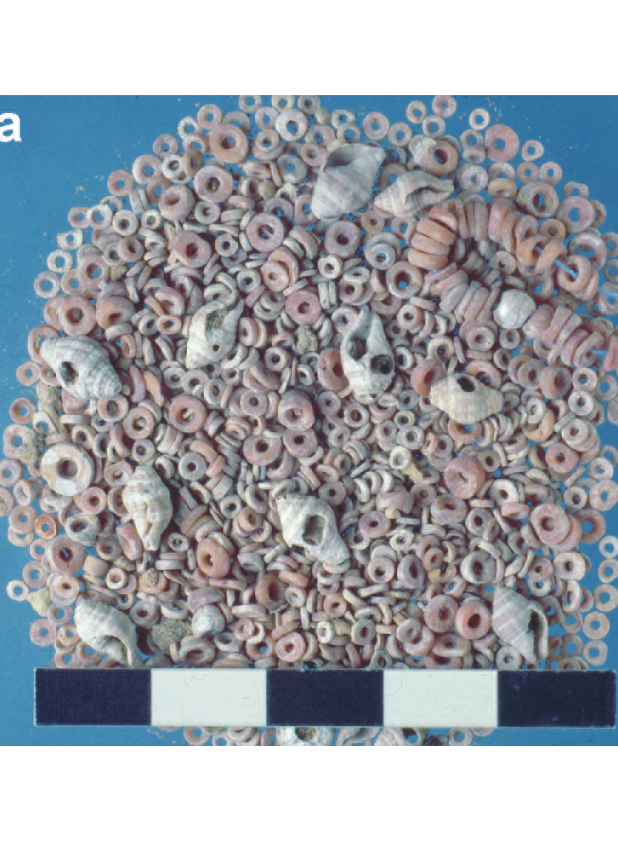
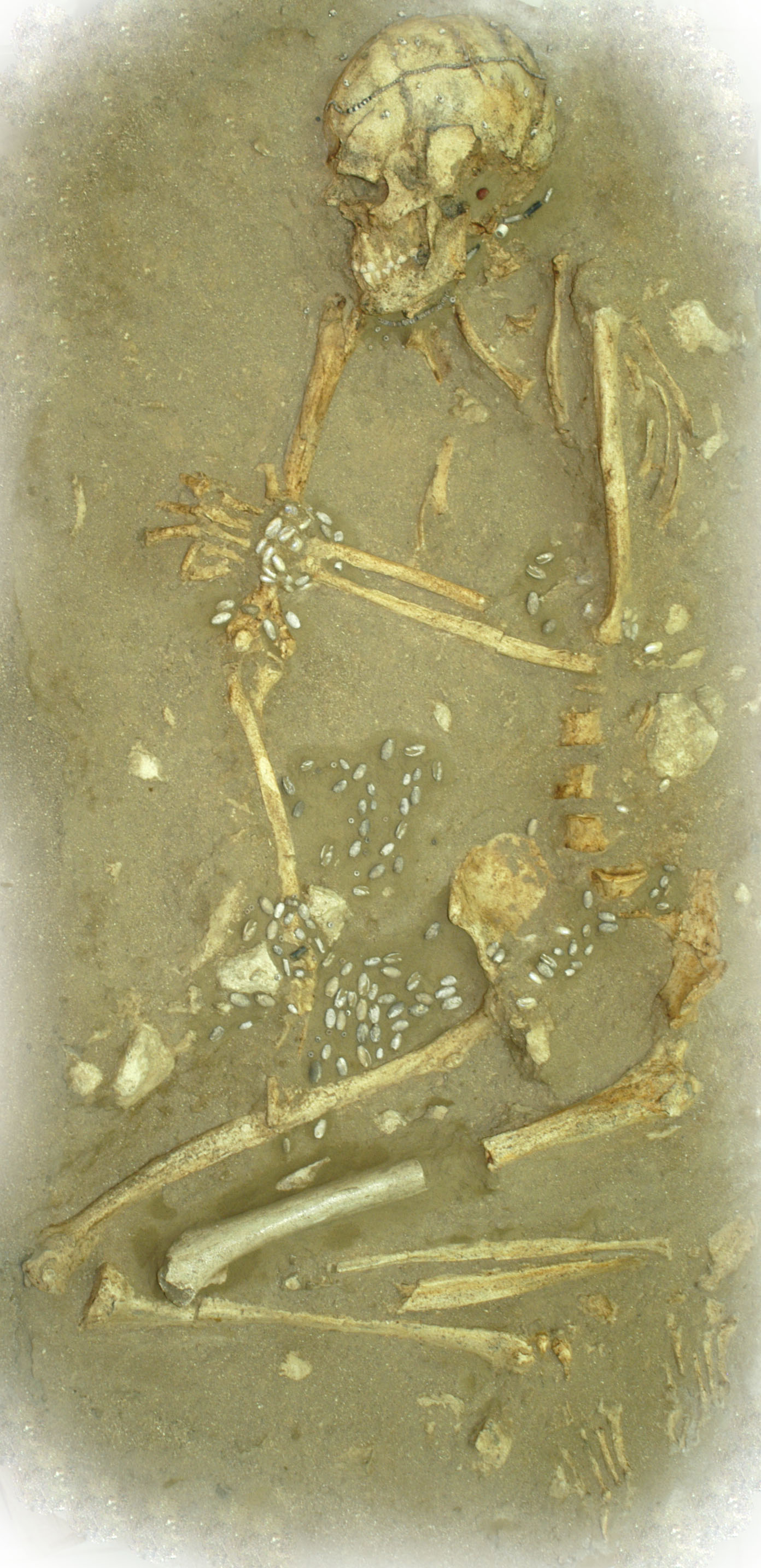
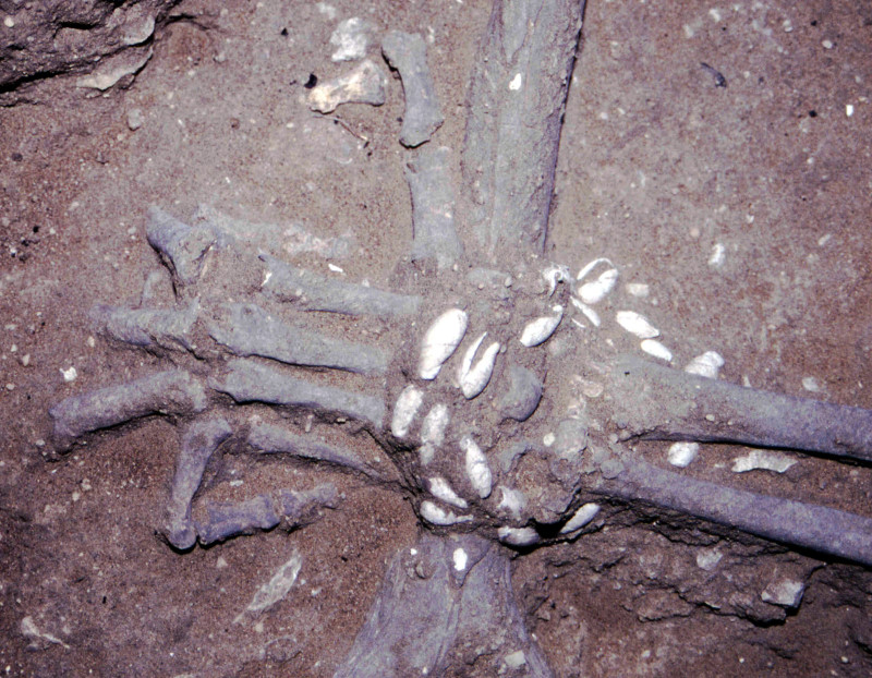
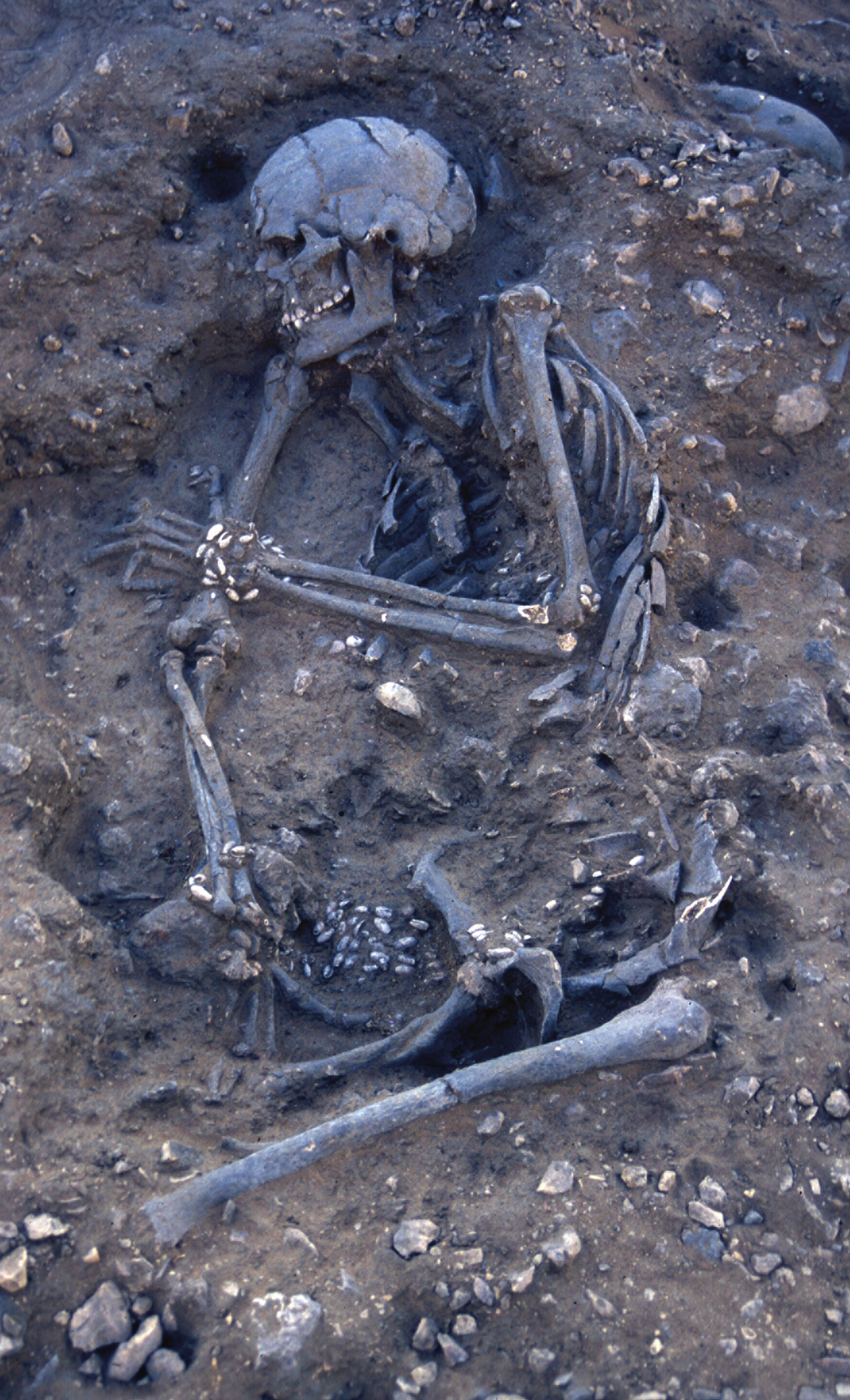
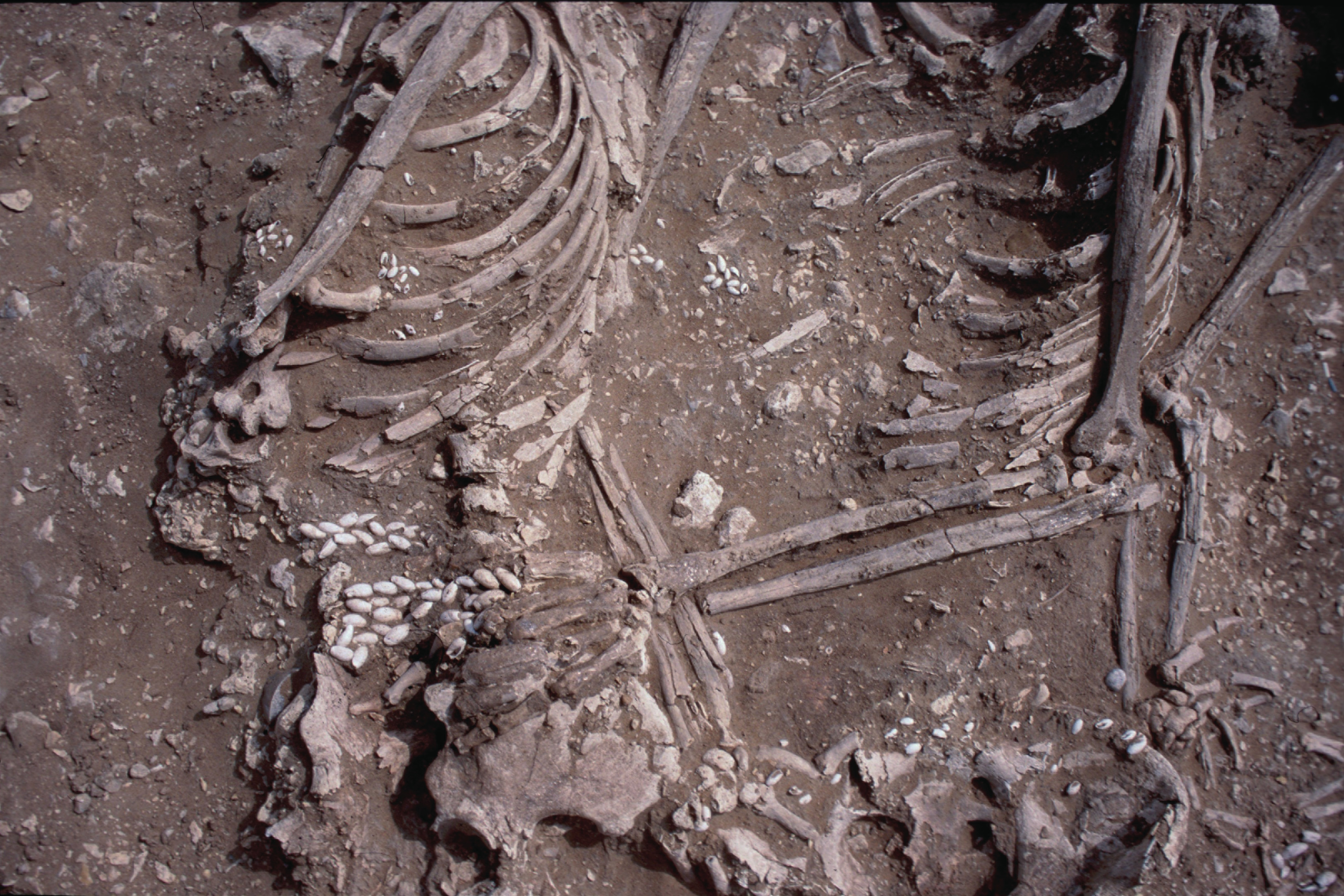
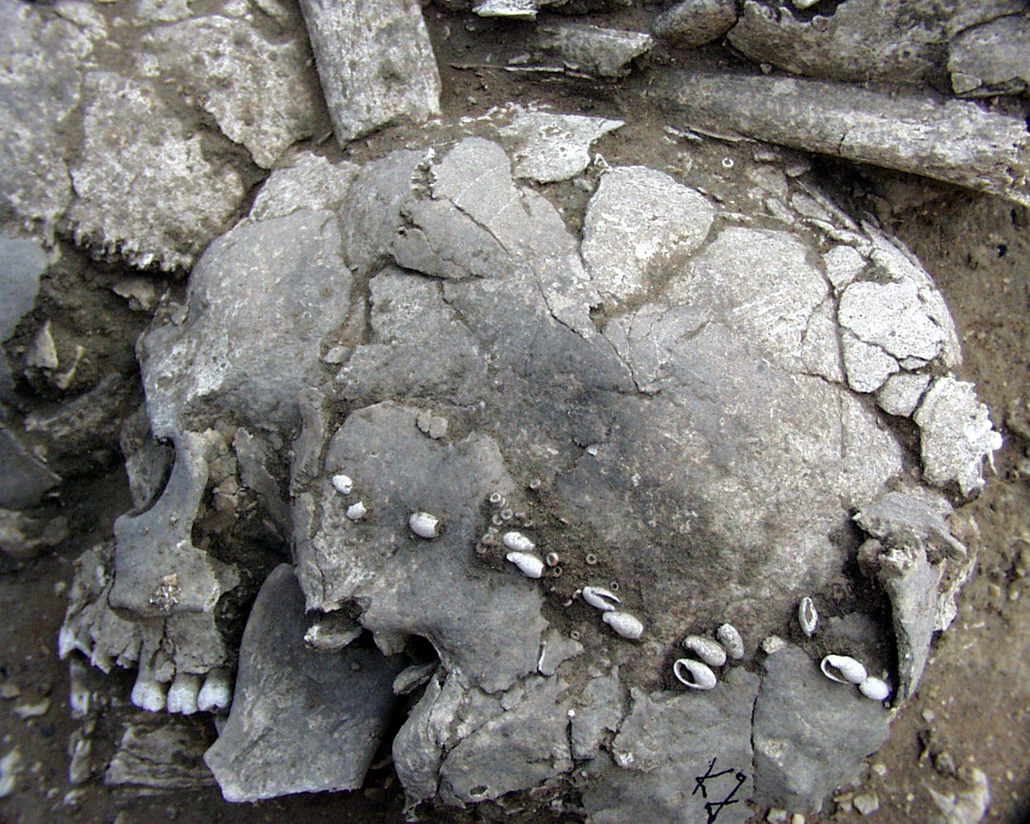
Inidvidual EE:
Adornments found around the skull of EE, including three pearls, pierced snails and disc beads.
Full Resolution
Full Resolution
Inidvidual EE:
Bracelet EE
Full Resolution
Full Resolution
Inidvidual EE:
Burial EE in situ
Full Resolution
Full Resolution
Inidvidual EG:
EG, cribra orbitalia
Full Resolution
Full Resolution
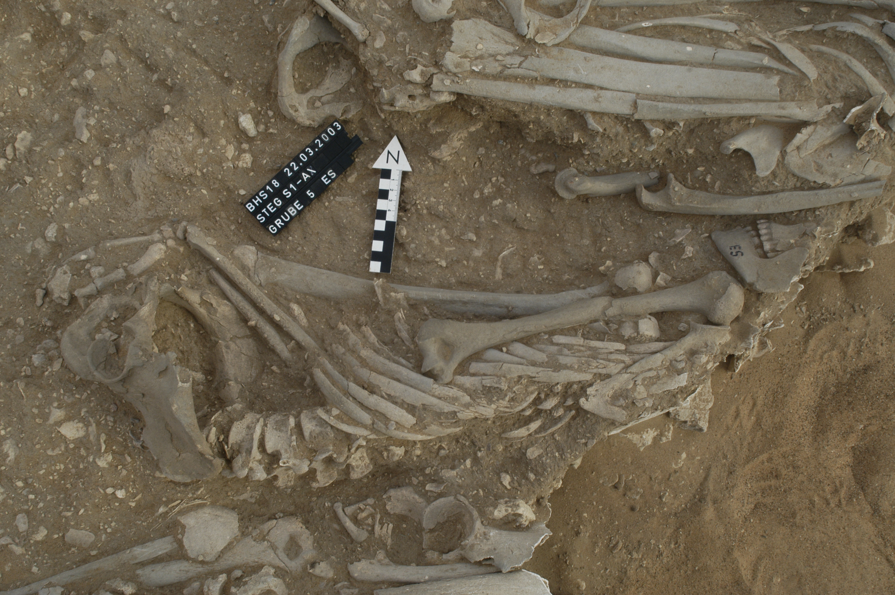
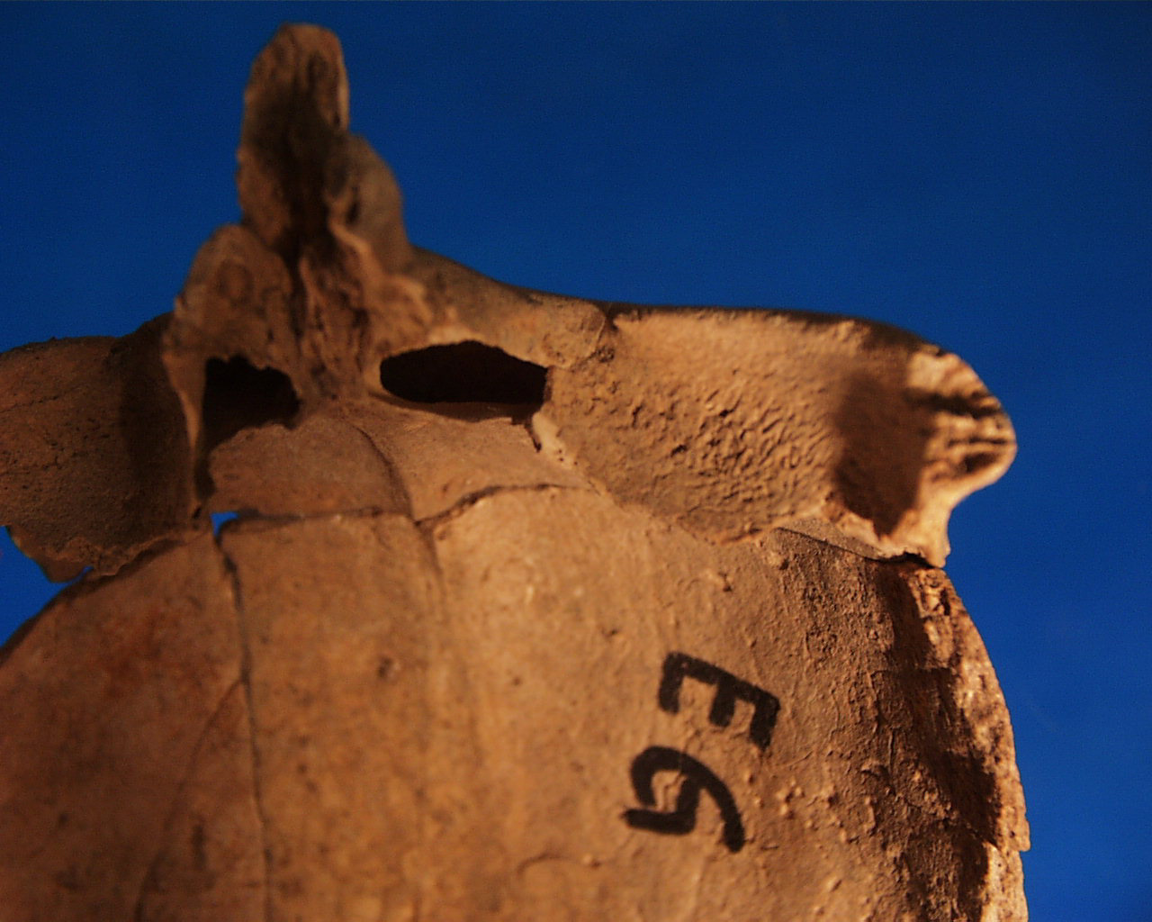
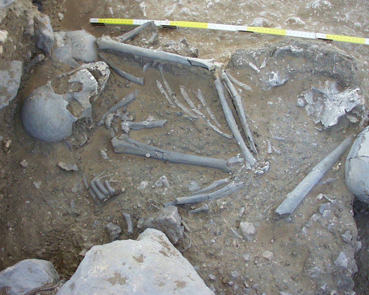
Inidvidual ES:
ES in situ, note position of femur
Full Resolution
Full Resolution
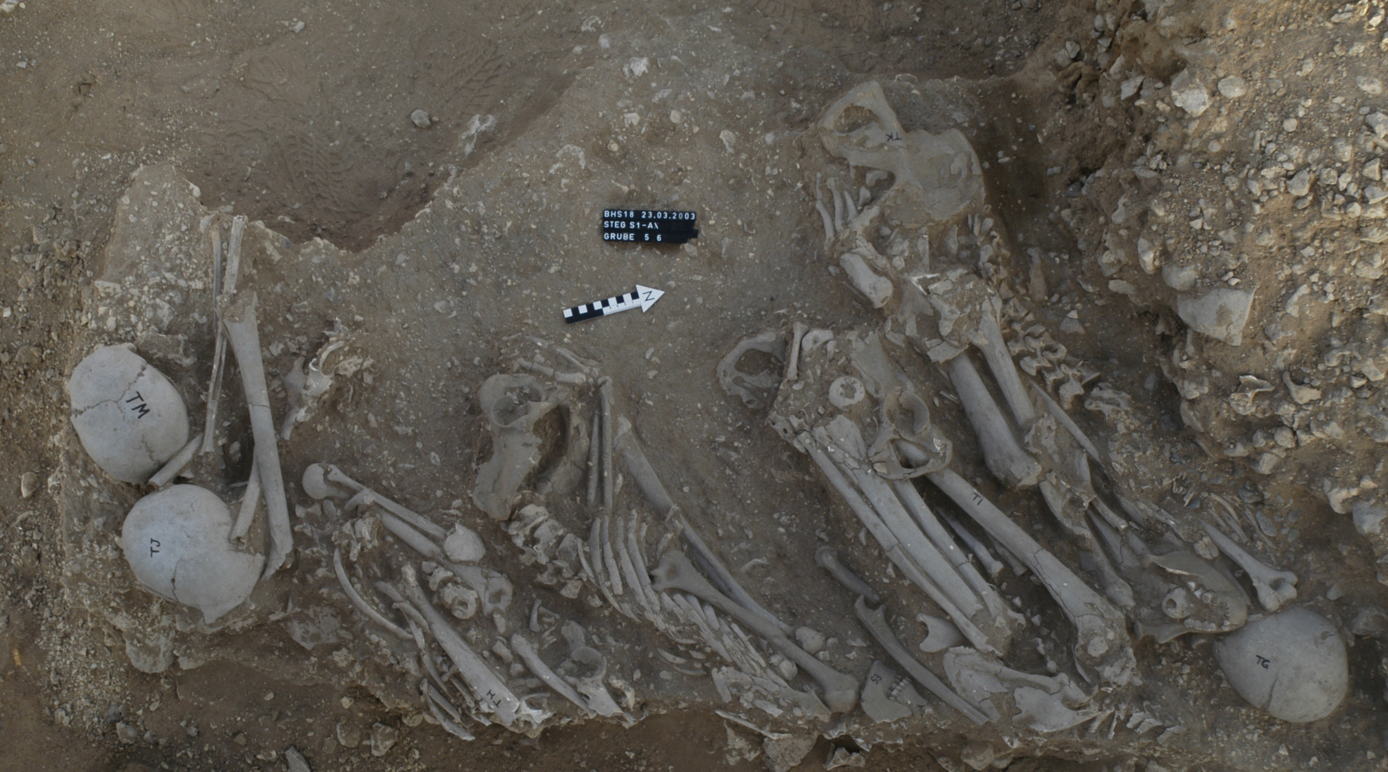
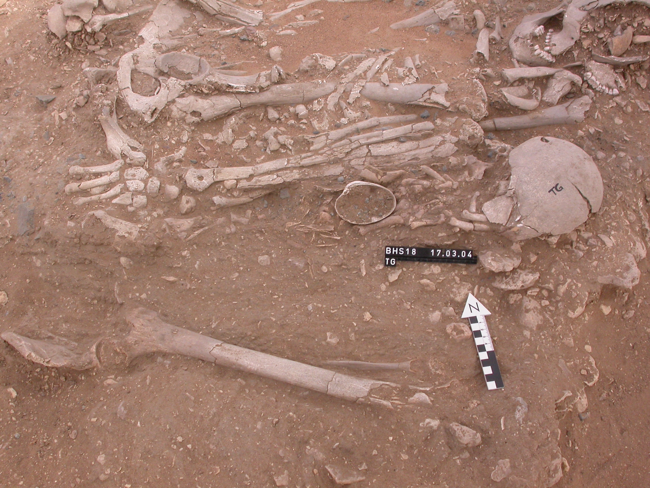
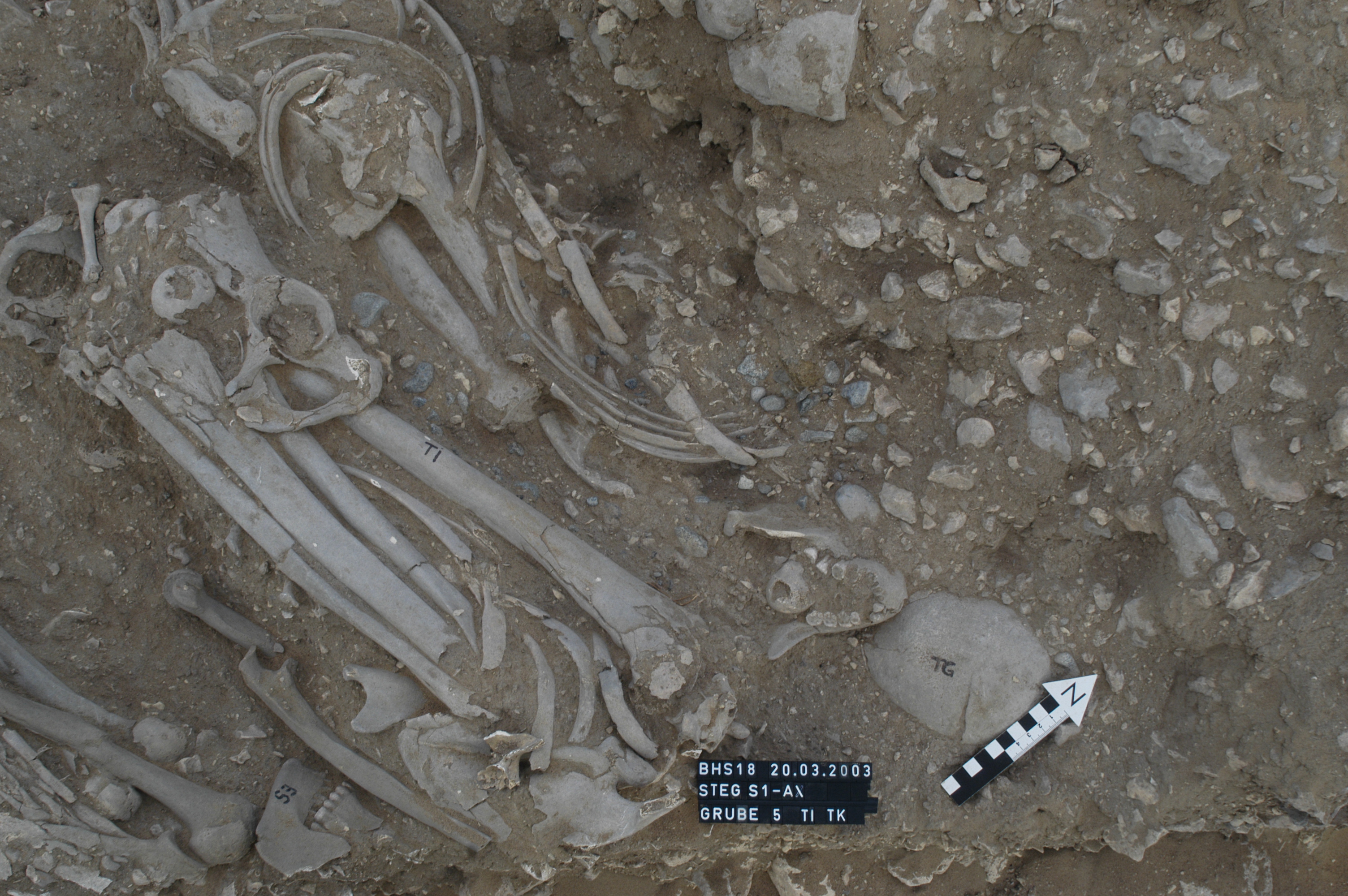
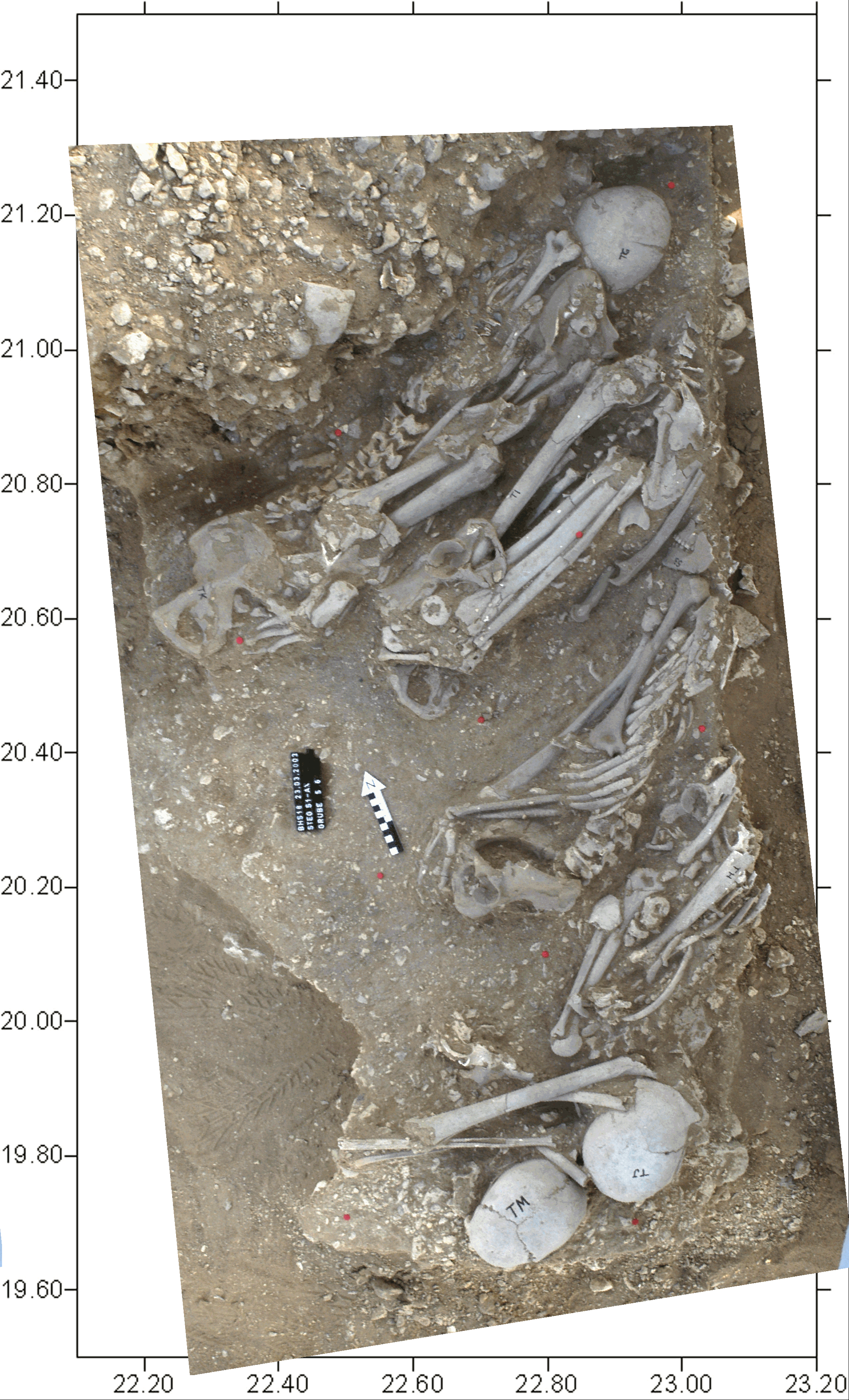
Inidvidual ES:
In situ along with TG, TI, TJ, TM, TH
Full Resolution
Full Resolution
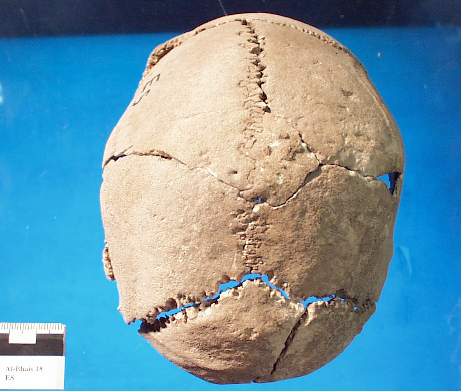
Inidvidual ES:
Skull ES superior
Full Resolution
Full Resolution

Inidvidual ES:
ES and ET in situ
Full Resolution
Full Resolution
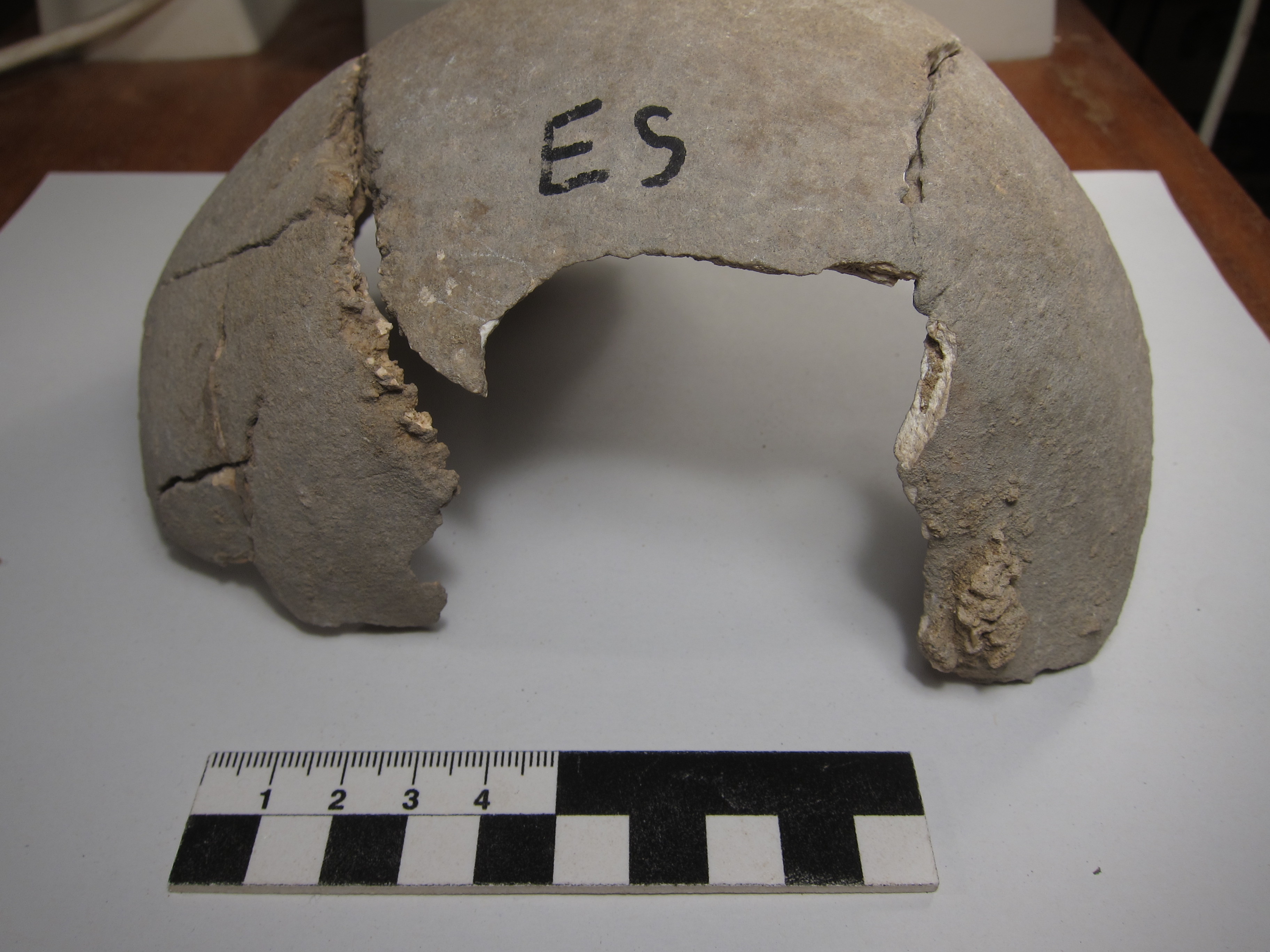
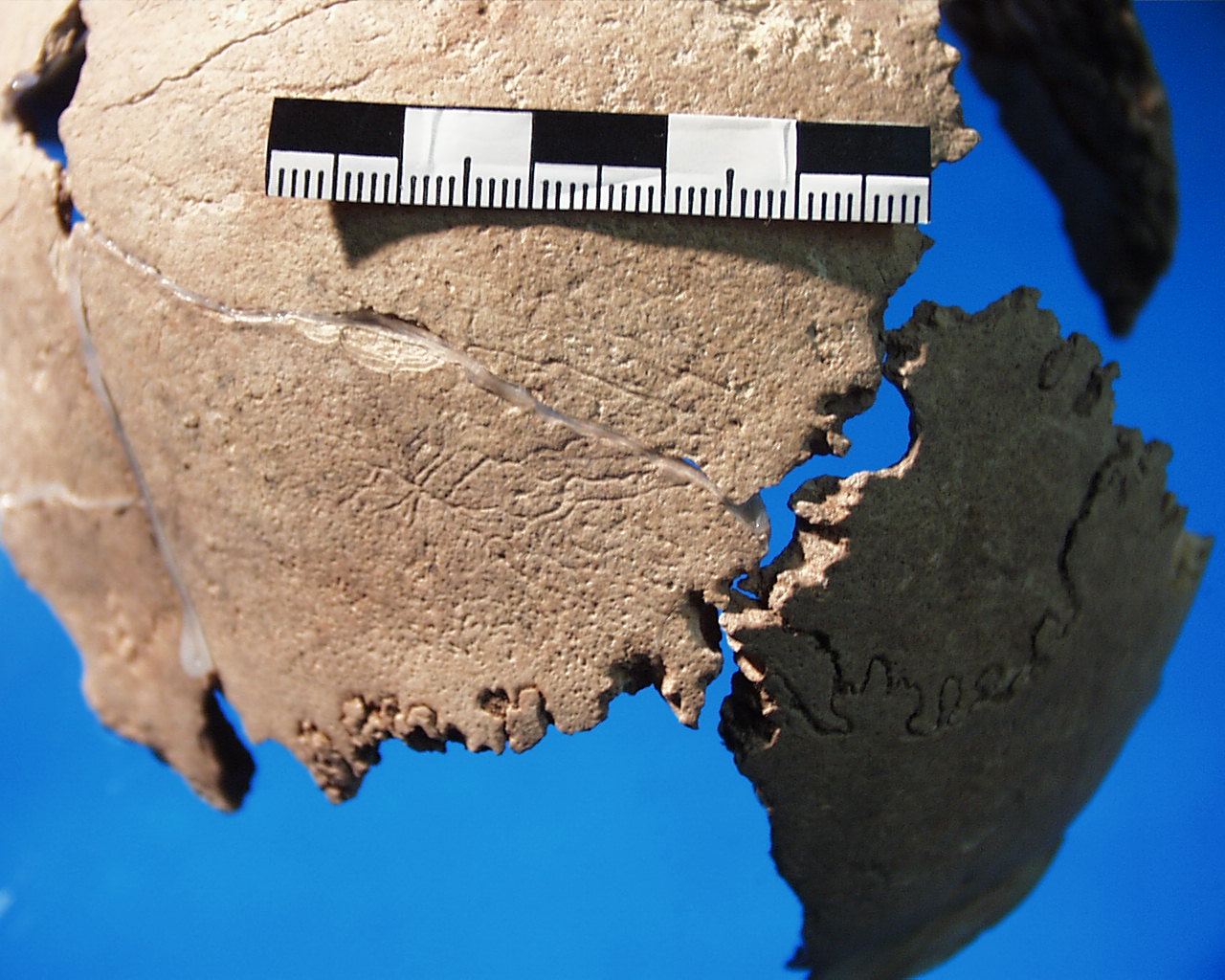
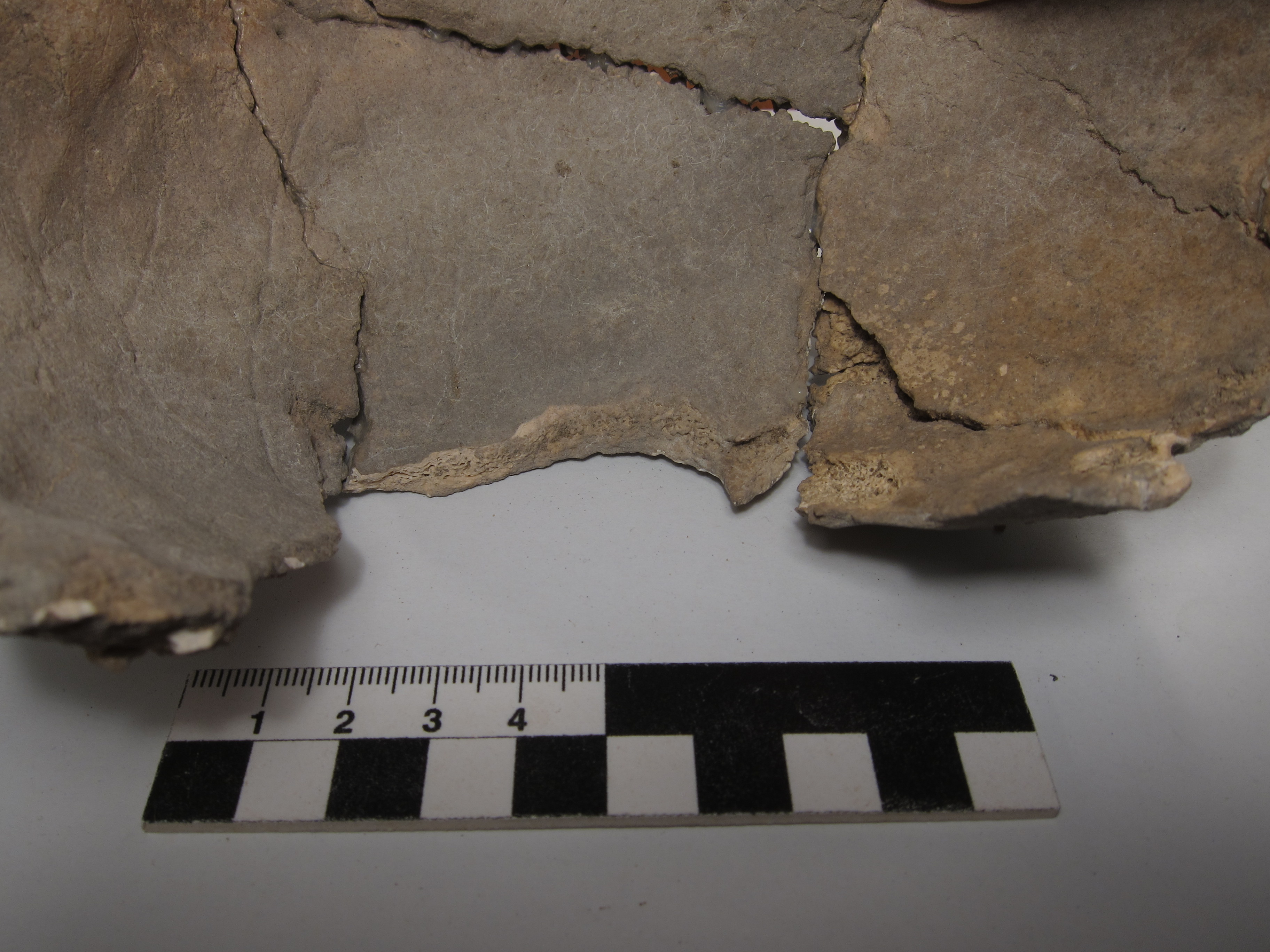
Inidvidual ES:
A sharp-edged round hole about 35 mm in diameter – most likely an unhealed pond fracture – is located on the left parietal bone close to the coronal
suture.
Full Resolution
Full Resolution
Inidvidual ES:
Fracture - Inner table of skull
Full Resolution
Full Resolution

Inidvidual ET:
ES and ET in situ
Full Resolution
Full Resolution
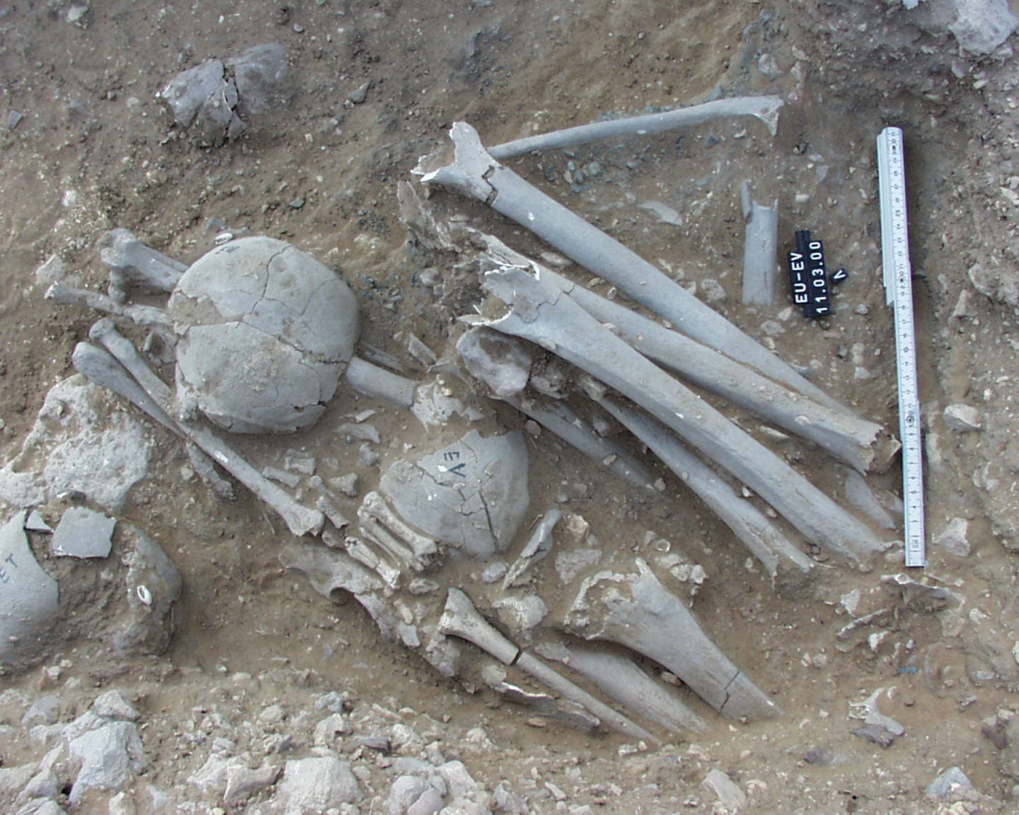
Inidvidual ET:
EU, EV and ET in situ
Full Resolution
Full Resolution
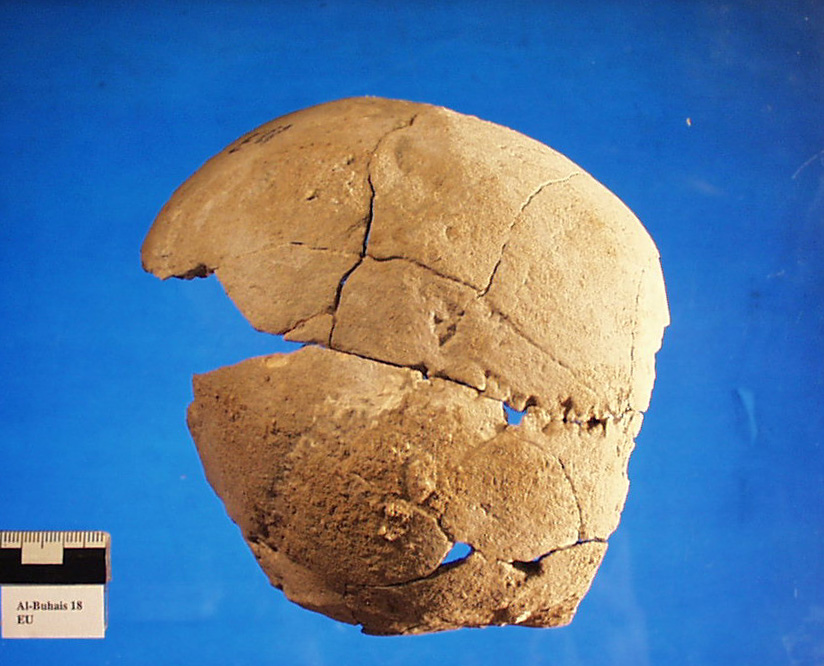
Inidvidual EU:
Skull EU
Full Resolution
Full Resolution

Inidvidual EU:
EU, EV and ET in situ
Full Resolution
Full Resolution
Inidvidual EU:
EU and EV with Ophiolite
Full Resolution
Full Resolution

Inidvidual EU:
LP, EU and EV in situ
Full Resolution
Full Resolution

Inidvidual EV:
EU, EV and ET in situ
Full Resolution
Full Resolution

Inidvidual EV:
EU and EV with Ophiolite
Full Resolution
Full Resolution

Inidvidual EV:
LP, EU and EV in situ
Full Resolution
Full Resolution
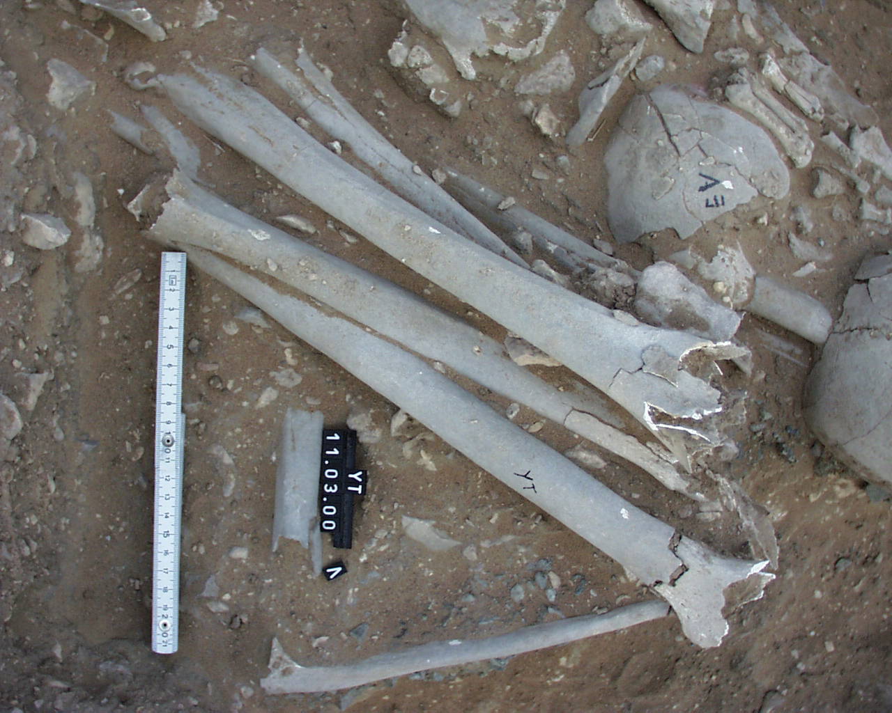
Inidvidual EV:
YT and EV in situ
Full Resolution
Full Resolution

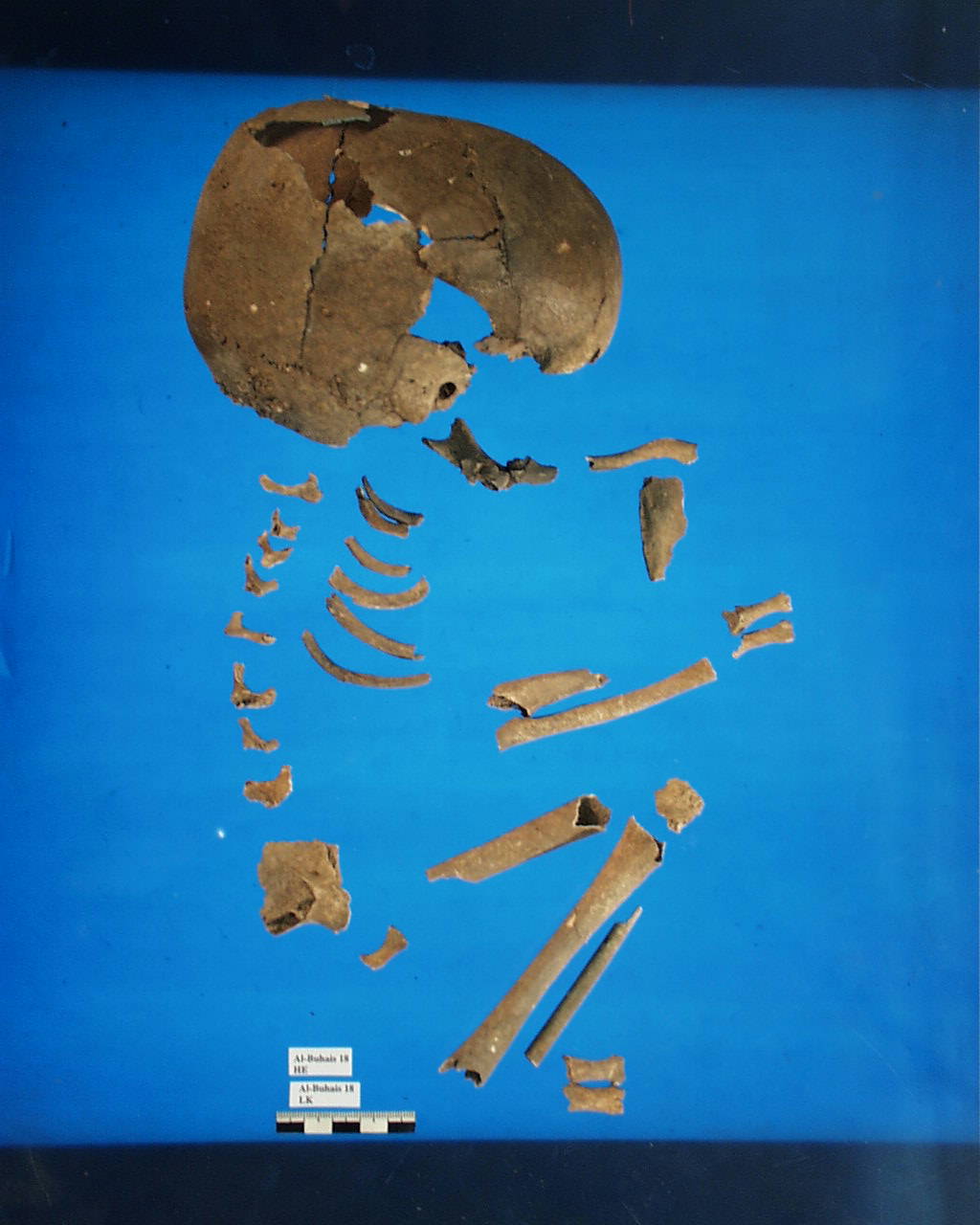
Inidvidual EX:
HD, HE, EX and LK, in situ
Full Resolution
Full Resolution
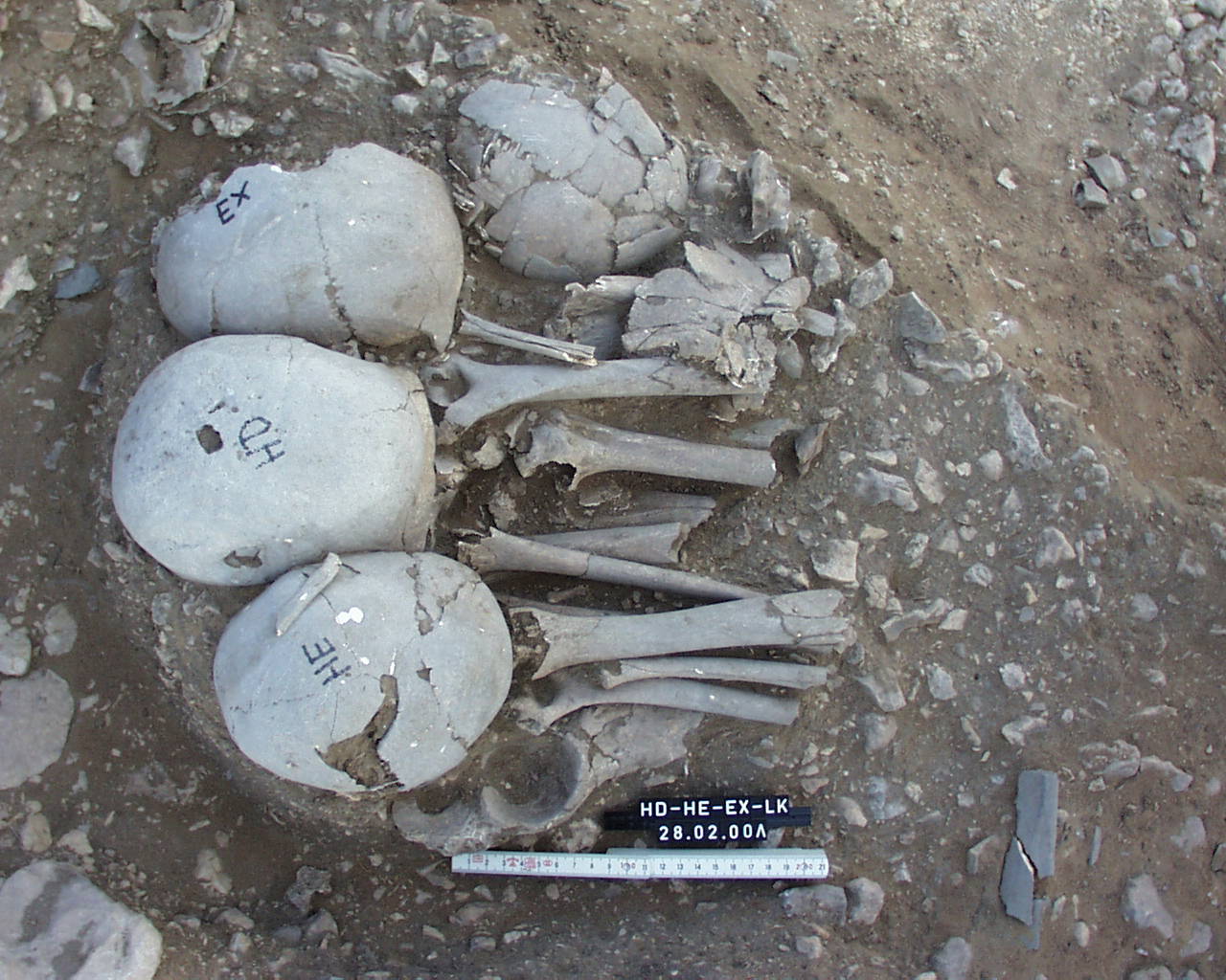
Inidvidual EX:
HD, HE, EX and LK in situ
Full Resolution
Full Resolution
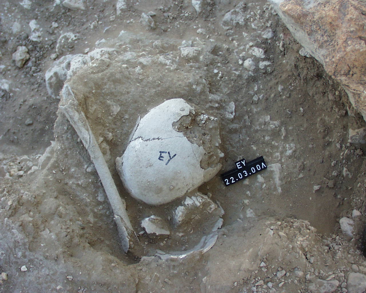
Inidvidual EY:
EY in situ
Full Resolution
Full Resolution

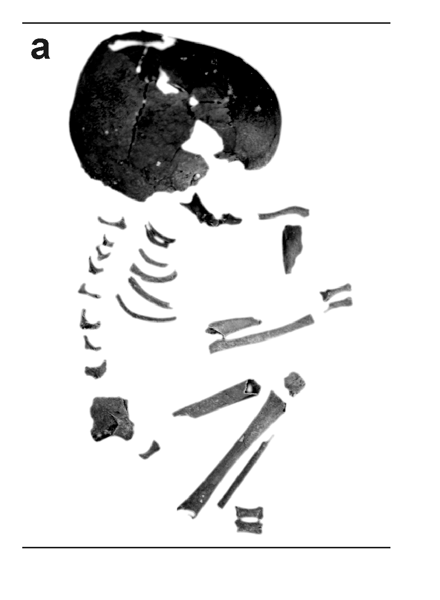
Inidvidual FA:
Spina bifida, HW1, individual FA
Full Resolution
Full Resolution

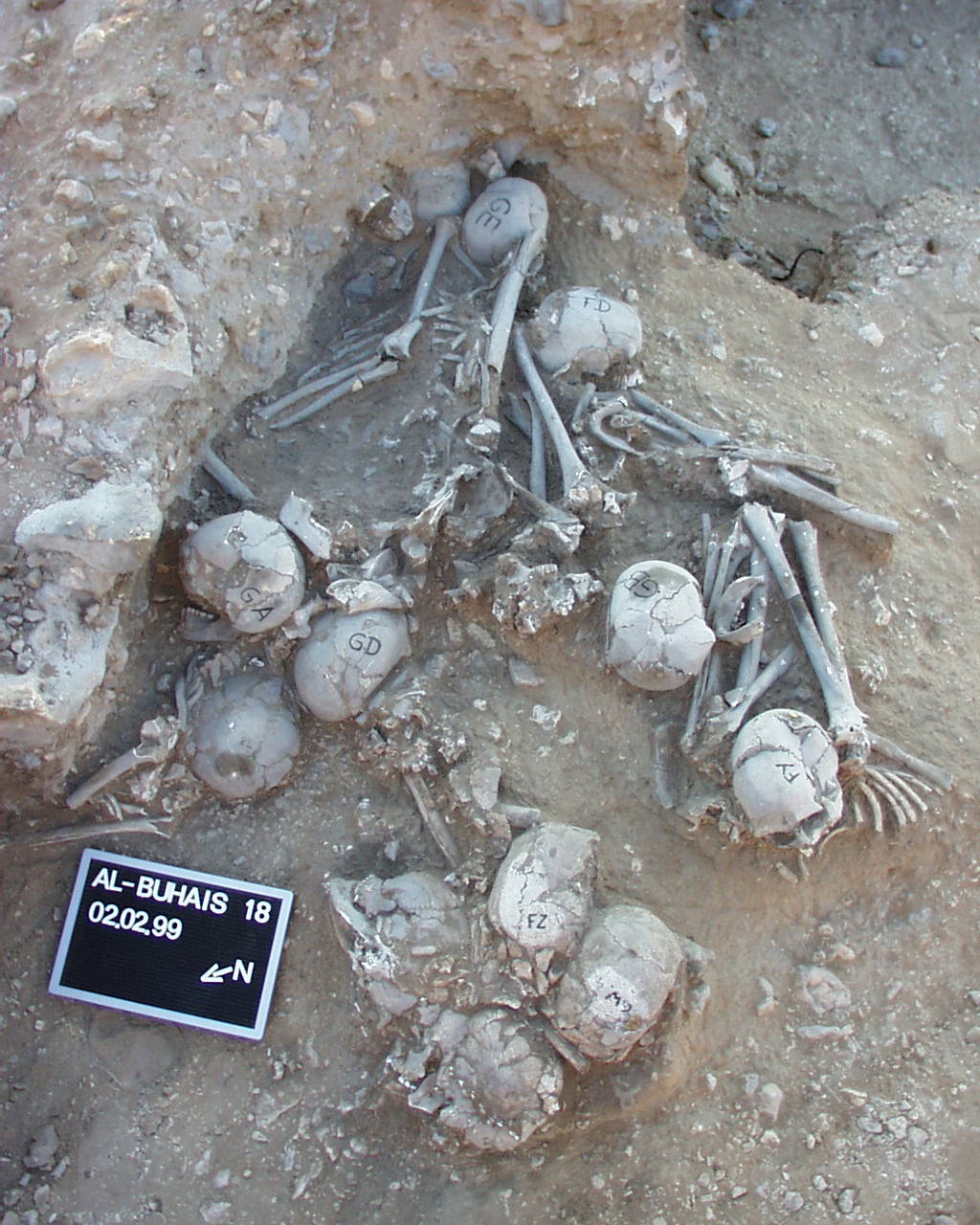
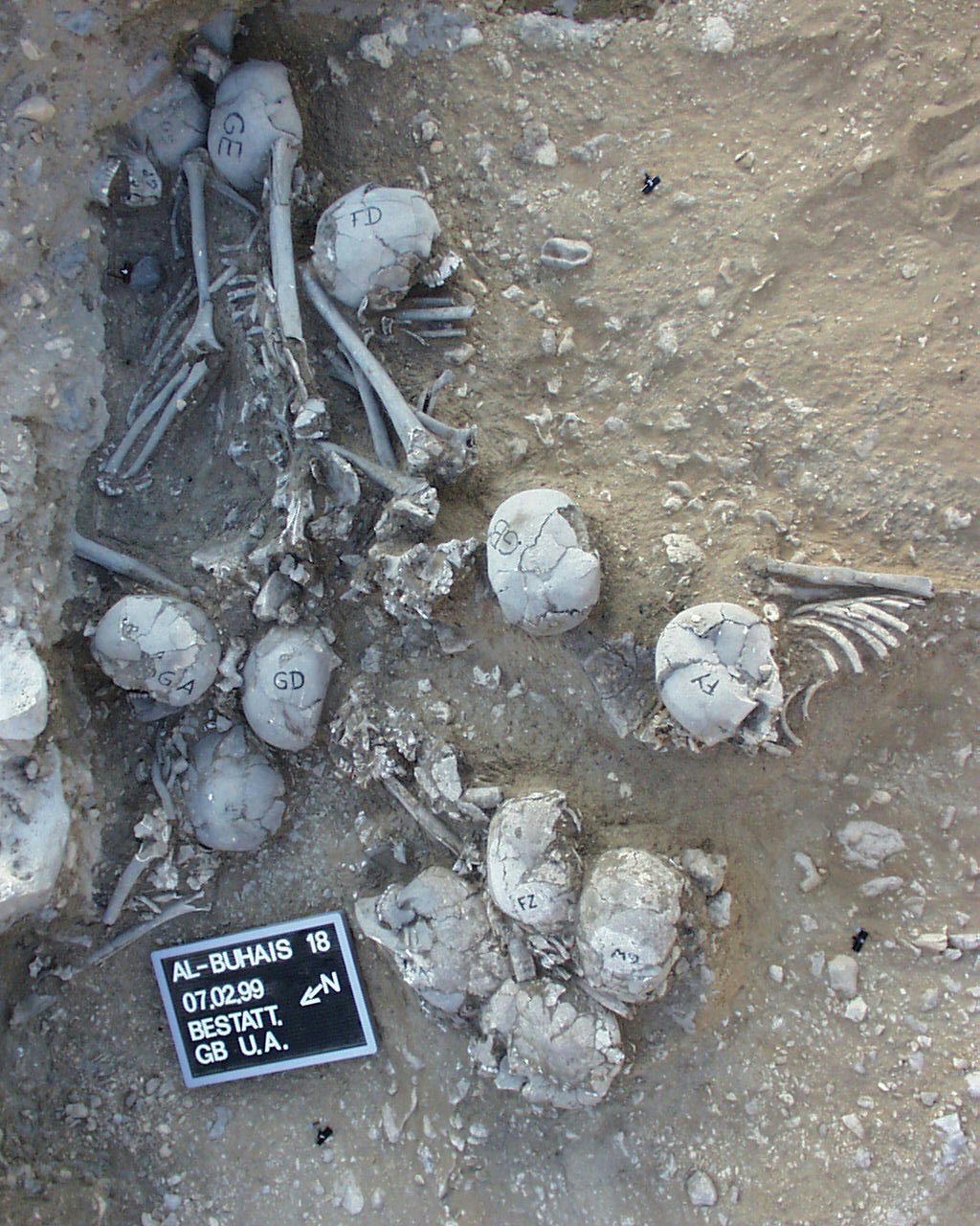
Inidvidual FD:
FD, FY, FZ, GA, GB, GD, GE, GO, GP, GW, HA, KJ, KL in situ
Full Resolution
Full Resolution

Inidvidual FD:
FD, FY, FZ, GA, GB, GD, GE, GO, GP, GW, KM, KL in situ
Full Resolution
Full Resolution

Inidvidual FD:
FD, FY, GA, GB, GD, GE, GP, KL, YA in situ
Full Resolution
Full Resolution

Inidvidual FD:
FD, FY, GA, GB, GD, GE, GP, KL, YA in situ
Full Resolution
Full Resolution
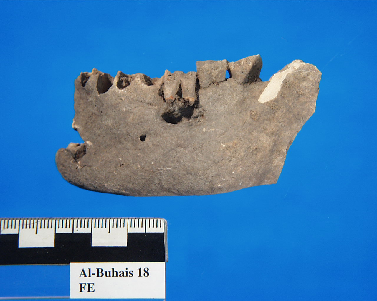
Inidvidual FE:
FE, mandible with cyst
Full Resolution
Full Resolution
Inidvidual FE:
FE, deepened meningeal impressions
Full Resolution
Full Resolution
Inidvidual FE:
unhealed fracture (blunt force injury) in the middle of the left parietal
Full Resolution
Full Resolution
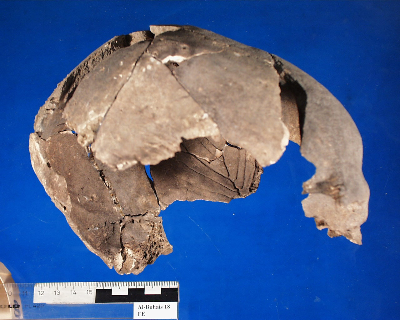
Inidvidual FE:
Skull
Full Resolution
Full Resolution
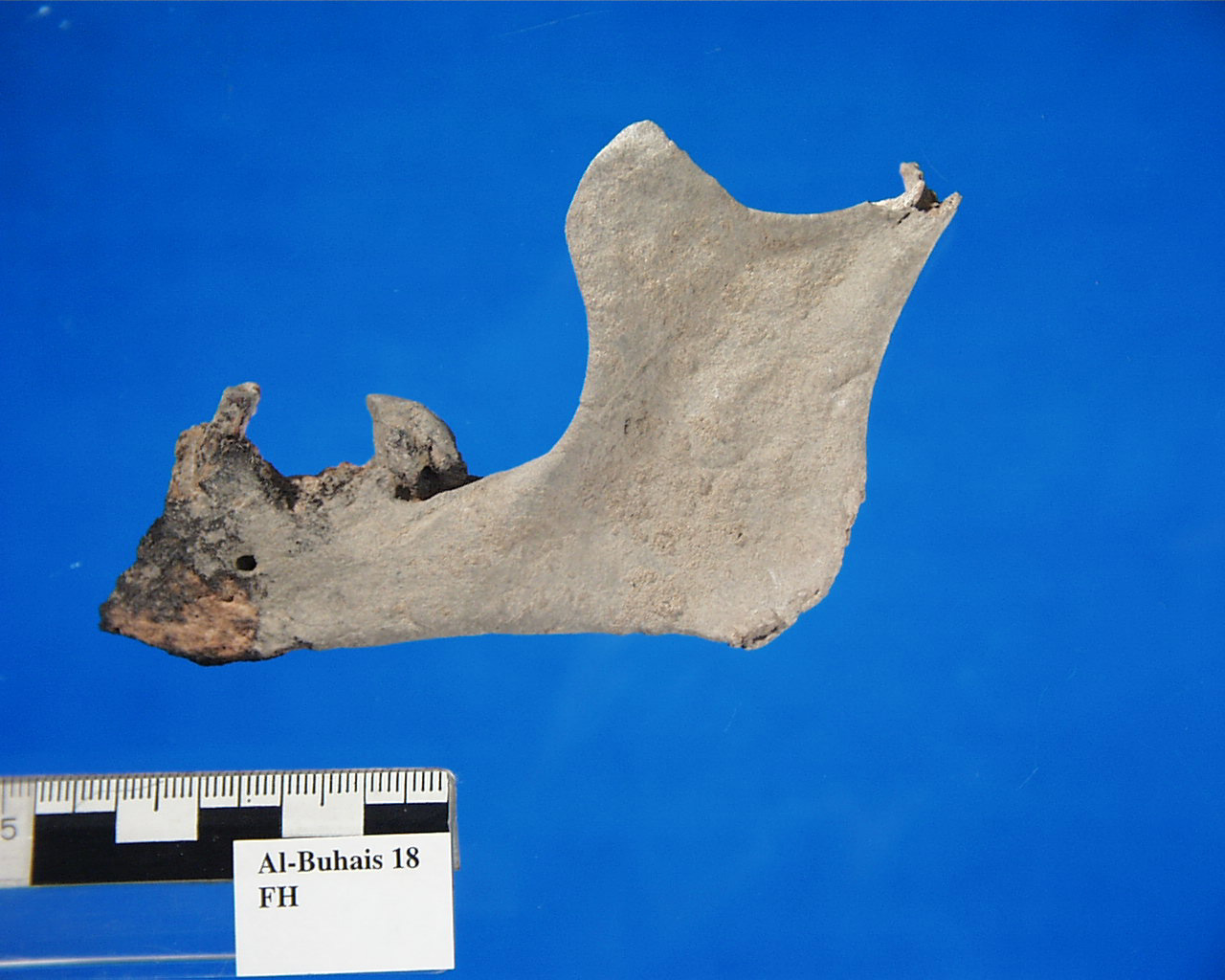
Inidvidual FH:
FH, mandible with intra-vitem tooth loss (Greisenkiefer)
Full Resolution
Full Resolution
Inidvidual FI:
Cast of child FI
Full Resolution
Full Resolution

Inidvidual FI:
FI i situ
Full Resolution
Full Resolution
Inidvidual FK:
Individual FK in situ
Full Resolution
Full Resolution
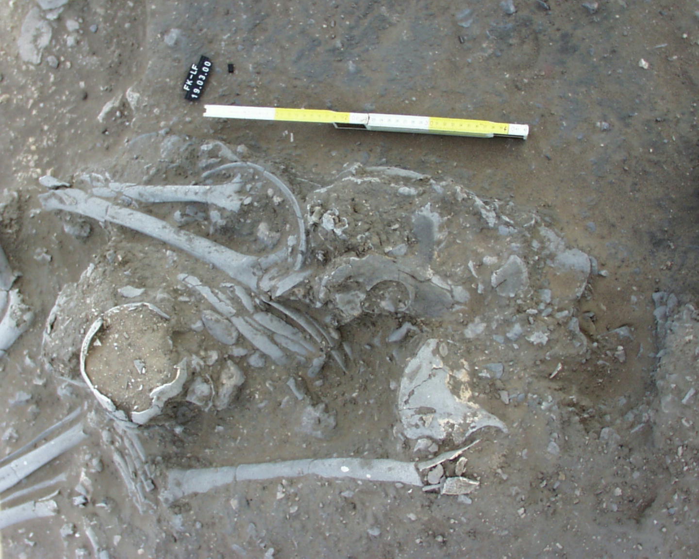
Inidvidual FK:
FK and LF in situ
Full Resolution
Full Resolution
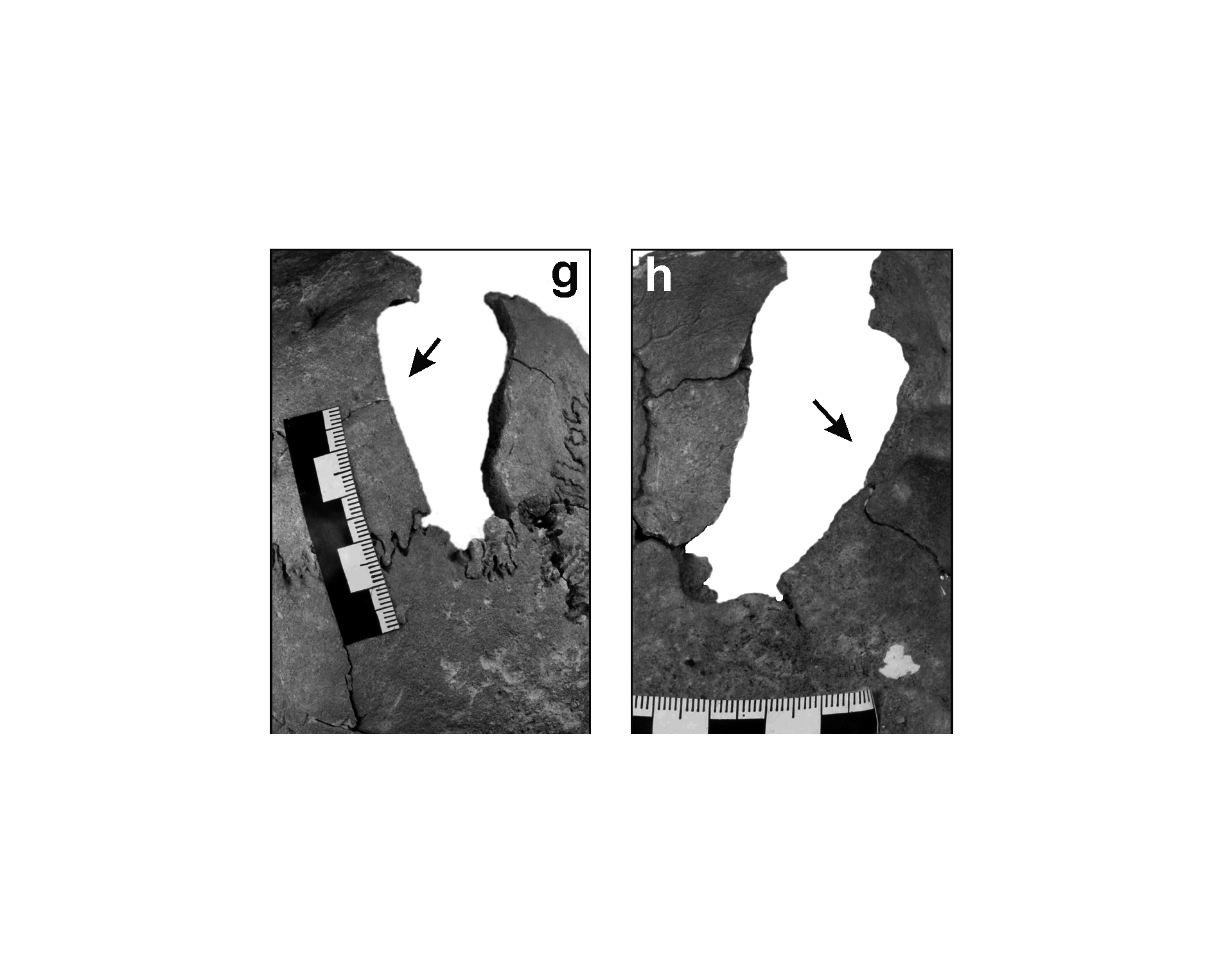
Inidvidual FL:
Chop wound of the occipital bone, skull DW. Exocranial (g) end endocranial (h) view.
Full Resolution
Full Resolution
Inidvidual FM:
The right part of the frontal bone with extensive fracture caused by blunt force
Full Resolution
Full Resolution
Inidvidual FO:
Multiple burial
Full Resolution
Full Resolution

Inidvidual FY:
FD, FY, FZ, GA, GB, GD, GE, GO, GP, GW, HA, KJ, KL in situ
Full Resolution
Full Resolution

Inidvidual FY:
FD, FY, FZ, GA, GB, GD, GE, GO, GP, GW, KM, KL in situ
Full Resolution
Full Resolution

Inidvidual FY:
FD, FY, GA, GB, GD, GE, GP, KL, YA in situ
Full Resolution
Full Resolution

Inidvidual FY:
FD, FY, GA, GB, GD, GE, GP, KL, YA in situ
Full Resolution
Full Resolution

Inidvidual FZ:
FD, FY, FZ, GA, GB, GD, GE, GO, GP, GW, HA, KJ, KL in situ
Full Resolution
Full Resolution

Inidvidual FZ:
FD, FY, FZ, GA, GB, GD, GE, GO, GP, GW, KM, KL in situ
Full Resolution
Full Resolution

Inidvidual GA:
FD, FY, FZ, GA, GB, GD, GE, GO, GP, GW, HA, KJ, KL in situ
Full Resolution
Full Resolution

Inidvidual GA:
FD, FY, FZ, GA, GB, GD, GE, GO, GP, GW, KM, KL in situ
Full Resolution
Full Resolution

Inidvidual GA:
FD, FY, GA, GB, GD, GE, GP, KL, YA in situ
Full Resolution
Full Resolution

Inidvidual GA:
FD, FY, GA, GB, GD, GE, GP, KL, YA in situ
Full Resolution
Full Resolution

Inidvidual GA:
GA, GP and KL in situ
Full Resolution
Full Resolution

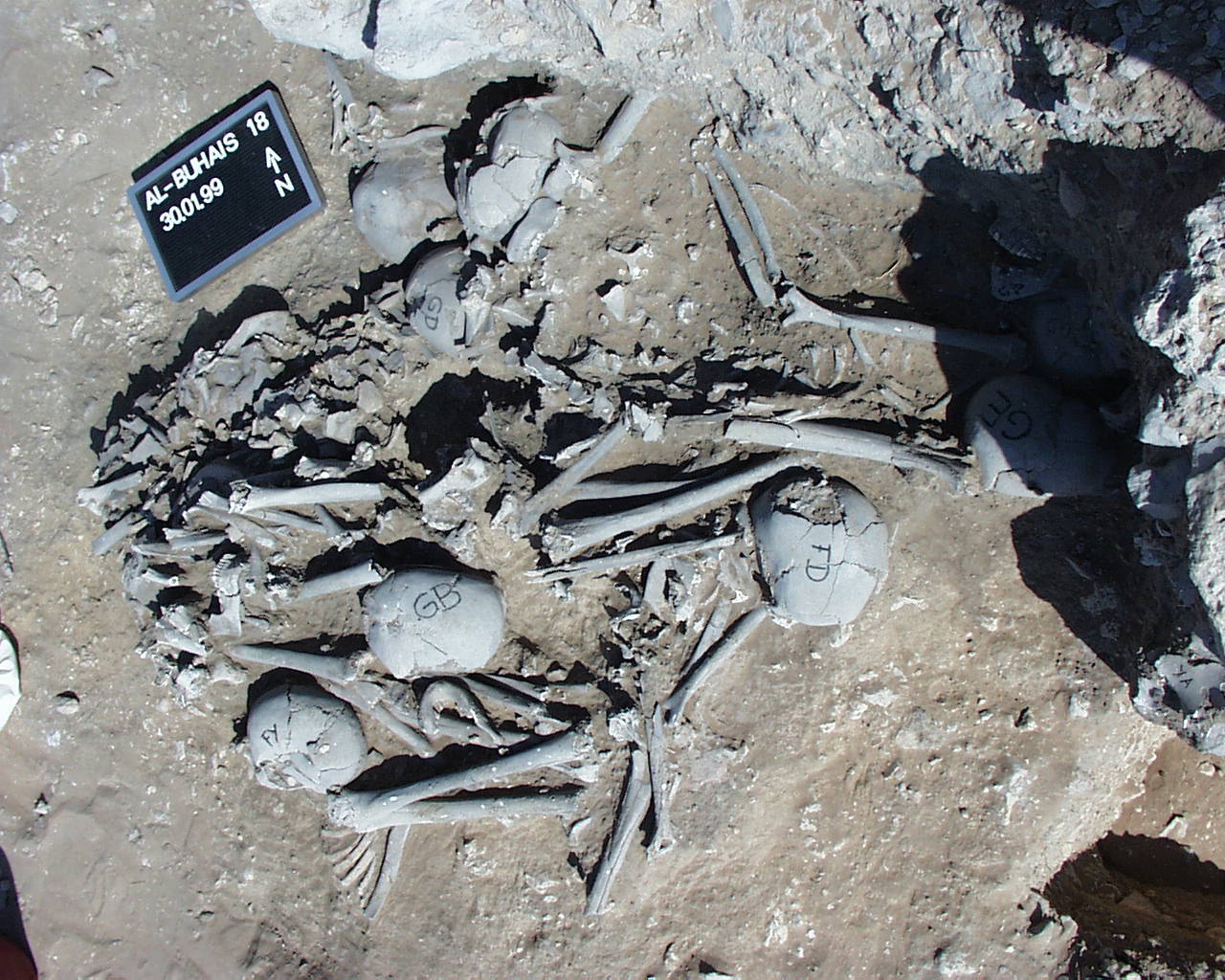
Inidvidual GB:
FD, FY, FZ, GA, GB, GD, GE, GO, GP, GW, HA, KJ, KL in situ
Full Resolution
Full Resolution

Inidvidual GB:
FD, FY, FZ, GA, GB, GD, GE, GO, GP, GW, KM, KL in situ
Full Resolution
Full Resolution

Inidvidual GB:
FD, FY, GA, GB, GD, GE, GP, KL, YA in situ
Full Resolution
Full Resolution
Inidvidual GB:
FD, FY, GA, GB, GD, GE, GP, KL, YA in situ
Full Resolution
Full Resolution
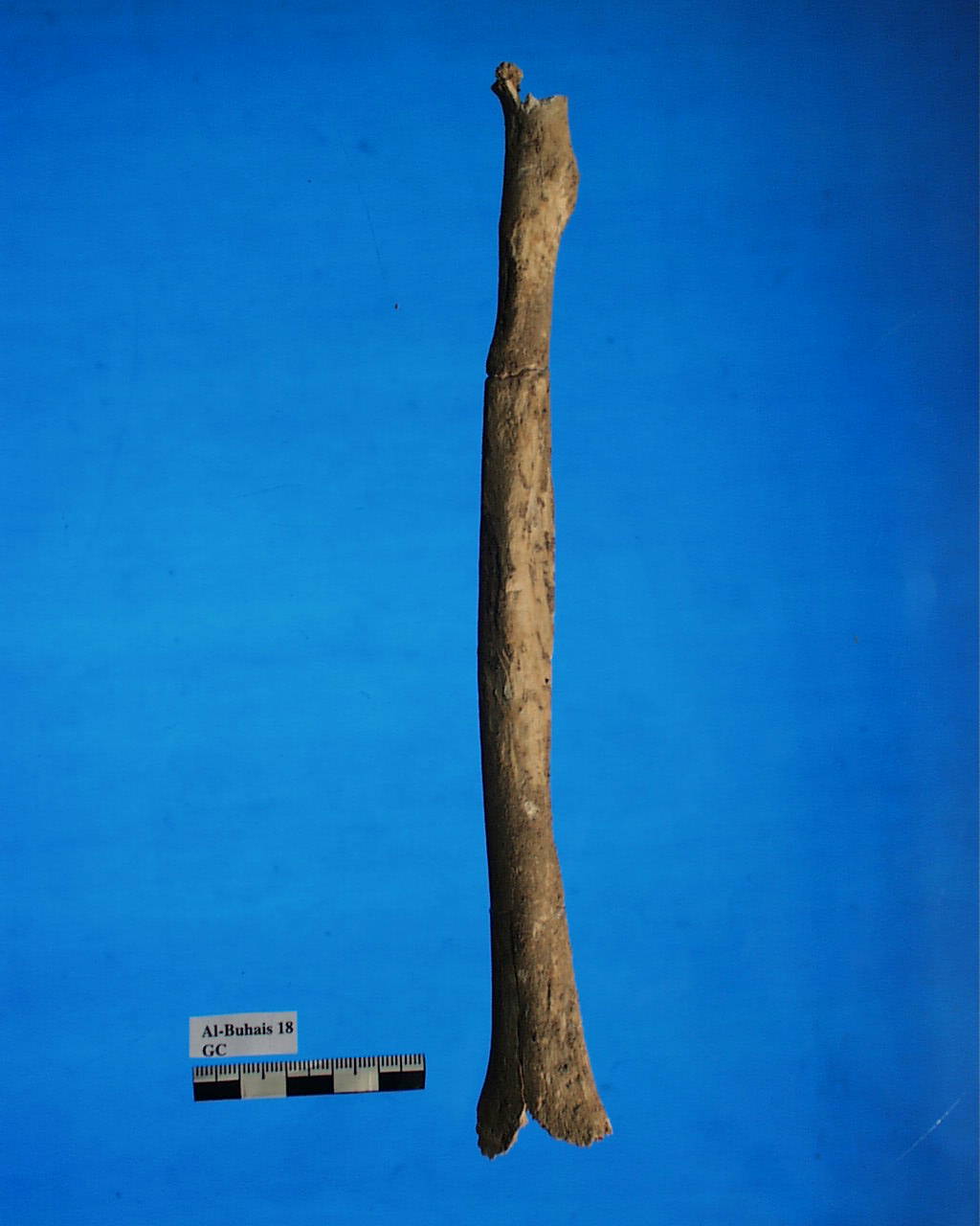
Inidvidual GC:
GC, left radius with atrophy
Full Resolution
Full Resolution
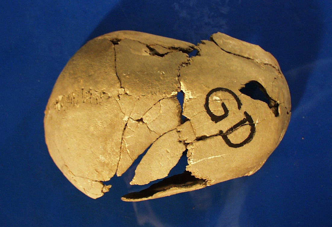
Inidvidual GD:
Skull GD with lethal head lesion
Full Resolution
Full Resolution

Inidvidual GD:
GD, lethal wound in skull, interior view
Full Resolution
Full Resolution

Inidvidual GD:
FD, FY, FZ, GA, GB, GD, GE, GO, GP, GW, HA, KJ, KL in situ
Full Resolution
Full Resolution

Inidvidual GD:
FD, FY, FZ, GA, GB, GD, GE, GO, GP, GW, KM, KL in situ
Full Resolution
Full Resolution
Inidvidual GD:
FD, FY, GA, GB, GD, GE, GP, KL, YA in situ
Full Resolution
Full Resolution

Inidvidual GD:
FD, FY, GA, GB, GD, GE, GP, KL, YA in situ
Full Resolution
Full Resolution
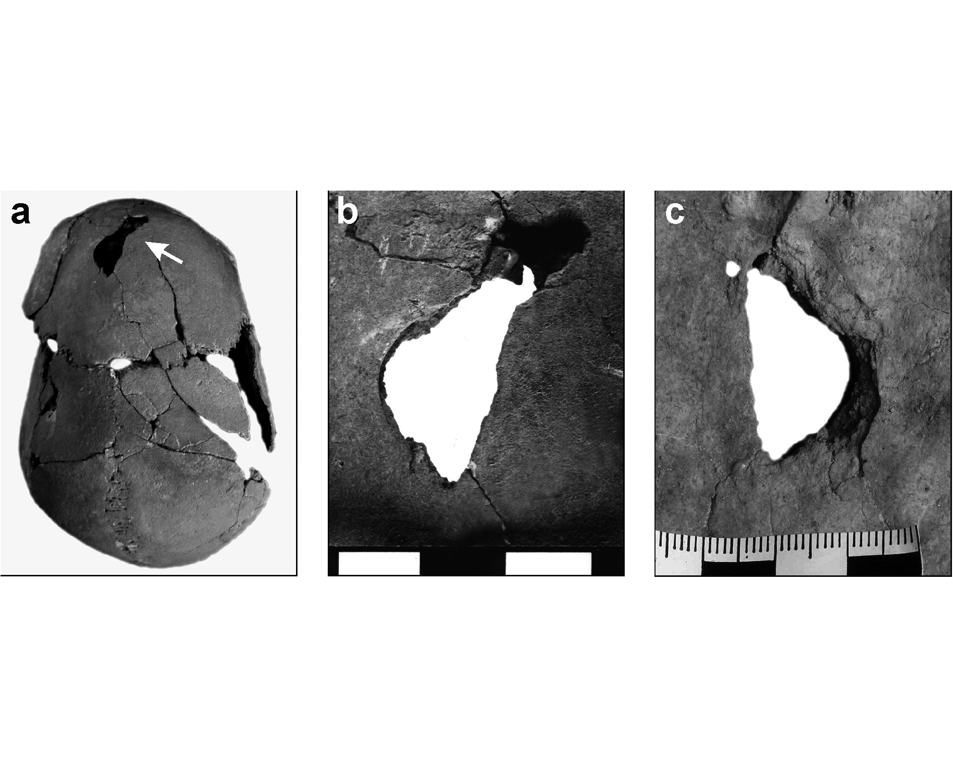
Inidvidual GD:
Skull injury (Fig. 6.2. in Kiesewetter 2006, Archaeology of Jebel al-Buhais, Volume 1)
Full Resolution
Full Resolution
Inidvidual GE:
FD, FY, FZ, GA, GB, GD, GE, GO, GP, GW, HA, KJ, KL in situ
Full Resolution
Full Resolution

Inidvidual GE:
FD, FY, FZ, GA, GB, GD, GE, GO, GP, GW, KM, KL in situ
Full Resolution
Full Resolution

Inidvidual GE:
FD, FY, GA, GB, GD, GE, GP, KL, YA in situ
Full Resolution
Full Resolution

Inidvidual GE:
FD, FY, GA, GB, GD, GE, GP, KL, YA in situ
Full Resolution
Full Resolution

Inidvidual GG:
Multiple burial
Full Resolution
Full Resolution
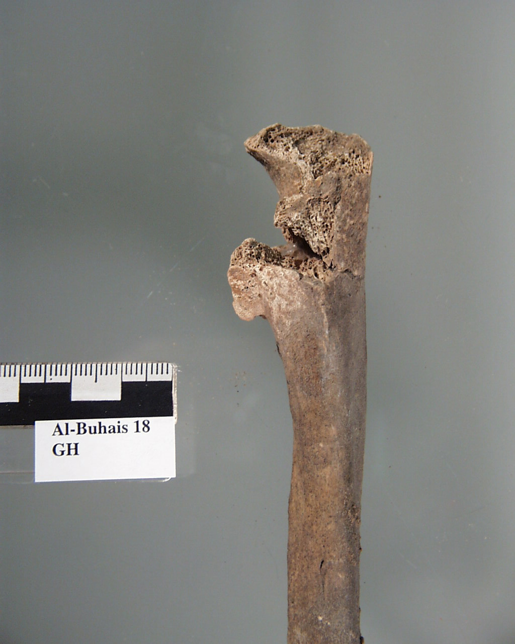
Inidvidual GH:
GH, right Ulna with osteophytes
Full Resolution
Full Resolution

Inidvidual GI:
Multiple burial
Full Resolution
Full Resolution

Inidvidual GO:
FD, FY, FZ, GA, GB, GD, GE, GO, GP, GW, HA, KJ, KL in situ
Full Resolution
Full Resolution

Inidvidual GO:
FD, FY, FZ, GA, GB, GD, GE, GO, GP, GW, KM, KL in situ
Full Resolution
Full Resolution

Inidvidual GP:
FD, FY, GA, GB, GD, GE, GP, KL, YA in situ
Full Resolution
Full Resolution

Inidvidual GP:
FD, FY, GA, GB, GD, GE, GP, KL, YA in situ
Full Resolution
Full Resolution

Inidvidual GP:
GA, GP and KL in situ
Full Resolution
Full Resolution
Inidvidual GP:
Individual GP in situ
Full Resolution
Full Resolution
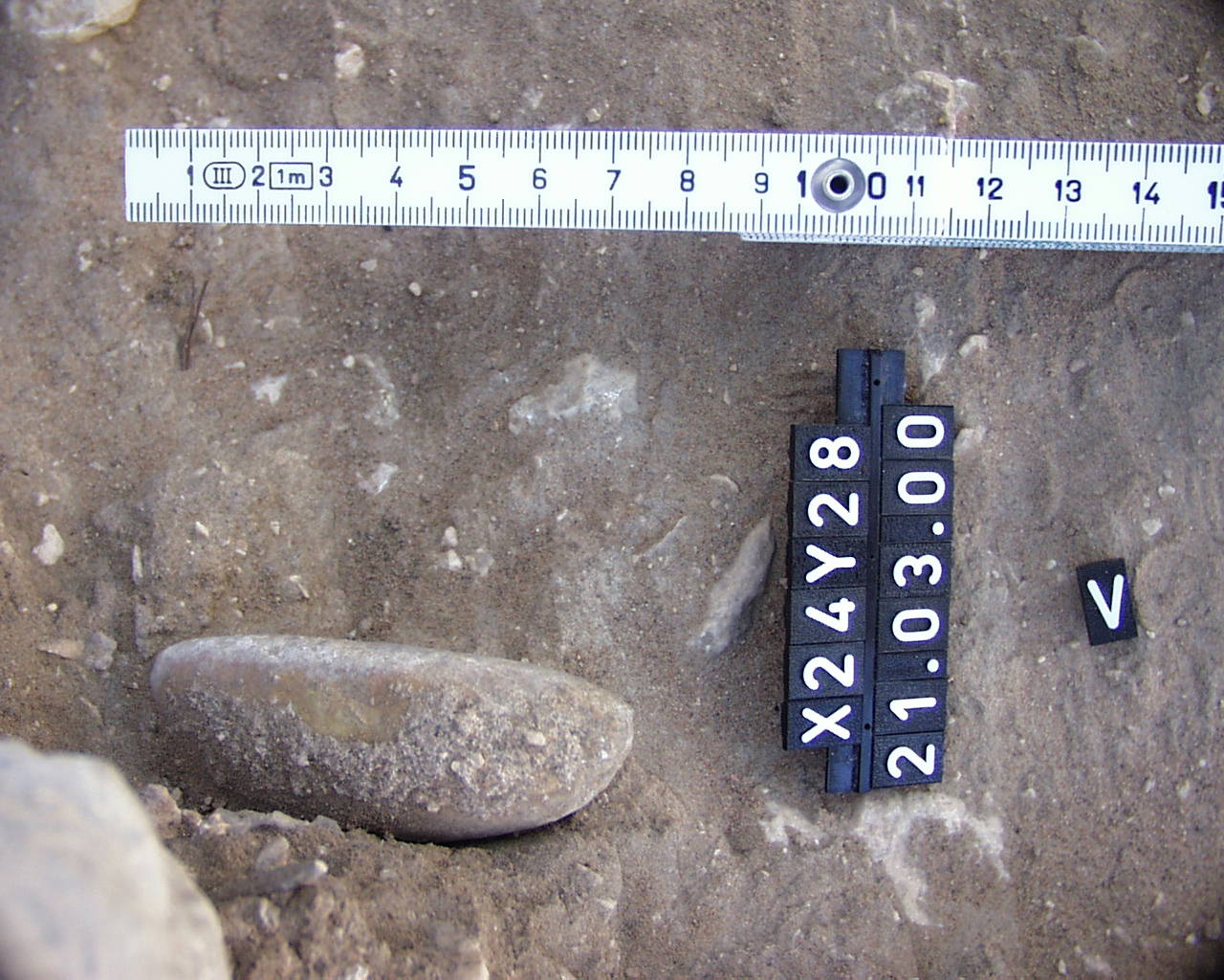
Inidvidual GP:
Stone adze in situ
Full Resolution
Full Resolution
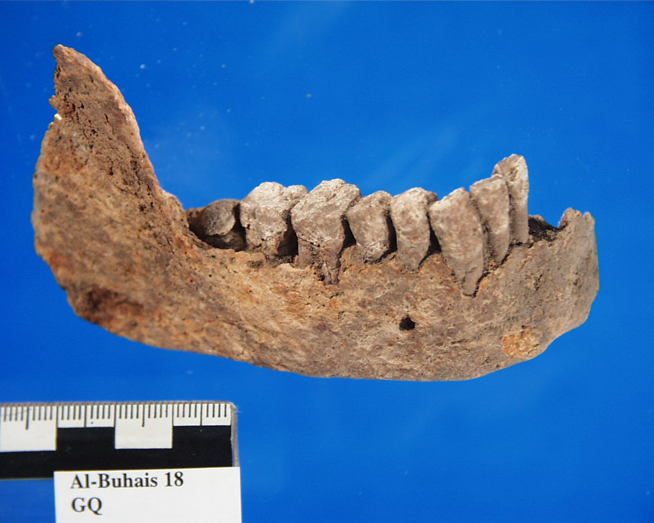
Inidvidual GQ:
mandible GQ with defective position of the wisdom tooth
Full Resolution
Full Resolution
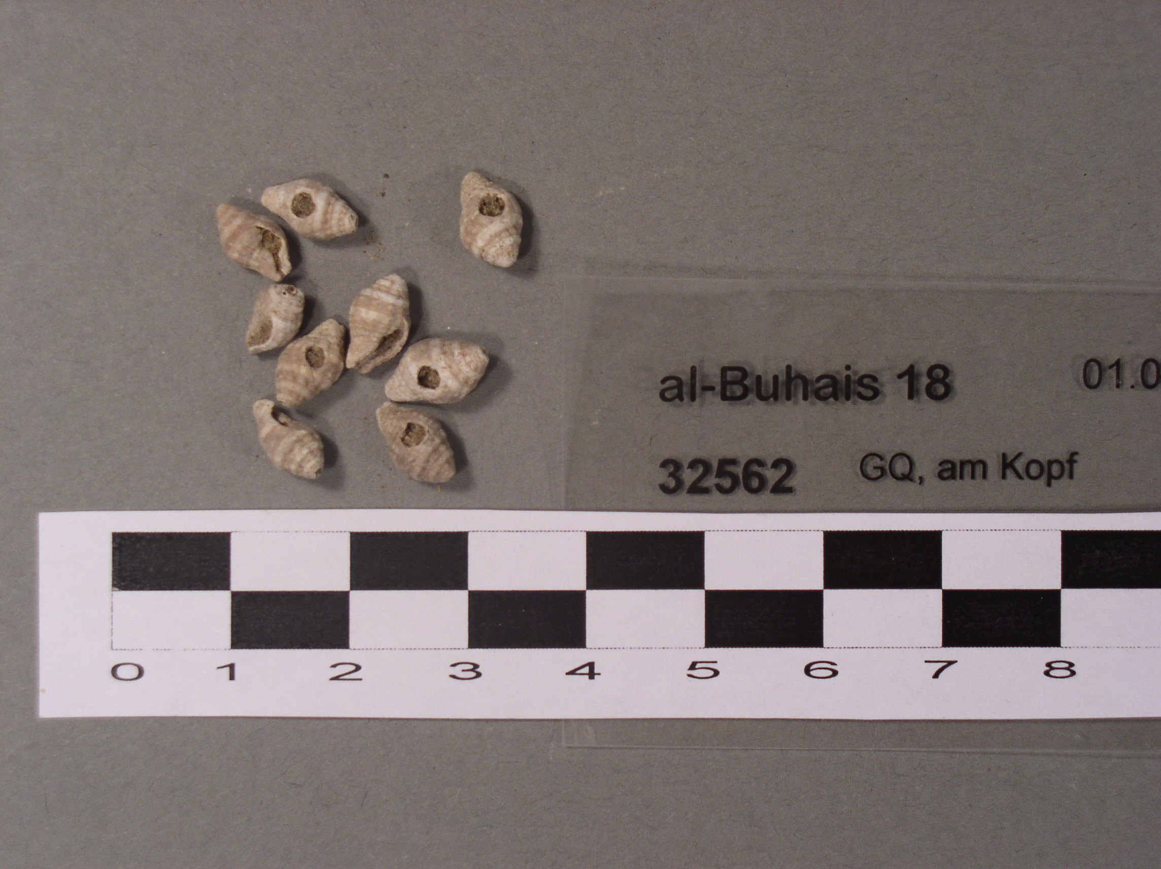
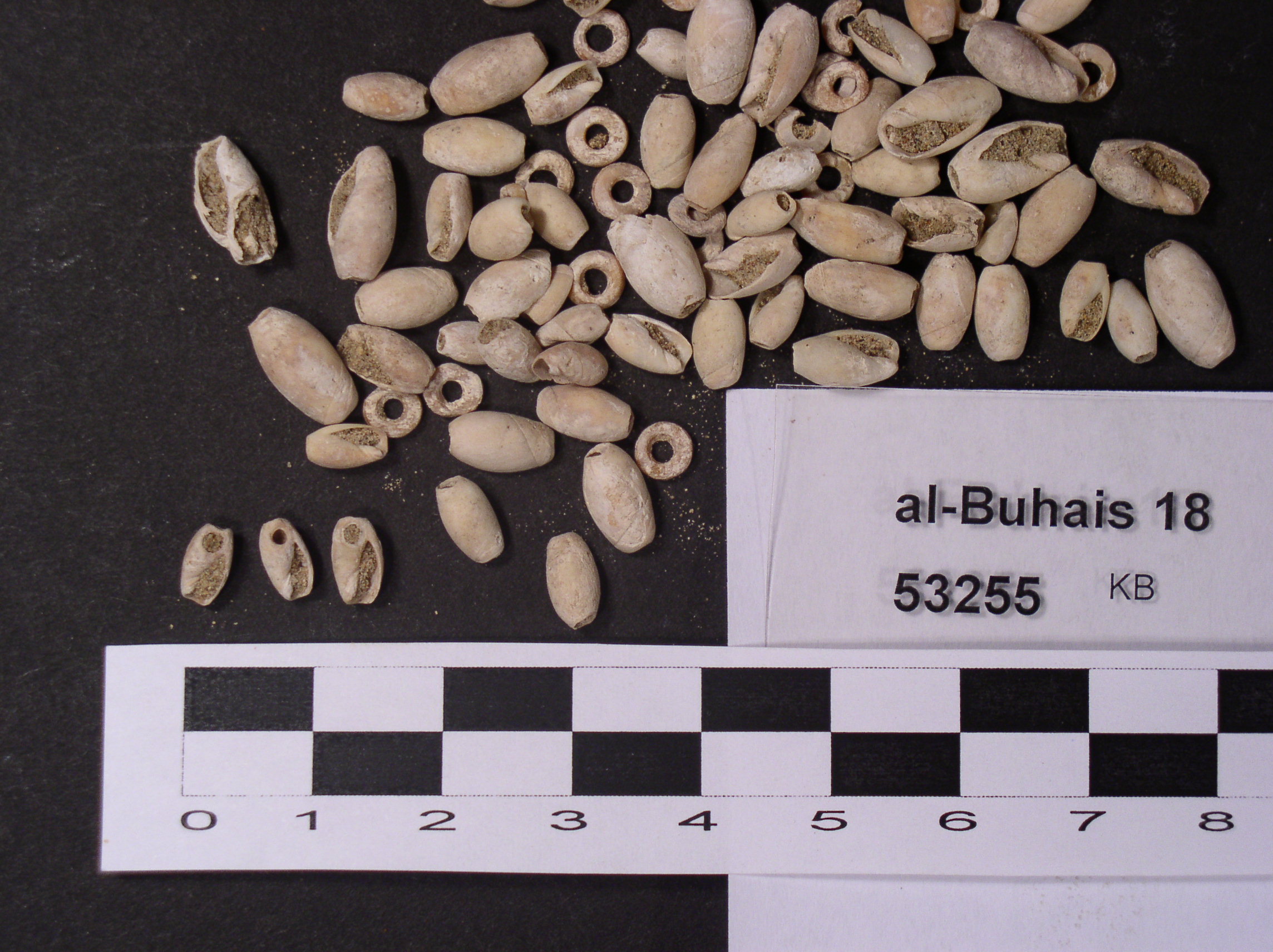
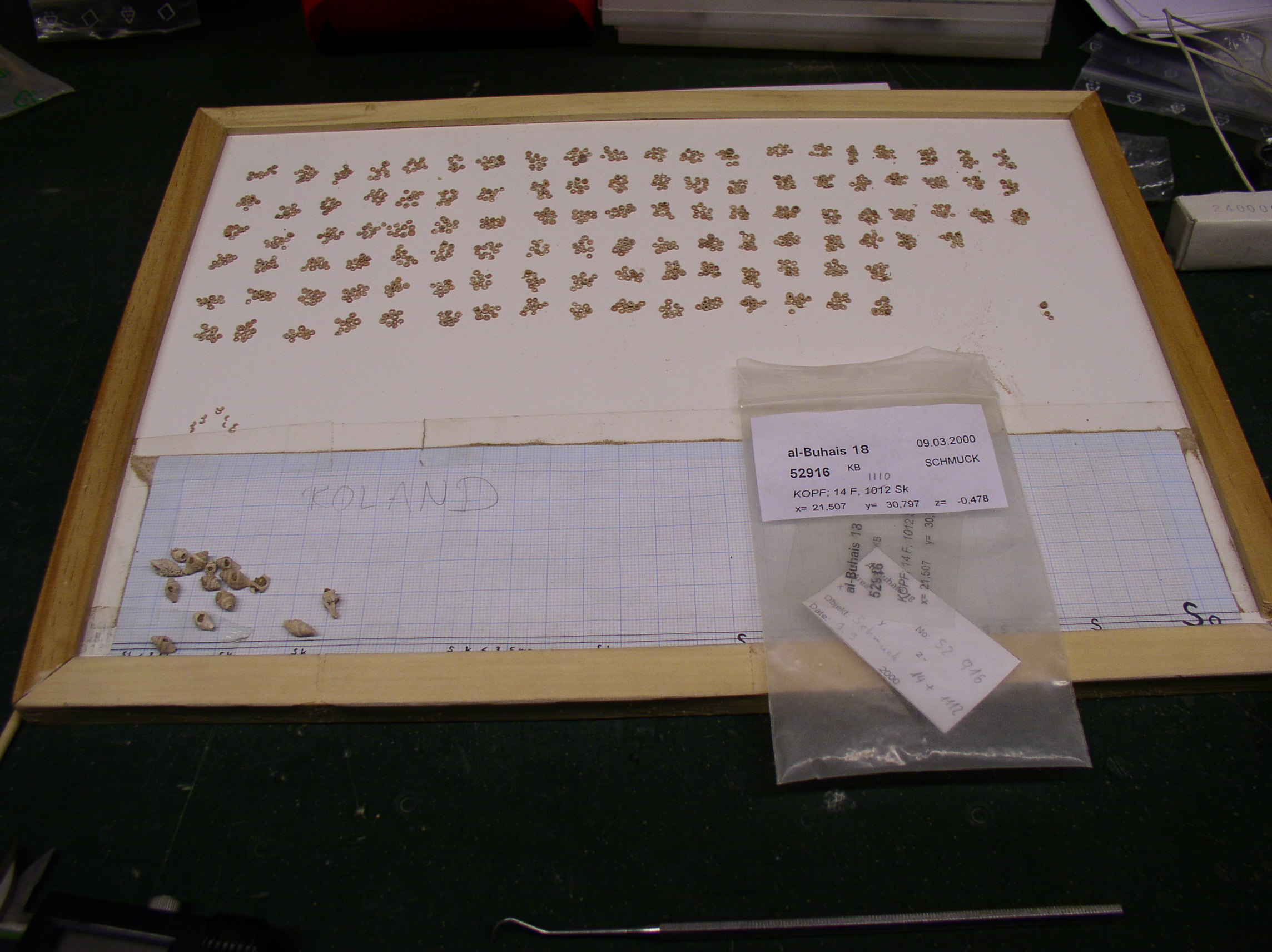
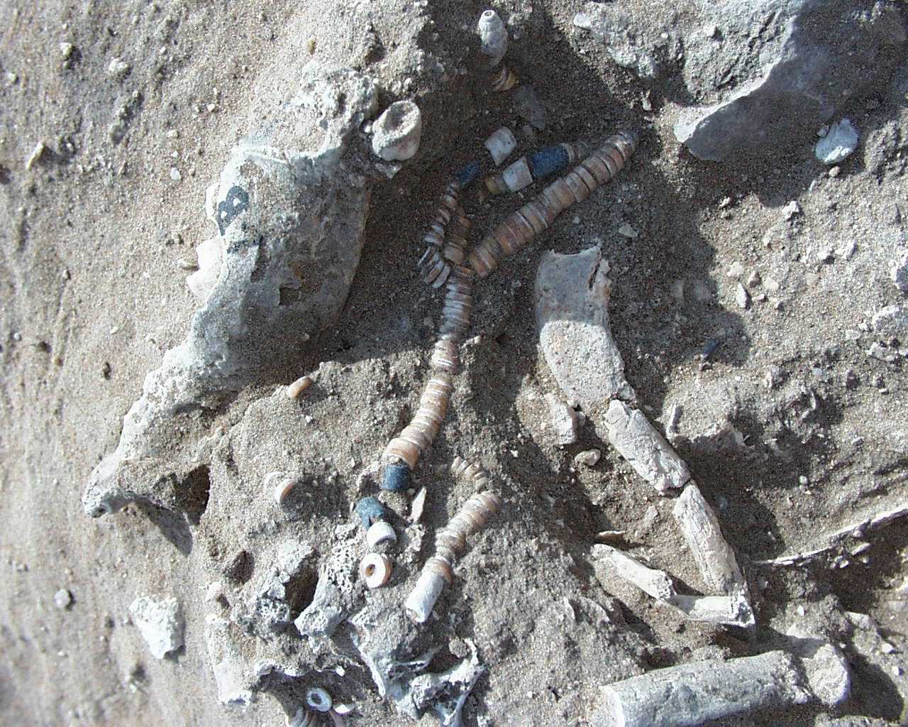
Inidvidual GQ:
Adornments head area
Full Resolution
Full Resolution
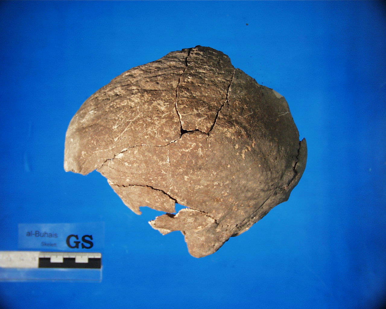
Inidvidual GS:
GS, linear impressions in frontal bone. cause?
Full Resolution
Full Resolution
Inidvidual GT:
Adornments
Full Resolution
Full Resolution
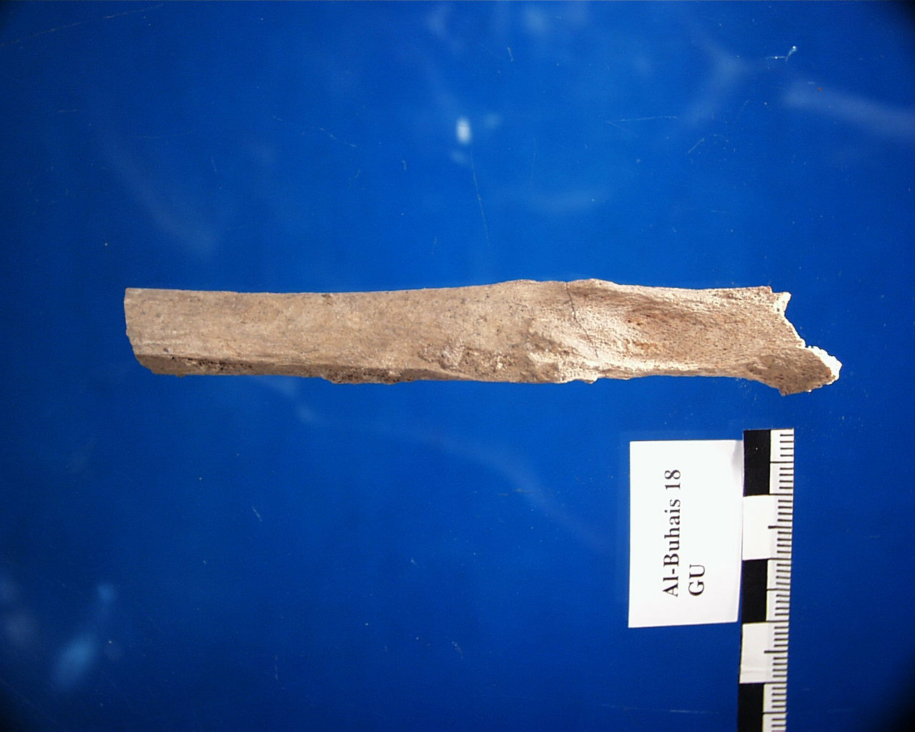
Inidvidual GU:
GU, fracture of fibula
Full Resolution
Full Resolution

Inidvidual GW:
FD, FY, FZ, GA, GB, GD, GE, GO, GP, GW, HA, KJ, KL in situ
Full Resolution
Full Resolution

Inidvidual GW:
FD, FY, FZ, GA, GB, GD, GE, GO, GP, GW, KM, KL in situ
Full Resolution
Full Resolution

Inidvidual HA:
FD, FY, FZ, GA, GB, GD, GE, GO, GP, GW, HA, KJ, KL in situ. HA and KJ are located in the lower most part of the picture.
Full Resolution
Full Resolution
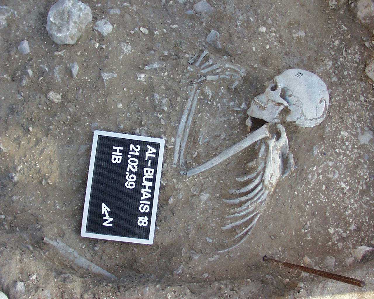
Inidvidual HB:
individual HB in situ
Full Resolution
Full Resolution

Inidvidual HB:
HB, HC and LC in situ
Full Resolution
Full Resolution
Inidvidual HB:
Pendant of HB during excavation
Full Resolution
Full Resolution
Inidvidual HC:
HB, HC and LC in situ
Full Resolution
Full Resolution

Inidvidual HC:
LC and HC in situ
Full Resolution
Full Resolution

Inidvidual HC:
LC and HC in situ
Full Resolution
Full Resolution
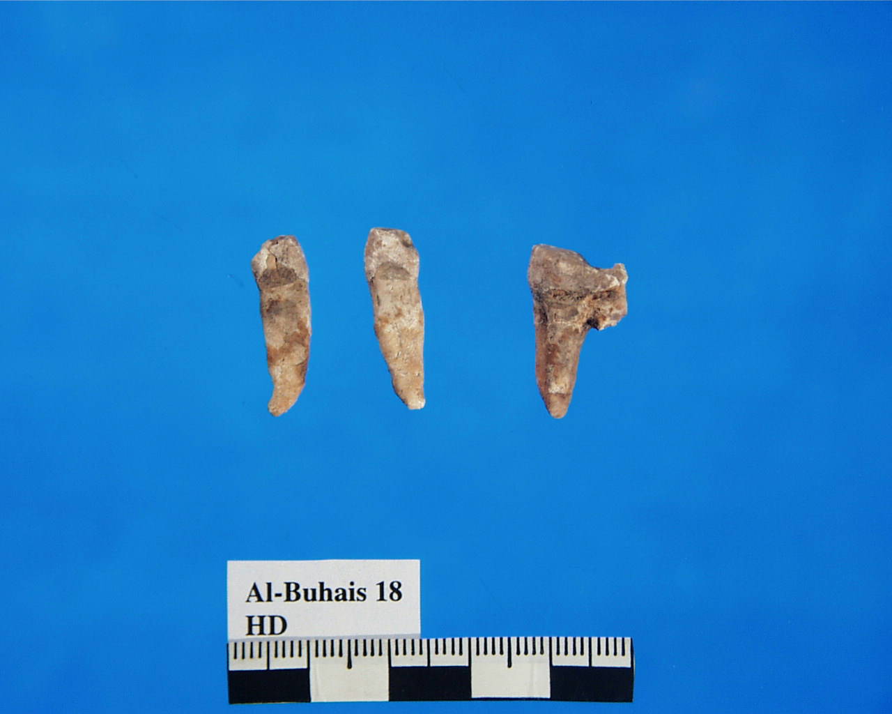
Inidvidual HD:
HD, teeth with tooth pick grooves
Full Resolution
Full Resolution
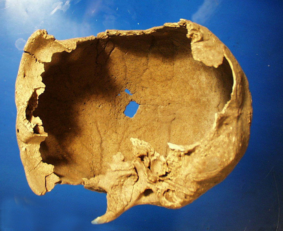
Inidvidual HD:
Skull HD interior
Full Resolution
Full Resolution

Inidvidual HD:
HD, HE, EX and LK, Depot. In situ
Full Resolution
Full Resolution

Inidvidual HD:
HD, HE, EX and LK, Depot in situ
Full Resolution
Full Resolution
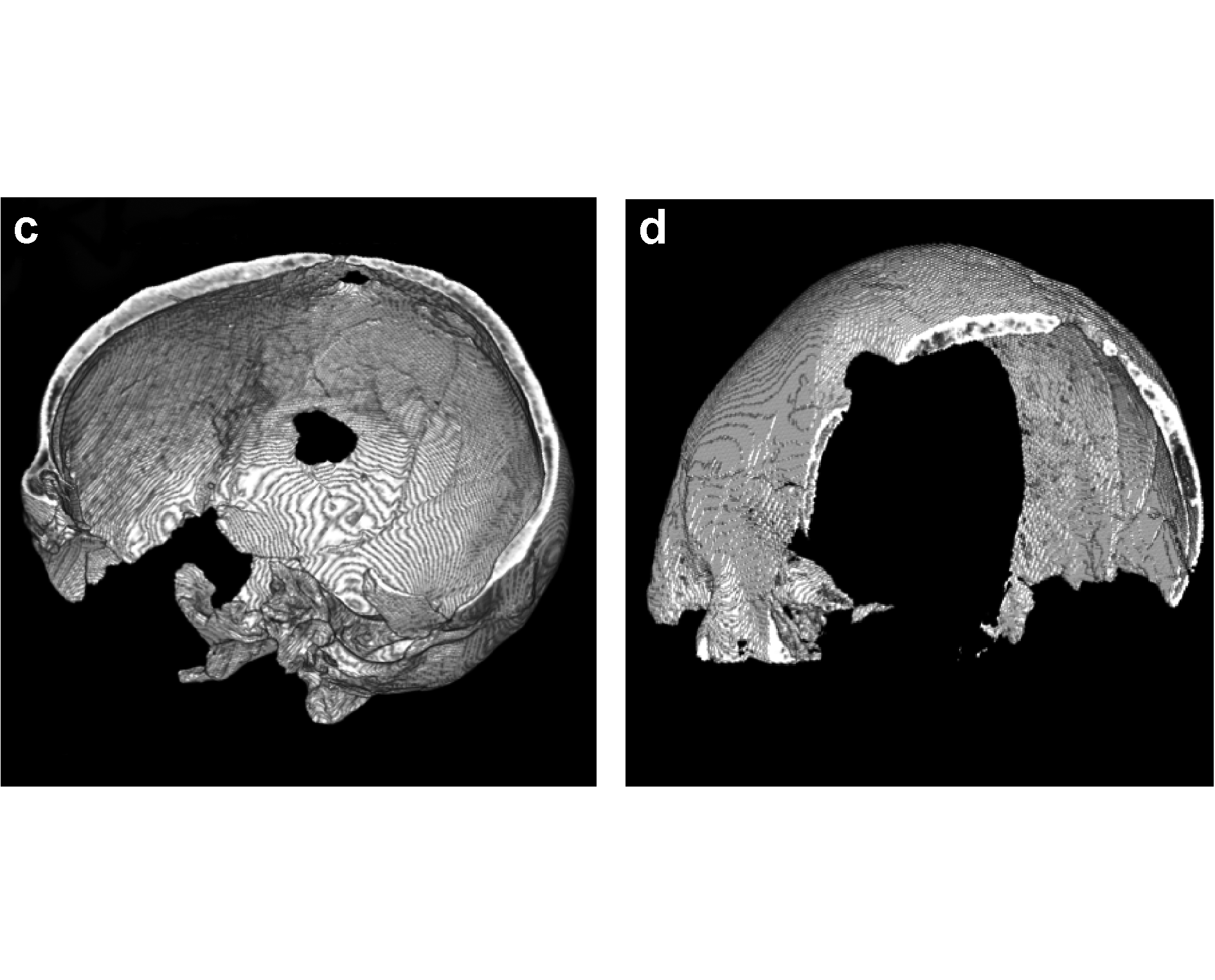
Inidvidual HD:
CT-scan, showing the trephined area, virtual cut (Kiesewetter 2006)
Full Resolution
Full Resolution

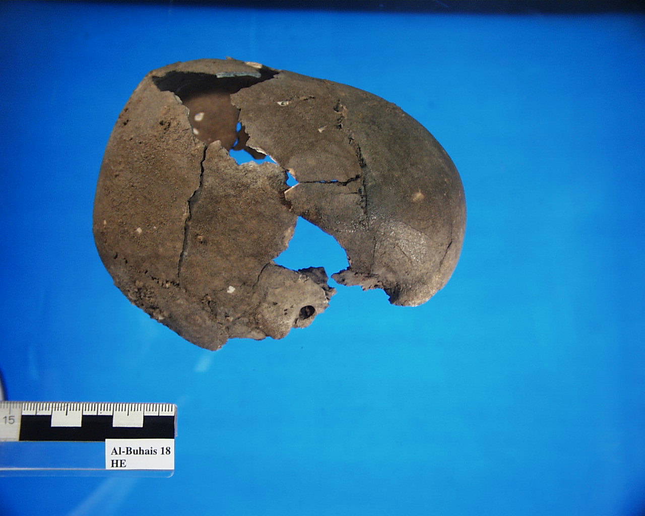
Inidvidual HD:
Cranium HD
Full Resolution
Full Resolution
Inidvidual HE:
HE, infant skull
Full Resolution
Full Resolution
Inidvidual HE:
HE, skeleton and skull. Postcranial bones could also belong to LK
Full Resolution
Full Resolution

Inidvidual HE:
HD, HE, EX and LK, Depot. In situ
Full Resolution
Full Resolution

Inidvidual HE:
HD, HE, EX and LK, Depot in situ
Full Resolution
Full Resolution

Inidvidual HN:
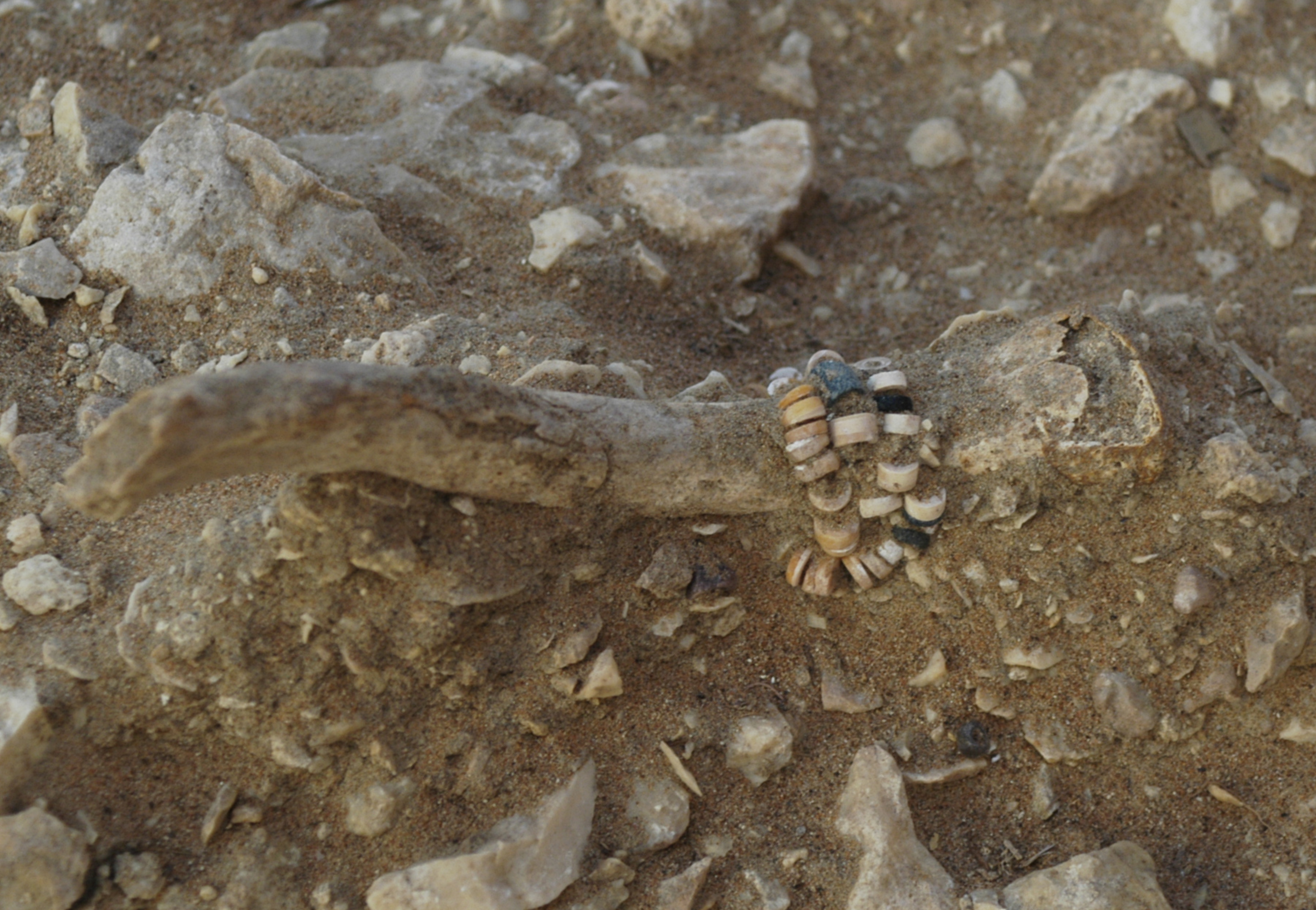
Adornments individual HN. Tubular beads, disc beads and molluscs
Full Resolution
Full Resolution

Inidvidual HP:
Individual HP in situ
Full Resolution
Full Resolution
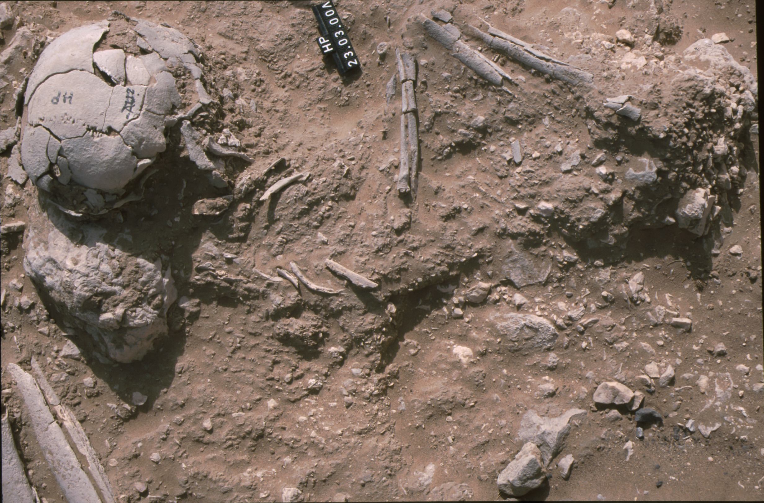
Inidvidual HP:
HP detail
Full Resolution
Full Resolution
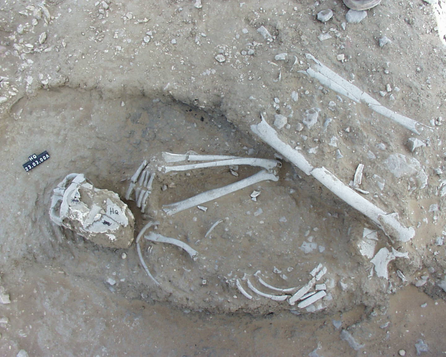
Inidvidual HQ:
individual HQ in situ
Full Resolution
Full Resolution

Inidvidual HS:
BX, BY, HS and CL in situ
Full Resolution
Full Resolution

Inidvidual HS:
BX, BY, HS, CL, KG and KE in situ
Full Resolution
Full Resolution

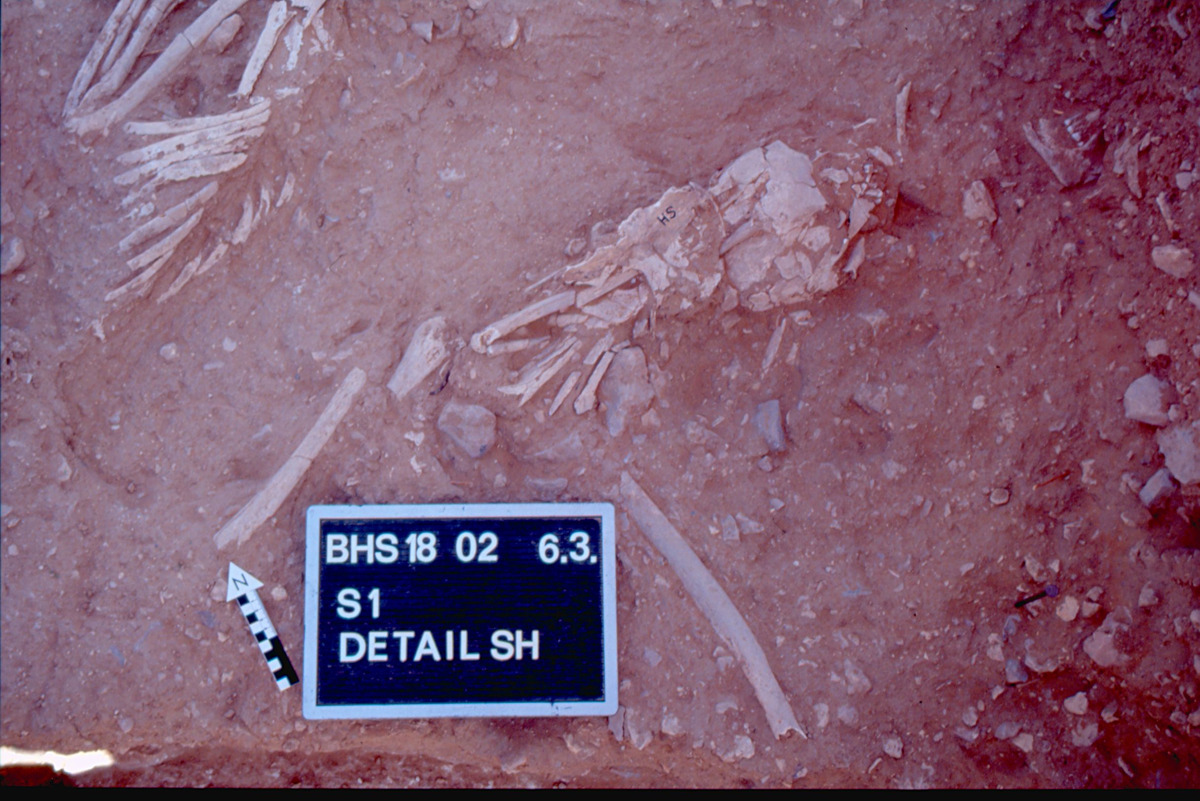
Inidvidual HS:
Detail HS
Full Resolution
Full Resolution
Inidvidual HS:
Detail HS and BY, adornments
Full Resolution
Full Resolution
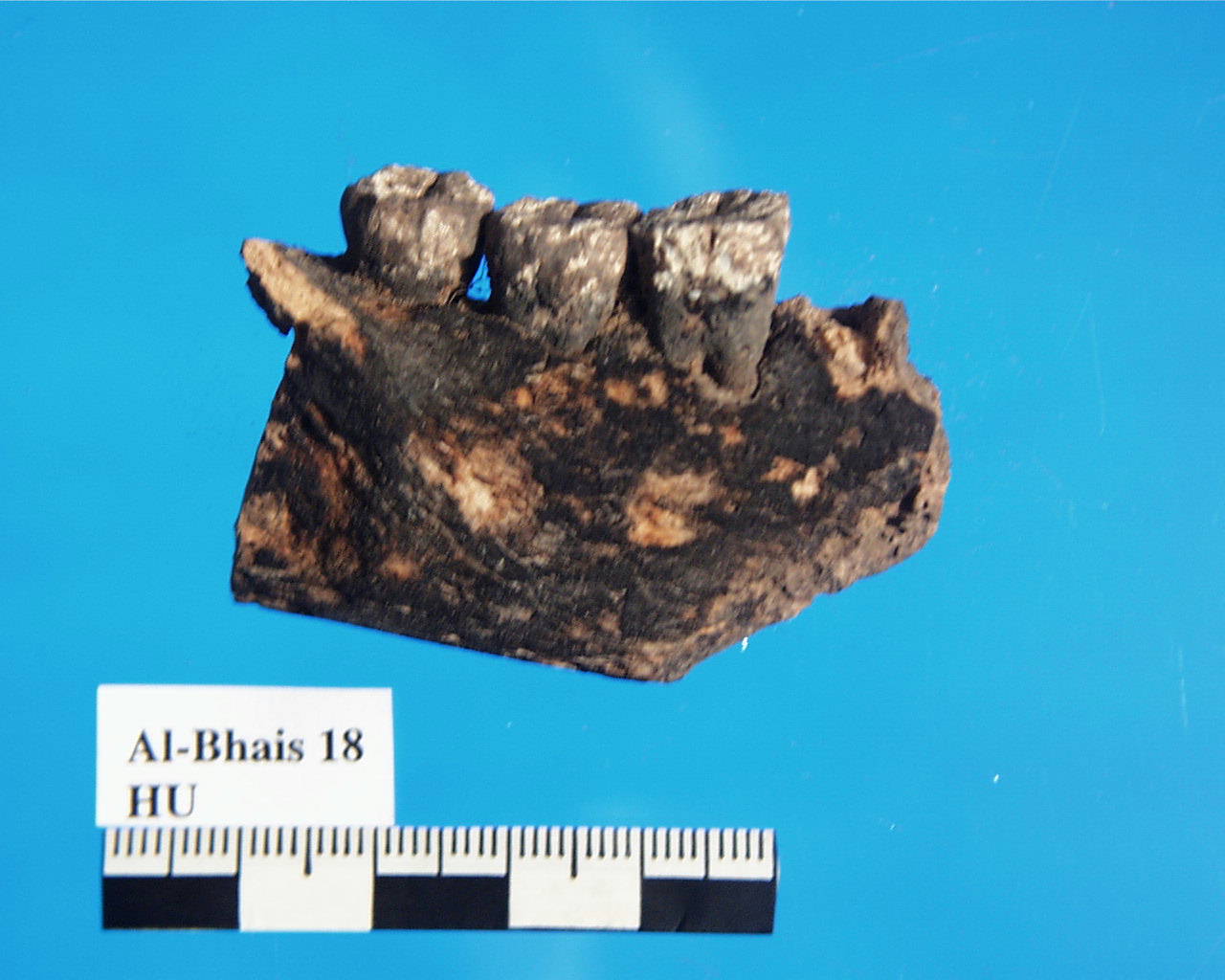
Inidvidual HU:
Mandible HU, burnt
Full Resolution
Full Resolution
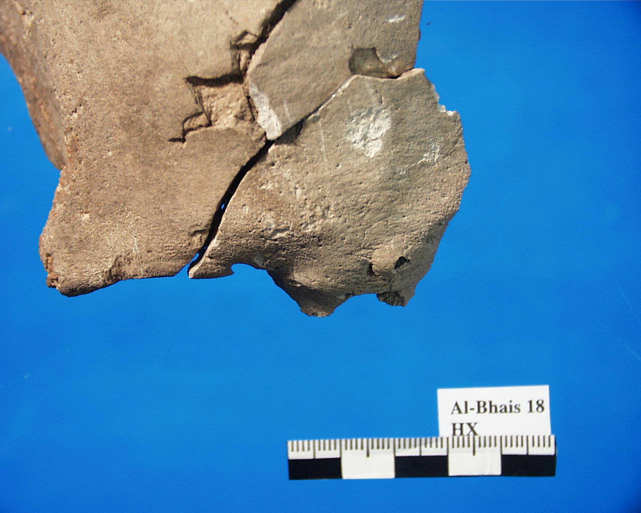
Inidvidual HX:
HX, Osteoma on frontal bone
Full Resolution
Full Resolution
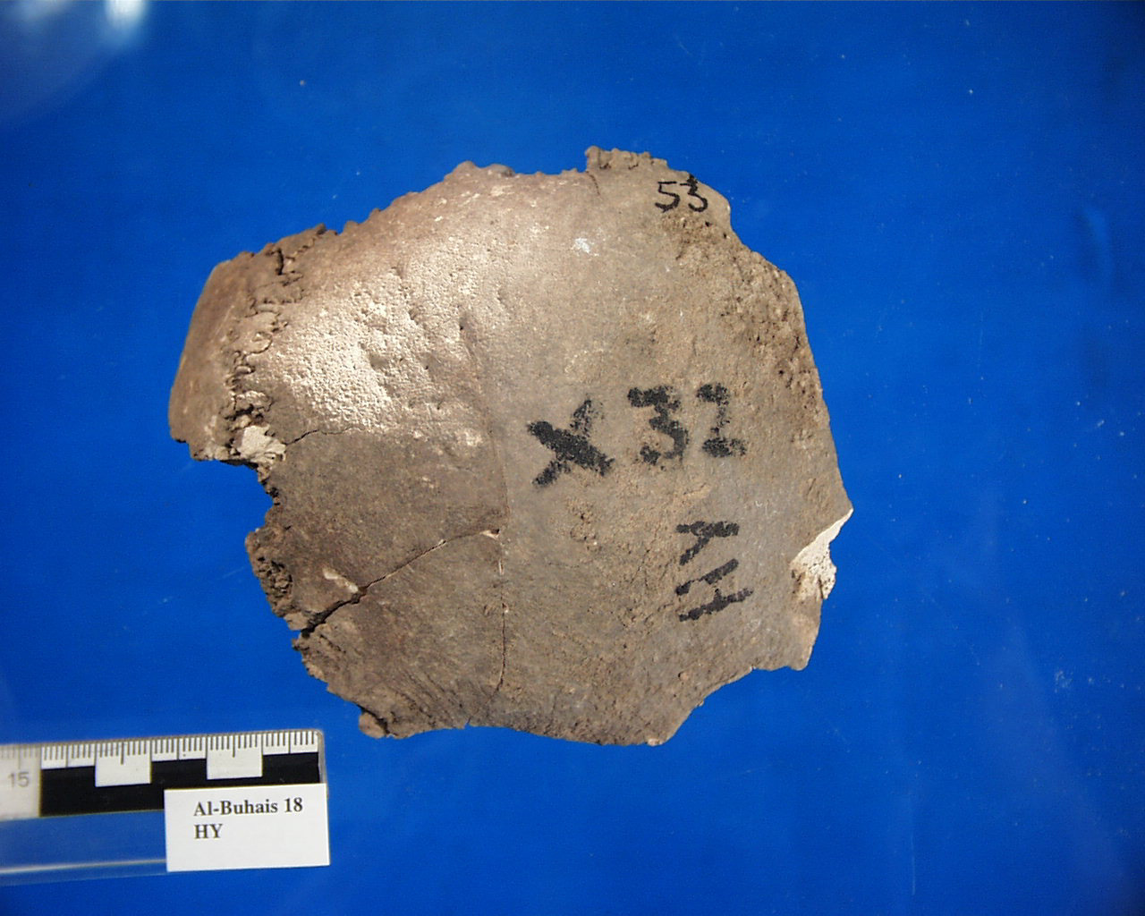
Inidvidual HY:
Skull fragment HY with signs of inflammation
Full Resolution
Full Resolution
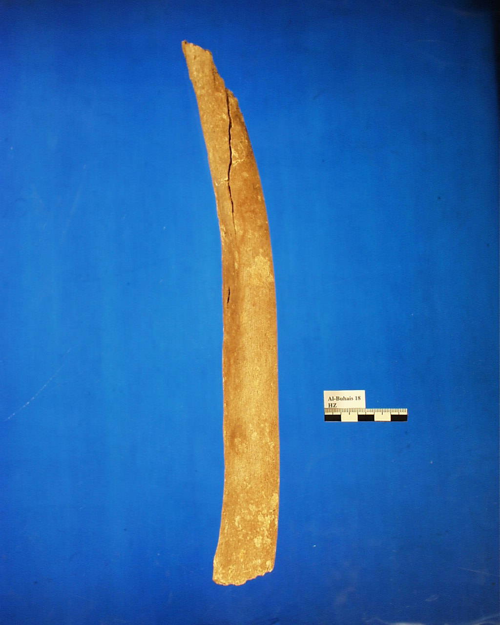
Inidvidual HZ:
Femur HZ, deformed
Full Resolution
Full Resolution
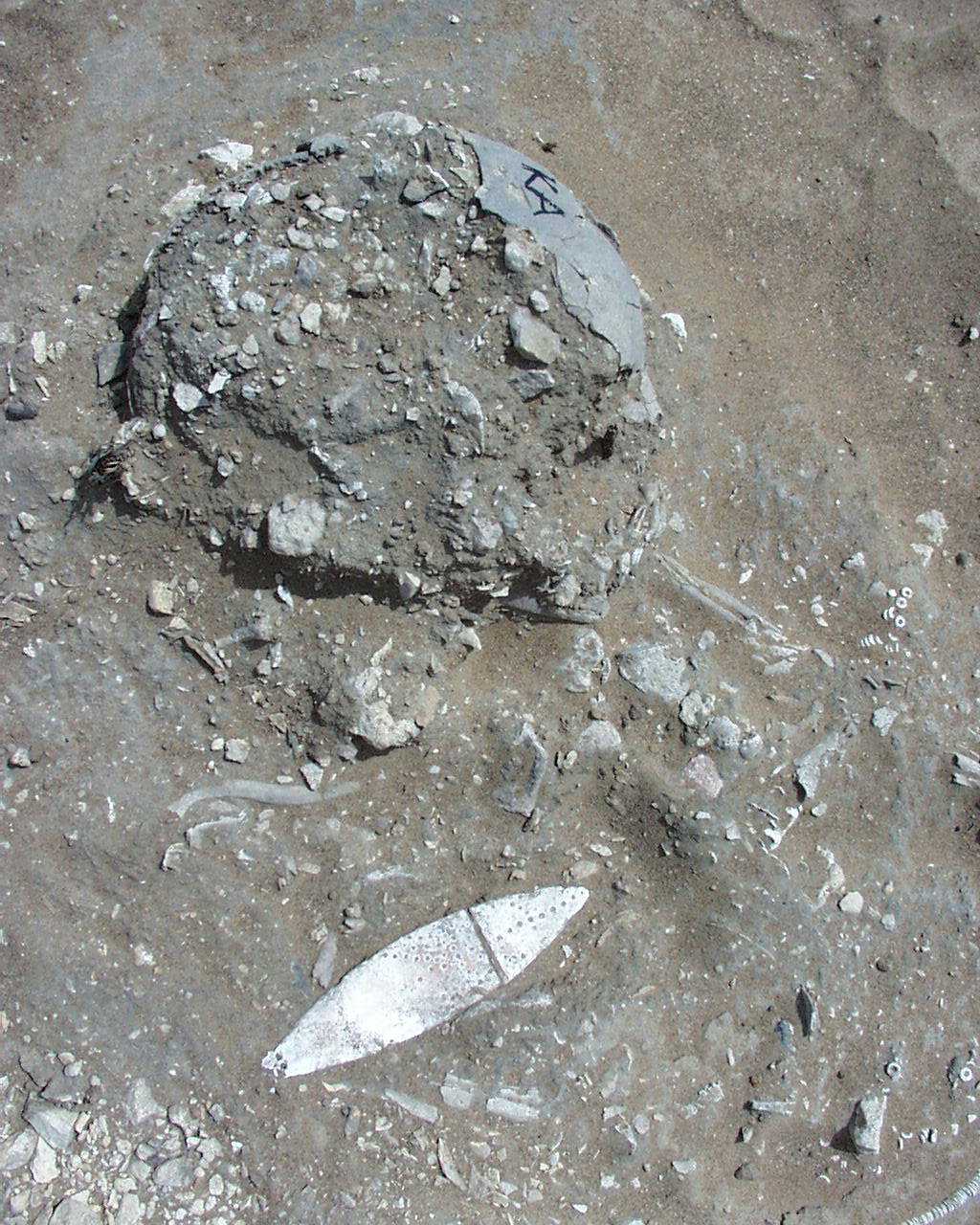
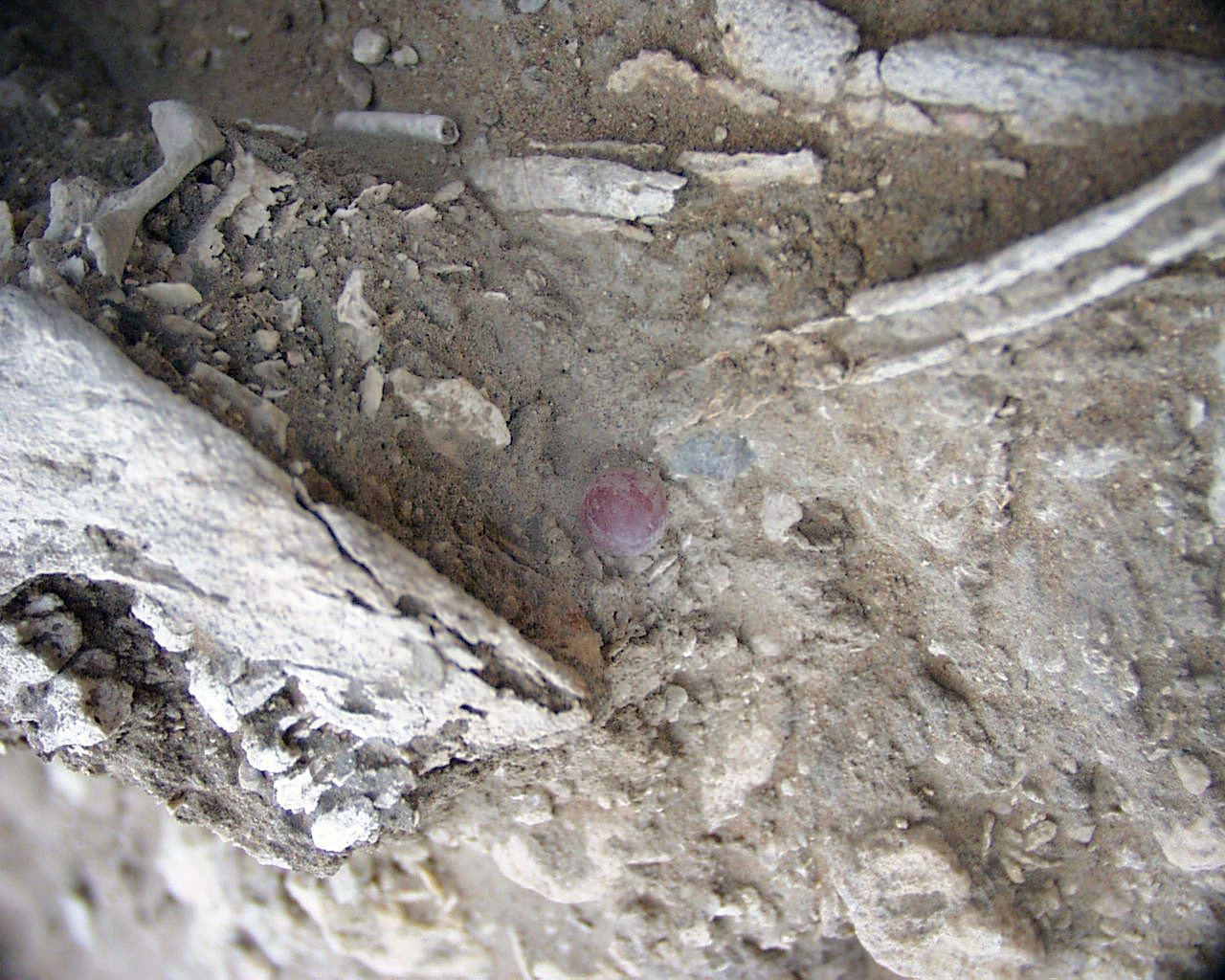
Inidvidual KA:
Individual KA with pendant
Full Resolution
Full Resolution

Inidvidual KA:
Pendant KA
Full Resolution
Full Resolution
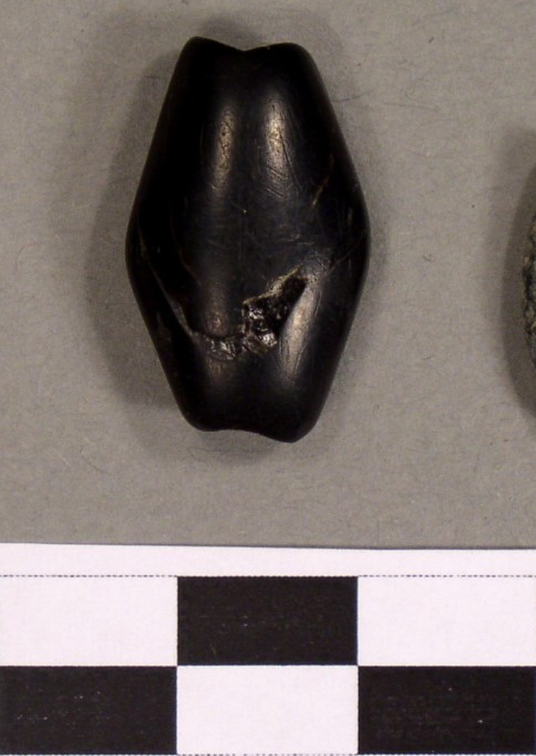
Inidvidual KA:
Black rhomboid bead KA
Full Resolution
Full Resolution
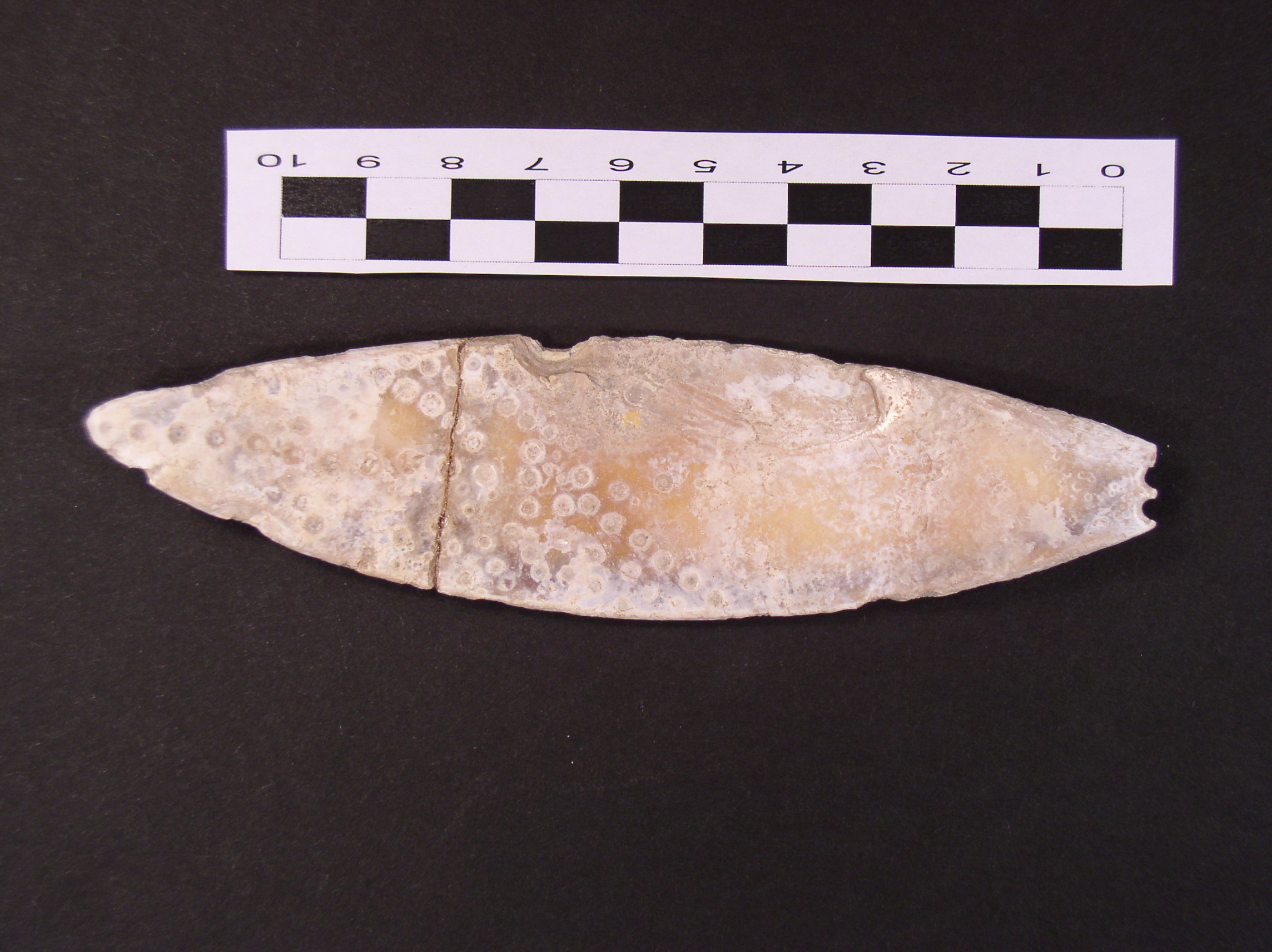
Inidvidual KA:
Pendant with scale KA
Full Resolution
Full Resolution

Inidvidual KB:
Skull KB with adornments in situ
Full Resolution
Full Resolution
Inidvidual KB:
Skull KB with adornments in situ, carnelian bead
Full Resolution
Full Resolution
Inidvidual KB:
KB adornments
Full Resolution
Full Resolution
Inidvidual KB:
Disc beads head KB
Full Resolution
Full Resolution
Inidvidual KE:
BX, BY, HS, CL, KG and KE in situ
Full Resolution
Full Resolution

Inidvidual KG:
BX, BY, HS, CL, KG and KE in situ
Full Resolution
Full Resolution
Inidvidual KG:
Parry fracture at left ulna, KG
Full Resolution
Full Resolution

Inidvidual KJ:
FD, FY, FZ, GA, GB, GD, GE, GO, GP, GW, HA, KJ, KL in situ
Full Resolution
Full Resolution
Inidvidual KJ:
Adornments KJ
Full Resolution
Full Resolution

Inidvidual KL:
FD, FY, FZ, GA, GB, GD, GE, GO, GP, GW, HA, KJ, KL in situ
Full Resolution
Full Resolution
Inidvidual KL:
FD, FY, FZ, GA, GB, GD, GE, GO, GP, GW, KM, KL in situ
Full Resolution
Full Resolution

Inidvidual KL:
FD, FY, GA, GB, GD, GE, GP, KL, YA in situ
Full Resolution
Full Resolution

Inidvidual KL:
FD, FY, GA, GB, GD, GE, GP, KL, YA in situ
Full Resolution
Full Resolution
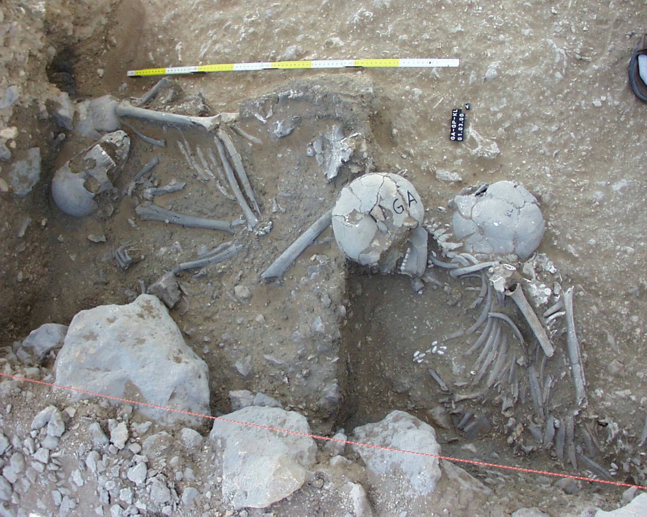
Inidvidual KL:
GA, GP and KL in situ
Full Resolution
Full Resolution

Inidvidual KM:
FD, FY, FZ, GA, GB, GD, GE, GO, GP, GW, KM, KL in situ
Full Resolution
Full Resolution
Inidvidual KS:
ED and KS in situ
Full Resolution
Full Resolution
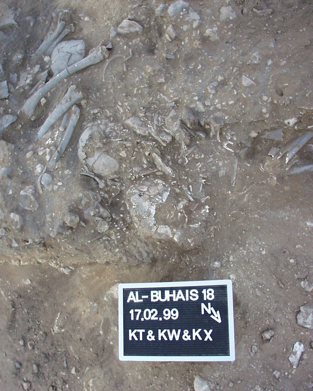
Inidvidual KT:
KT, KW, KX in situ
Full Resolution
Full Resolution
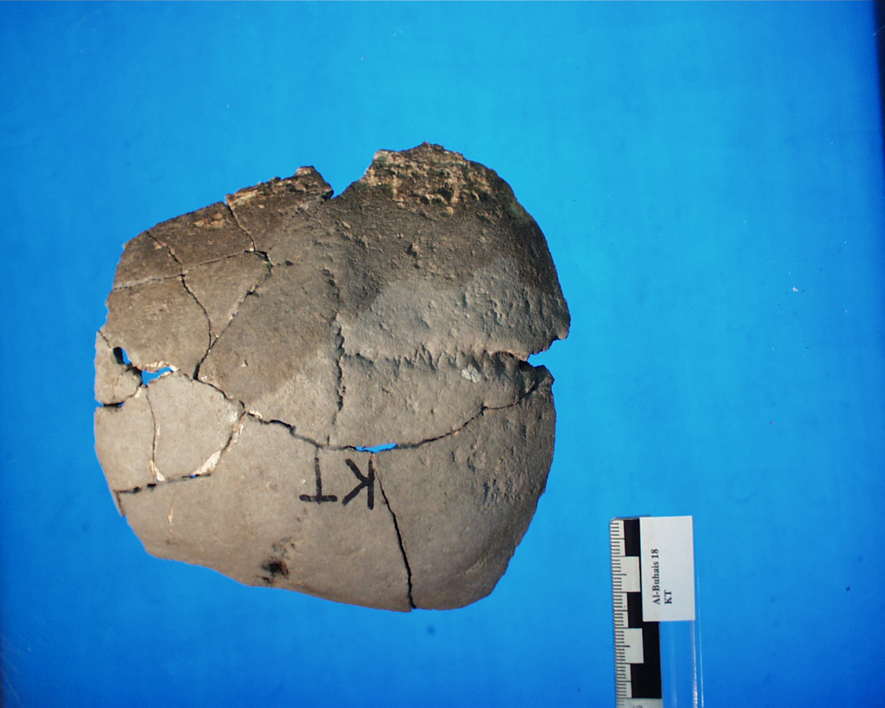
Inidvidual KT:
Skull KT superior
Full Resolution
Full Resolution
Inidvidual KV:
Adornments
Full Resolution
Full Resolution
Inidvidual KY:
Femura KY, deformed
Full Resolution
Full Resolution
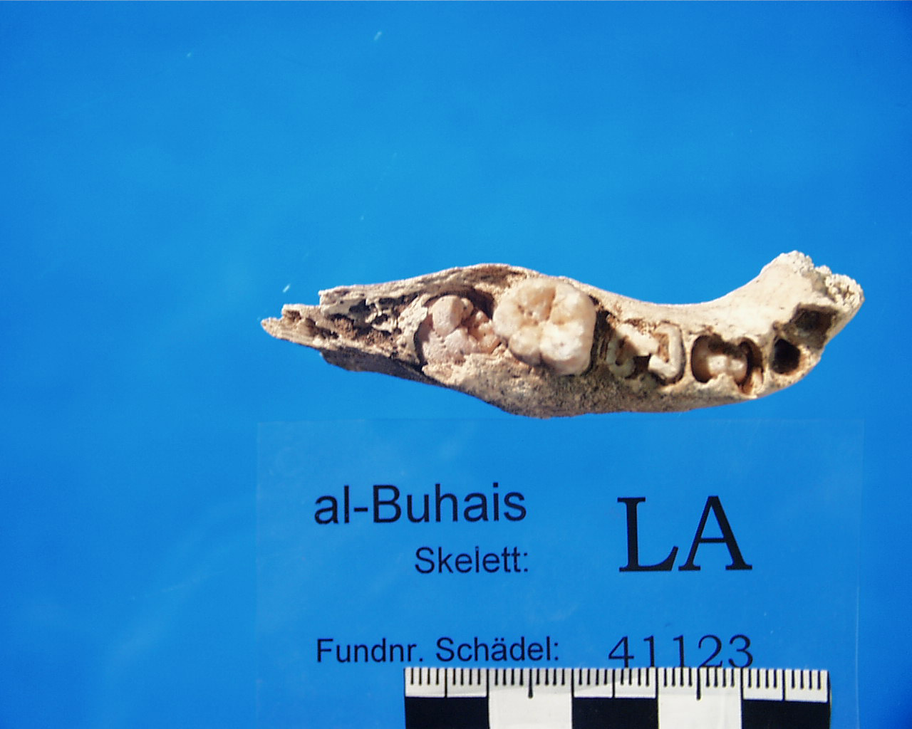
Inidvidual LA:
Mandible fragment LA, enamel hypoplasia is visible at the erupting tooth
Full Resolution
Full Resolution
Inidvidual LB:
Adornments LB, necklace
Full Resolution
Full Resolution
Inidvidual LB:
Adornments LB, necklace
Full Resolution
Full Resolution
Inidvidual LB:
Adornments LB, stomach area
Full Resolution
Full Resolution
Inidvidual LB:
LB, adornments neck, necklace
Full Resolution
Full Resolution

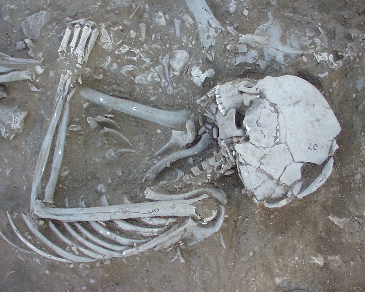
Inidvidual LC:
HB, HC and LC in situ
Full Resolution
Full Resolution
Inidvidual LC:
Burial LC, upper part of body. On the skull mollusk beads are visible.
Full Resolution
Full Resolution
Inidvidual LC:
Burial LC in situ
Full Resolution
Full Resolution
Inidvidual LC:
LC in situ with pearl in front of the nose
Full Resolution
Full Resolution
Inidvidual LC:
LC and HC in situ
Full Resolution
Full Resolution
Inidvidual LC:
LC and HC in situ
Full Resolution
Full Resolution
Inidvidual LC:
Skull individual LC in situ
Full Resolution
Full Resolution
Inidvidual LC:
LC in situ during excavation. Workman.
Full Resolution
Full Resolution
Inidvidual LC:
LC detail skull
Full Resolution
Full Resolution
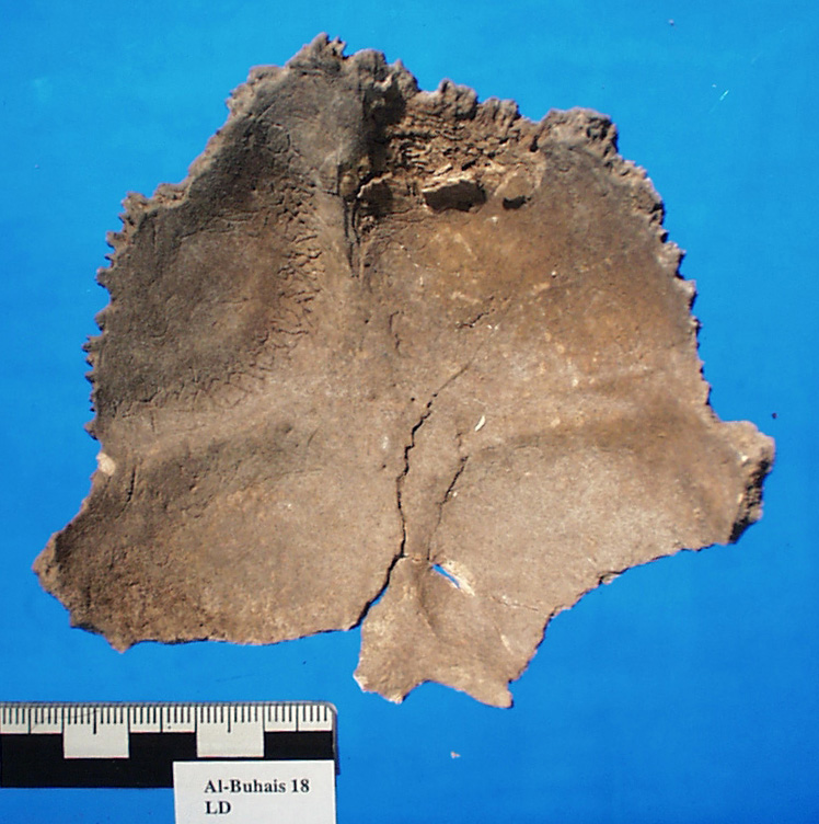
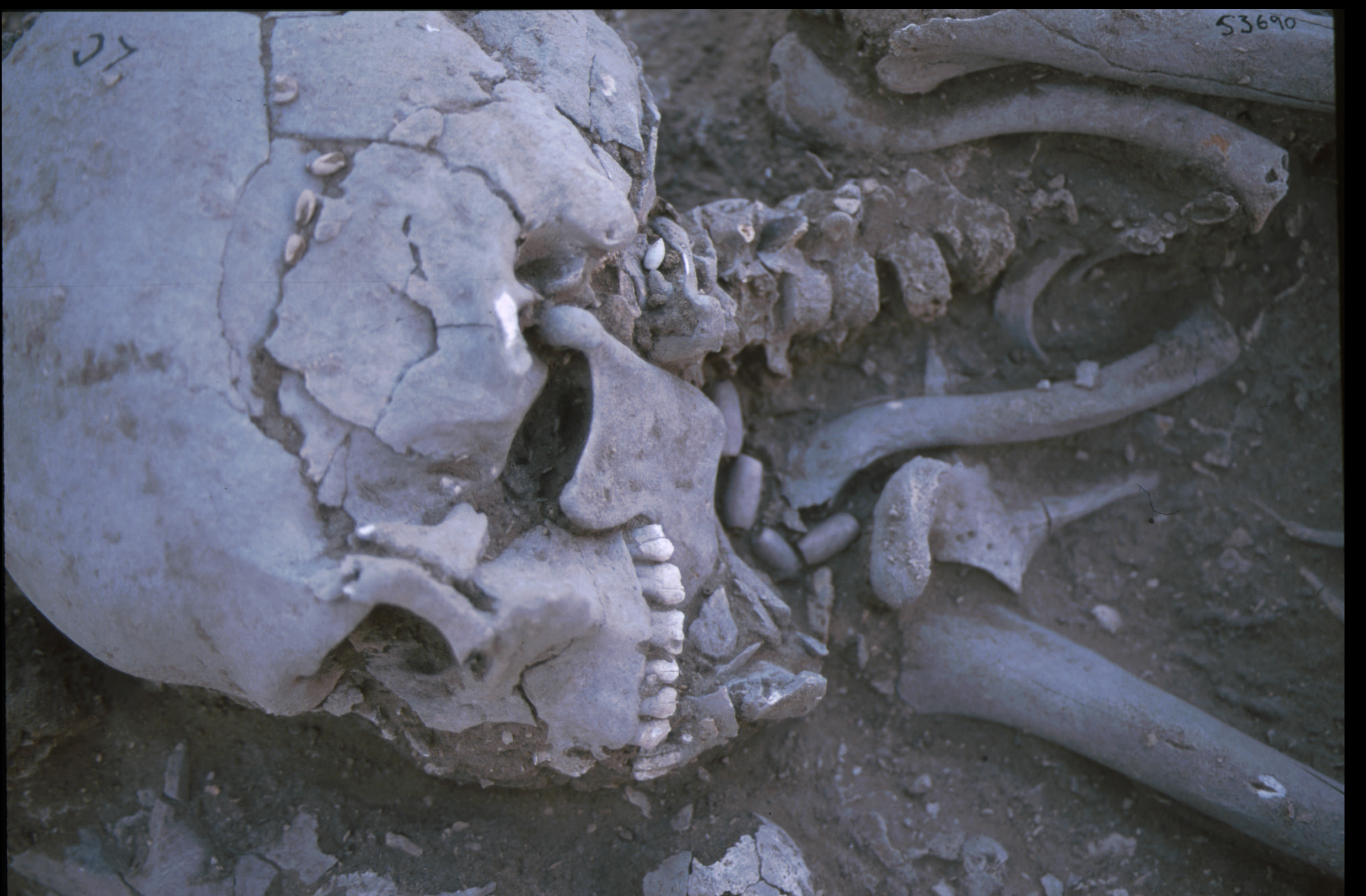
Inidvidual LD:
Occiput LD with perisinus abscess
Full Resolution
Full Resolution
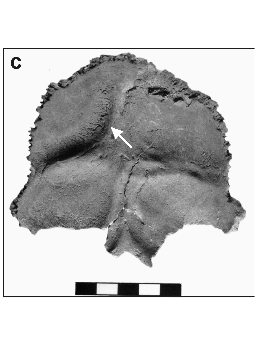
Inidvidual LD:
Occiput LD with persinius abscess, inflammation
Full Resolution
Full Resolution
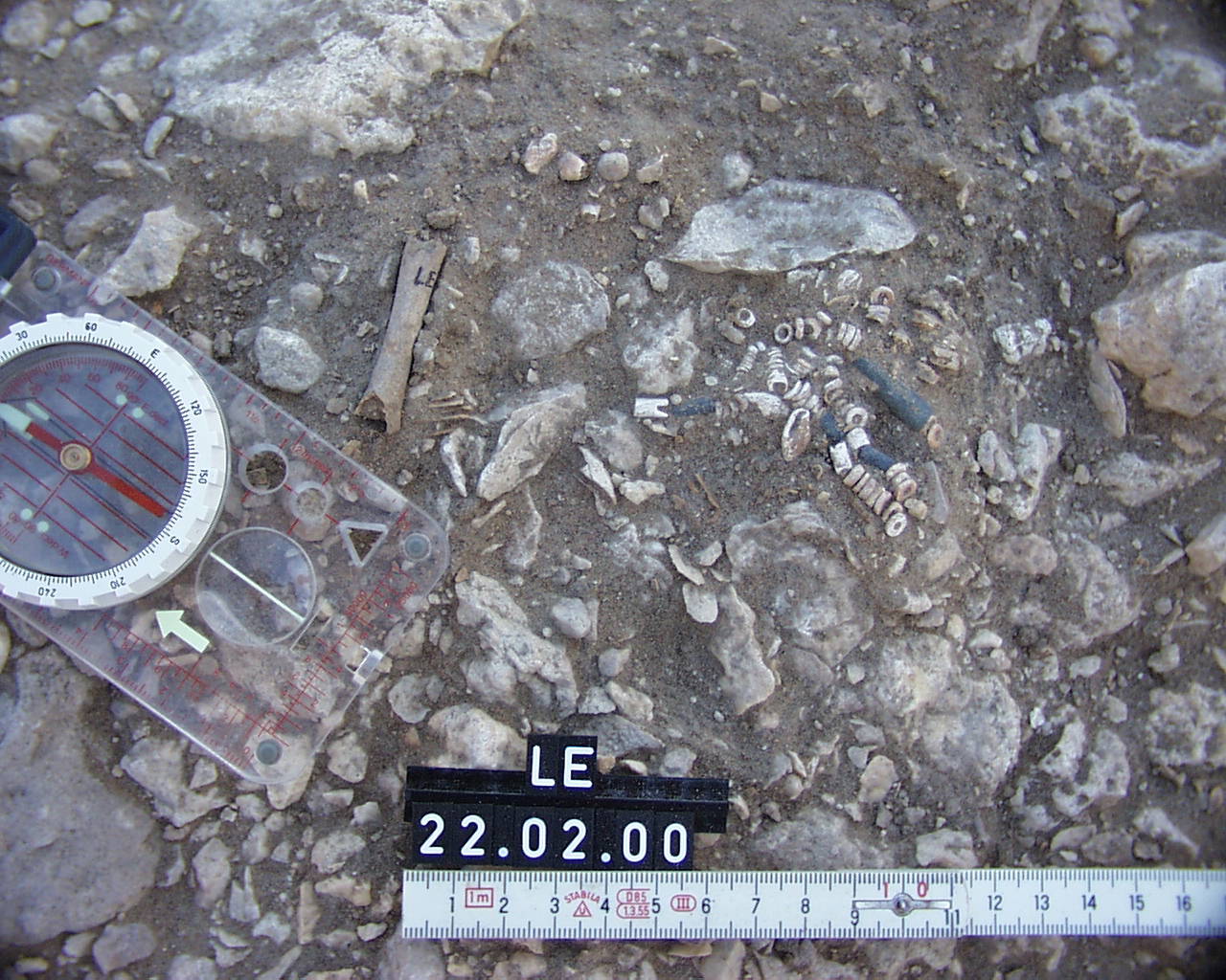
Inidvidual LE:
Adornments individual LE
Full Resolution
Full Resolution

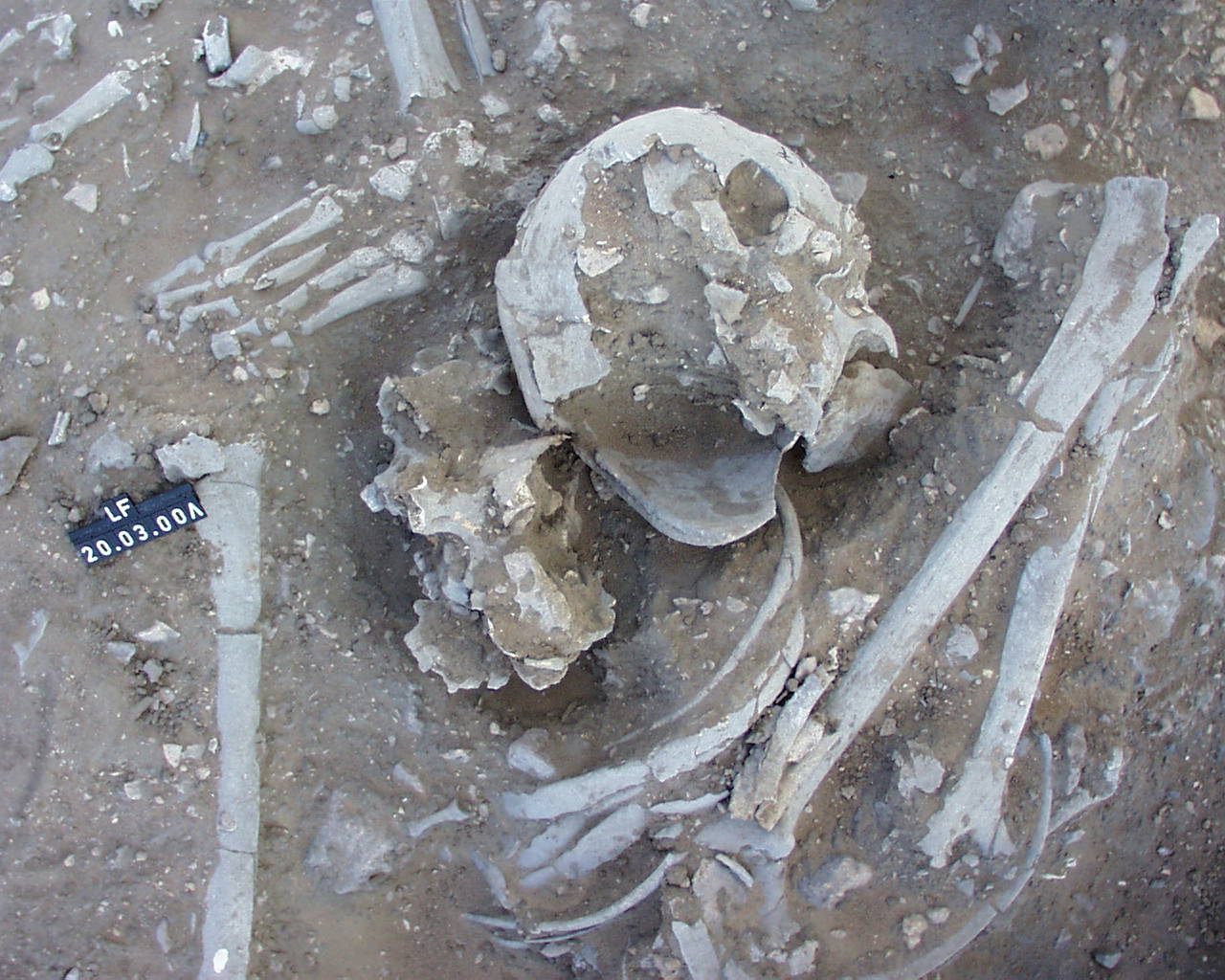
Inidvidual LF:
FK and LF in situ
Full Resolution
Full Resolution
Inidvidual LF:
LF in situ
Full Resolution
Full Resolution
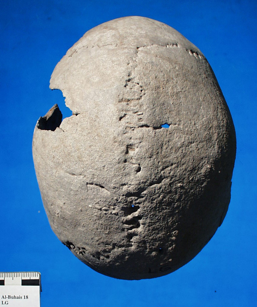
Inidvidual LG:
Skull LG superior
Full Resolution
Full Resolution
Inidvidual LG:
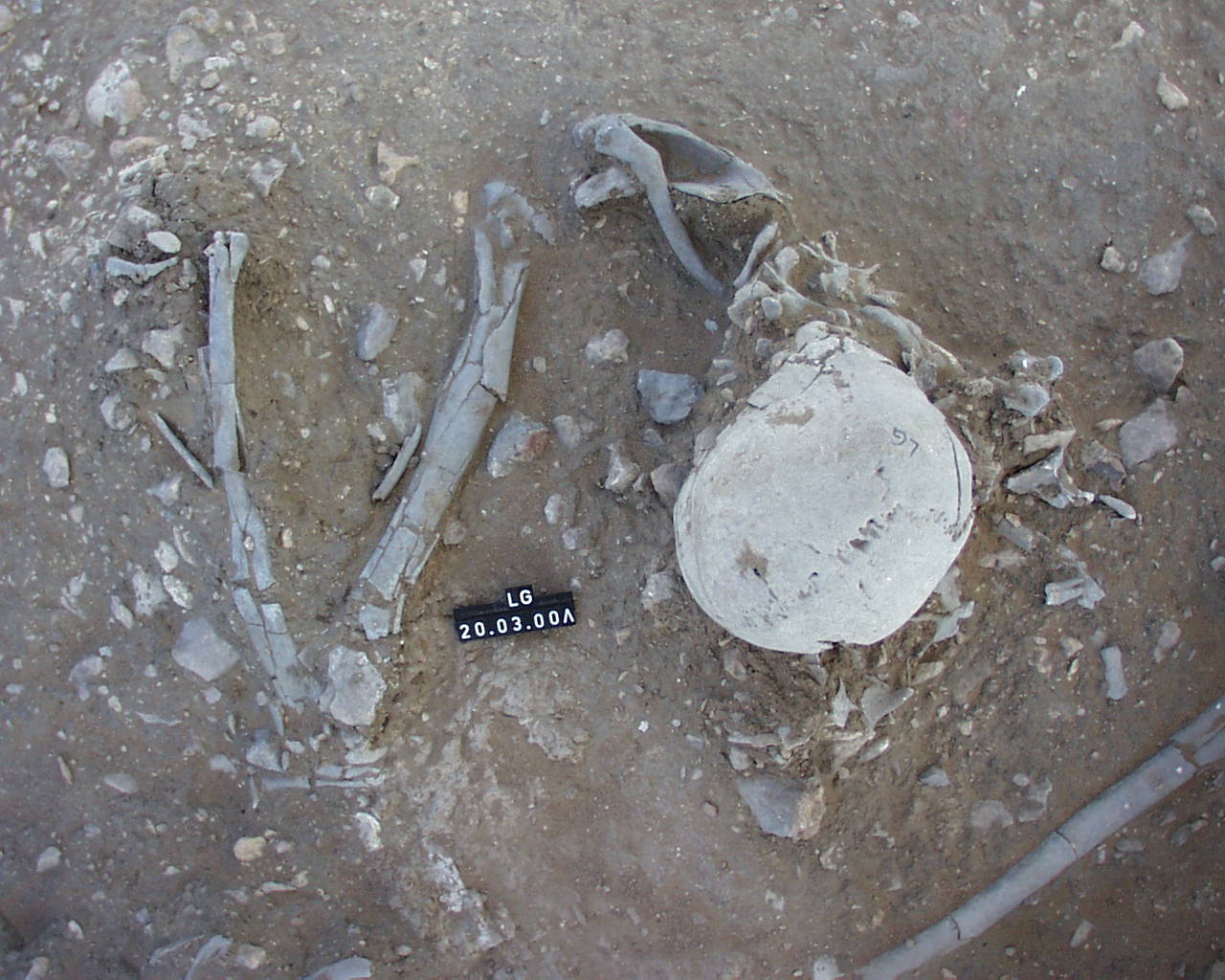
Individual LG in situ
Full Resolution
Full Resolution

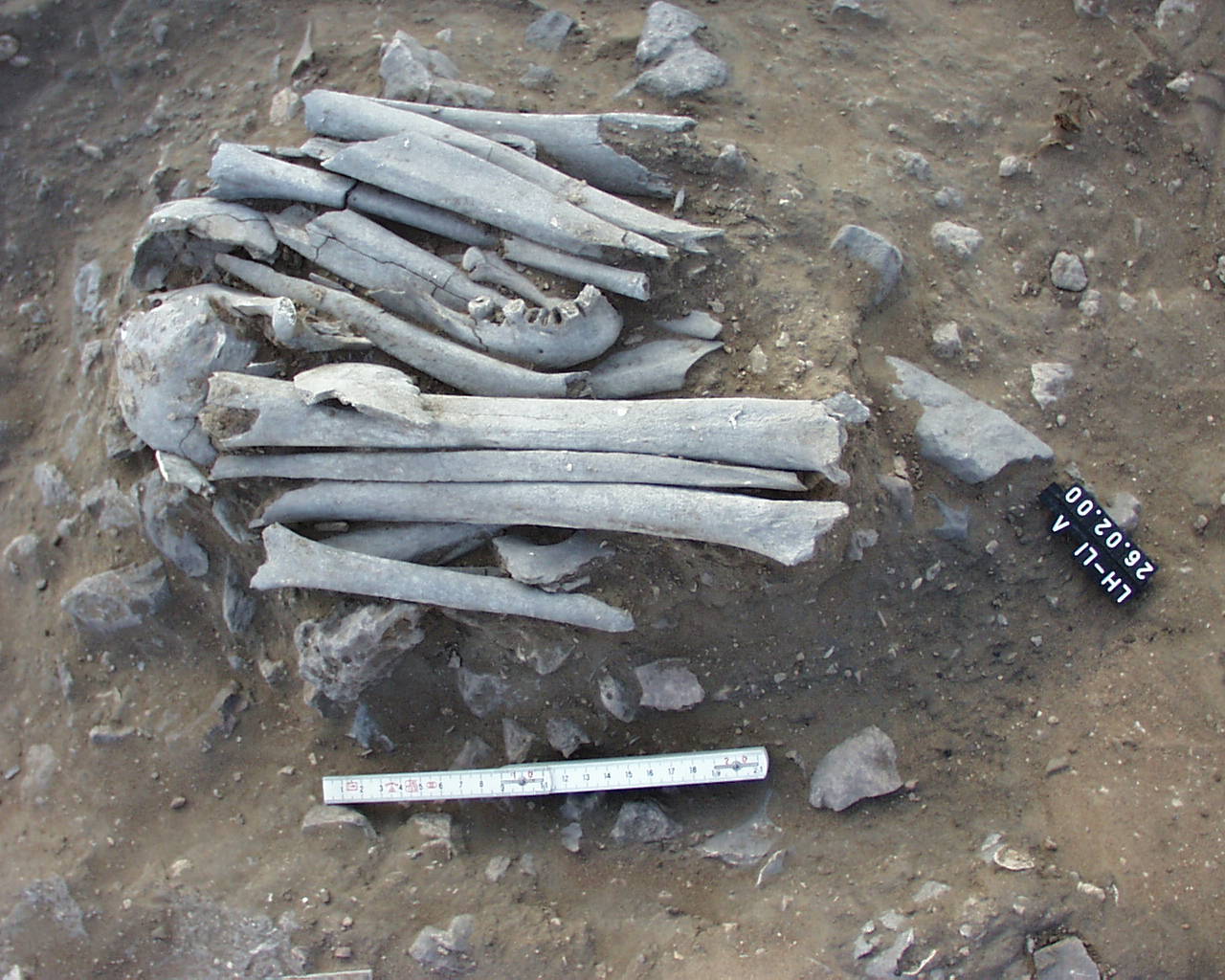
Inidvidual LH:
Depot LH/LI in situ
Full Resolution
Full Resolution
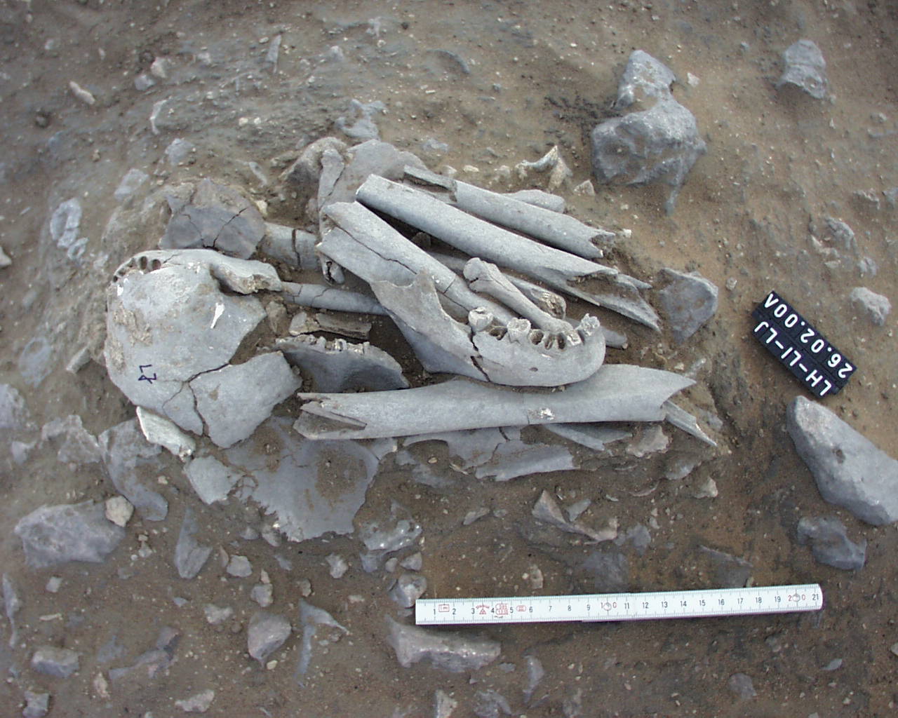
Inidvidual LH:
LH, LI, LJ in situ
Full Resolution
Full Resolution

Inidvidual LH:
LH, LI, LJ, LM in situ. Depot
Full Resolution
Full Resolution
Inidvidual LI:
Depot LH/LI in situ
Full Resolution
Full Resolution

Inidvidual LI:
LH, LI, LJ in situ
Full Resolution
Full Resolution
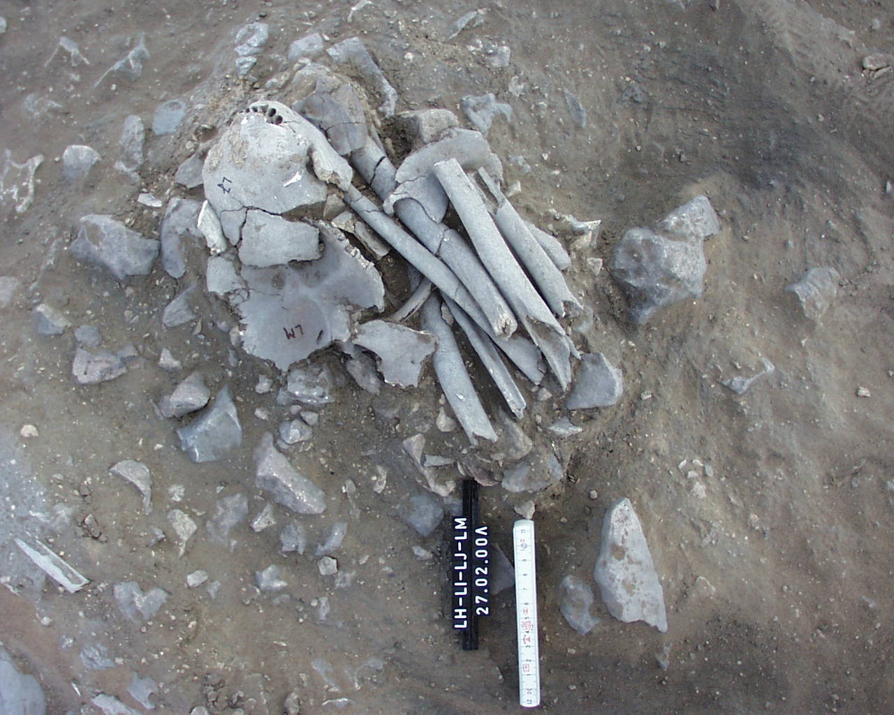
Inidvidual LI:
LH, LI, LJ, LM in situ. Depot
Full Resolution
Full Resolution

Inidvidual LJ:
LH, LI, LJ in situ
Full Resolution
Full Resolution

Inidvidual LJ:
LH, LI, LJ, LM in situ. Depot
Full Resolution
Full Resolution
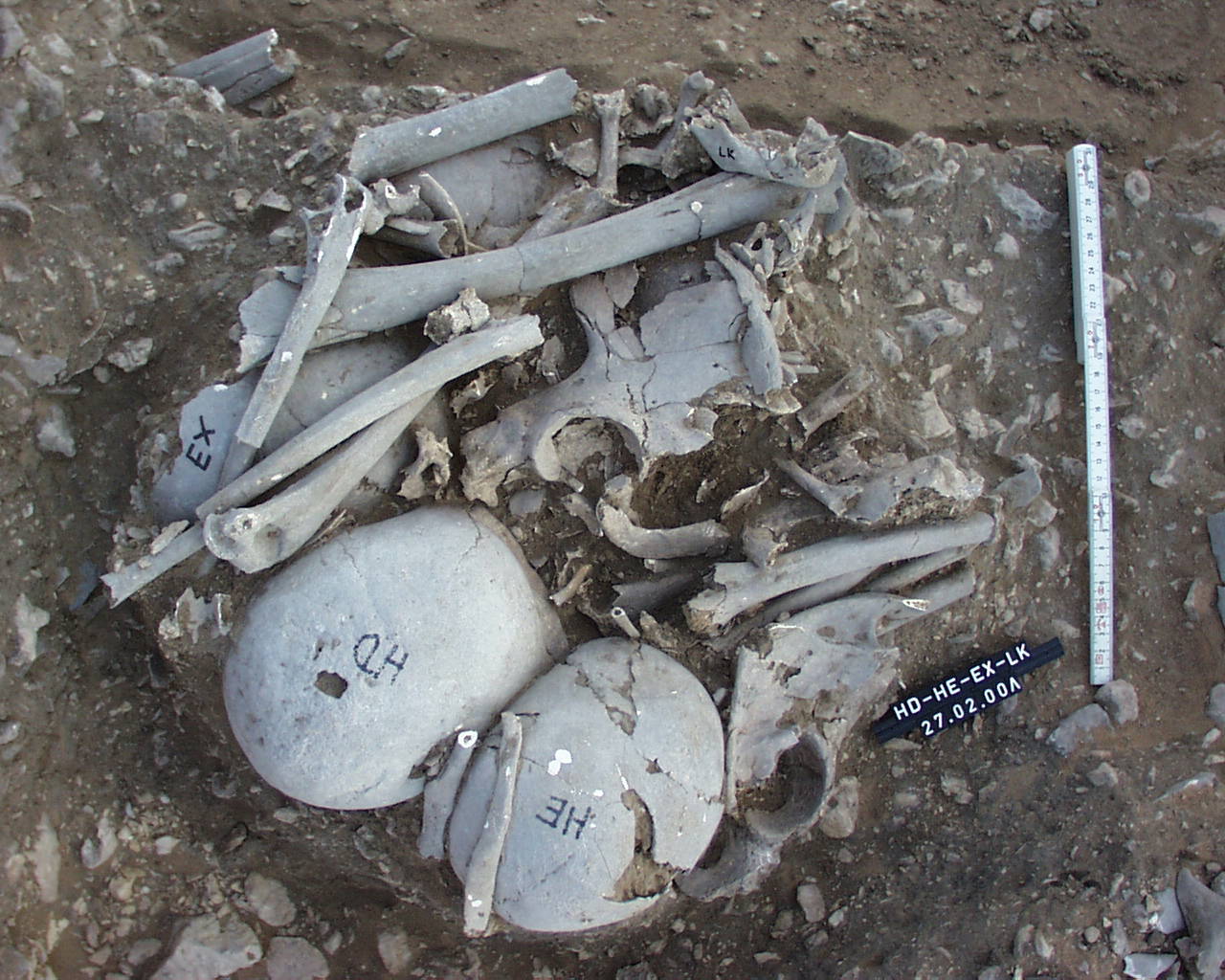
Inidvidual LK:
HD, HE, EX and LK, Depot. In situ
Full Resolution
Full Resolution

Inidvidual LK:
HD, HE, EX and LK, Depot in situ
Full Resolution
Full Resolution
Inidvidual LK:
Skeleton LK or HE
Full Resolution
Full Resolution
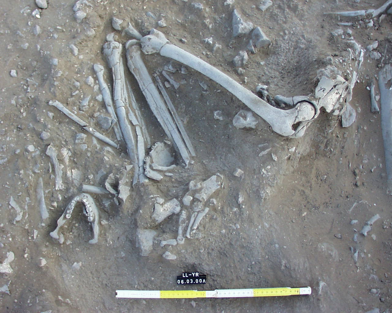
Inidvidual LL:
LL and YR in situ
Full Resolution
Full Resolution
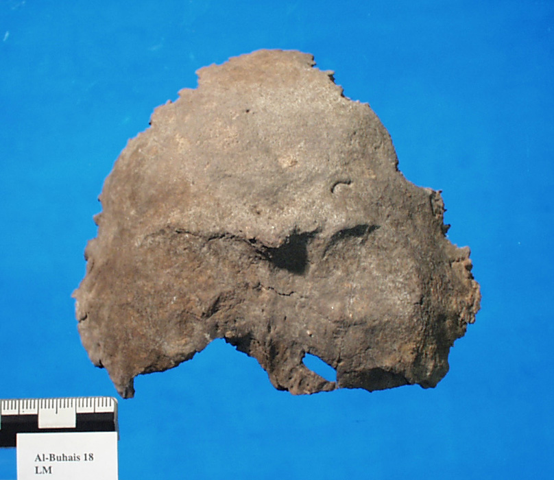
Inidvidual LM:
Occiput LM with prominent protuberantia occipitalis
Full Resolution
Full Resolution
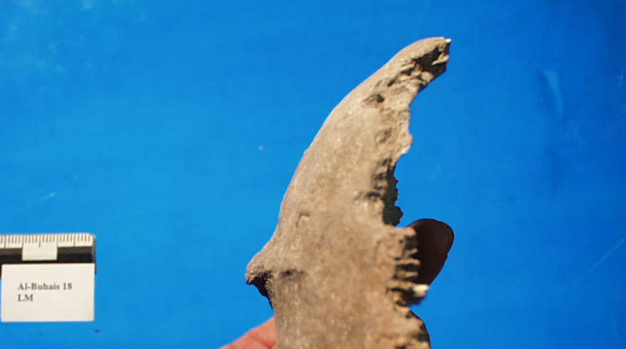
Inidvidual LM:
Occiput LM with prominent protuberantia occipitalis, seen from lateral
Full Resolution
Full Resolution

Inidvidual LM:
LH, LI, LJ, LM in situ. Depot
Full Resolution
Full Resolution
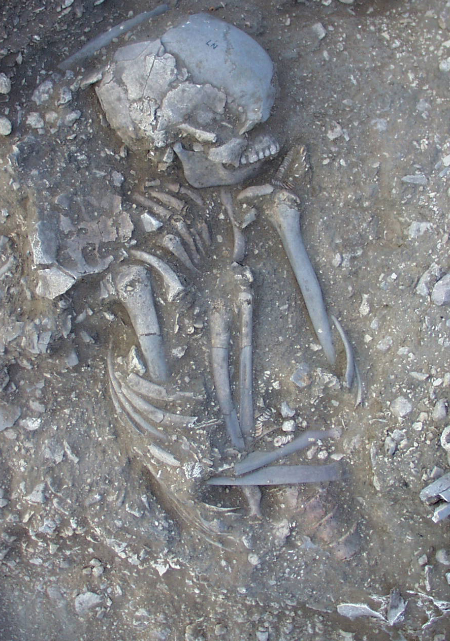
Inidvidual LN:
Individual LN, upper part of the body with adornments in the neck and elbow area
Full Resolution
Full Resolution
Inidvidual LN:
Individual LN, detail of adornments in neck area
Full Resolution
Full Resolution
Inidvidual LN:
Skull injury (Fig. 6.2. in Kiesewetter 2006, Archaeology of Jebel al-Buhais, Volume 1)
Full Resolution
Full Resolution
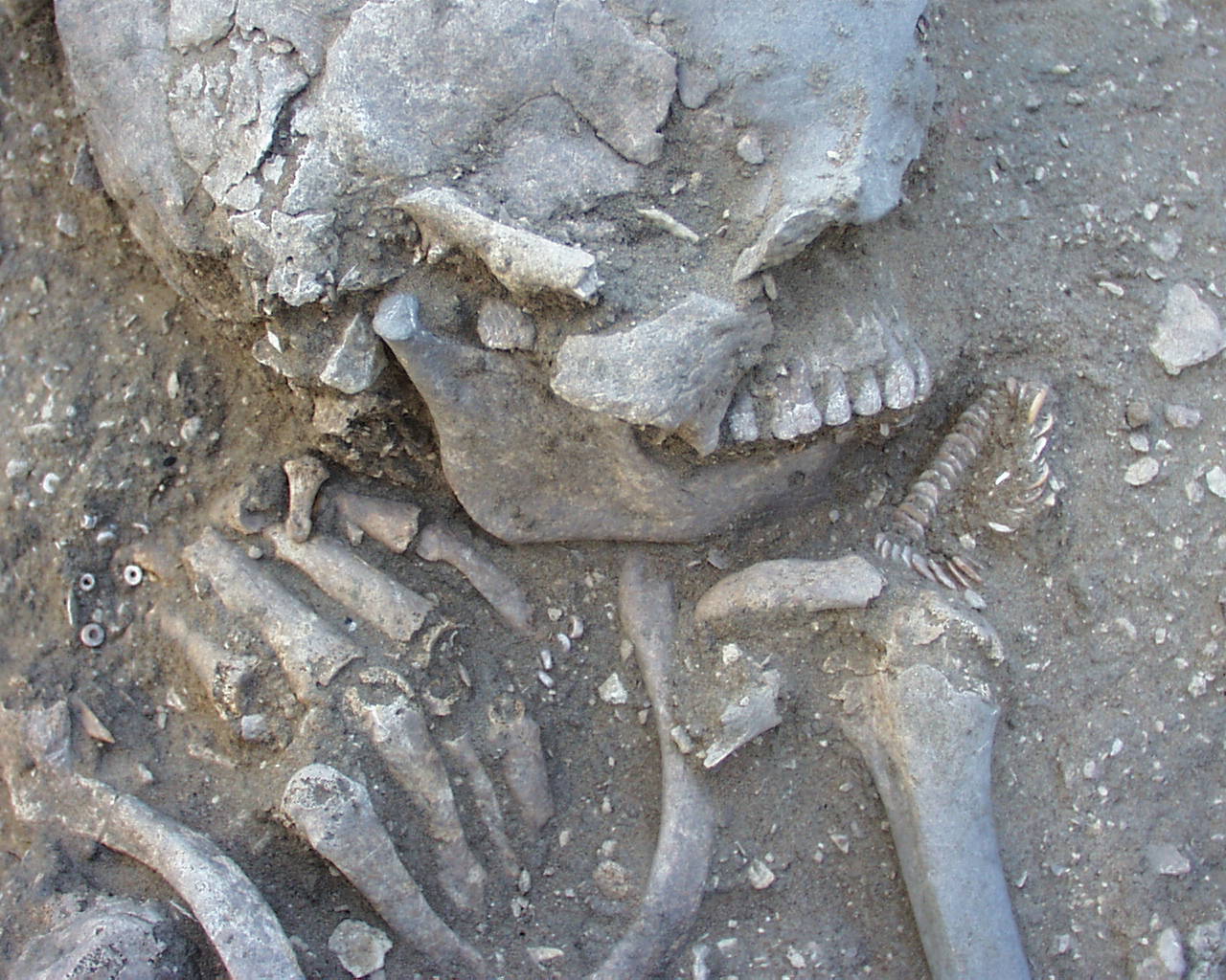
Inidvidual LO:
LO in situ
Full Resolution
Full Resolution

Inidvidual LP:
LP in situ
Full Resolution
Full Resolution
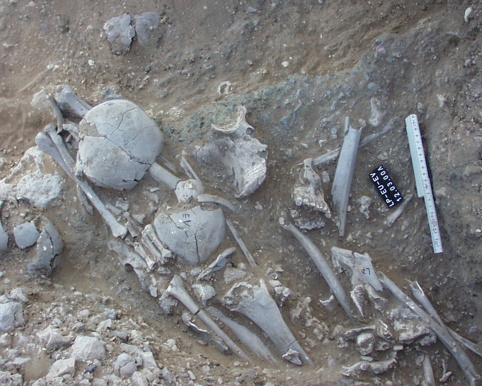
Inidvidual LP:
LP, EU and EV in situ
Full Resolution
Full Resolution
Inidvidual LQ:
Individual LQ in situ
Full Resolution
Full Resolution
Inidvidual LQ:
Individual LQ and longbones of unknown individual in situ
Full Resolution
Full Resolution
Inidvidual LQ:
LQ detail
Full Resolution
Full Resolution

Inidvidual LU:
skull LU and longbone fragments YX in situ
Full Resolution
Full Resolution
Inidvidual LY:
Skull LY
Full Resolution
Full Resolution
Inidvidual MA:
Mandible fracture
Full Resolution
Full Resolution

Inidvidual MA:
Fractured mandible
Full Resolution
Full Resolution
Inidvidual MF:
Deatil of skull MF with perforations
Full Resolution
Full Resolution
Inidvidual MG:
Peri-mortem fractures at the left side of skull MG
Full Resolution
Full Resolution
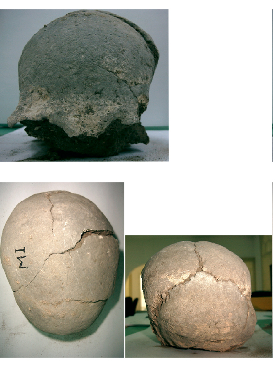
Inidvidual MI:
Skull MI
Full Resolution
Full Resolution
Inidvidual MJ:
Skull
Full Resolution
Full Resolution
Inidvidual MJ:
Skull
Full Resolution
Full Resolution
Inidvidual MM:
MM and MN in situ
Full Resolution
Full Resolution
Inidvidual MM:
MM and MN in situ
Full Resolution
Full Resolution

Inidvidual MN:
MM and MN in situ
Full Resolution
Full Resolution

Inidvidual MN:
MM and MN in situ
Full Resolution
Full Resolution
Inidvidual NA:
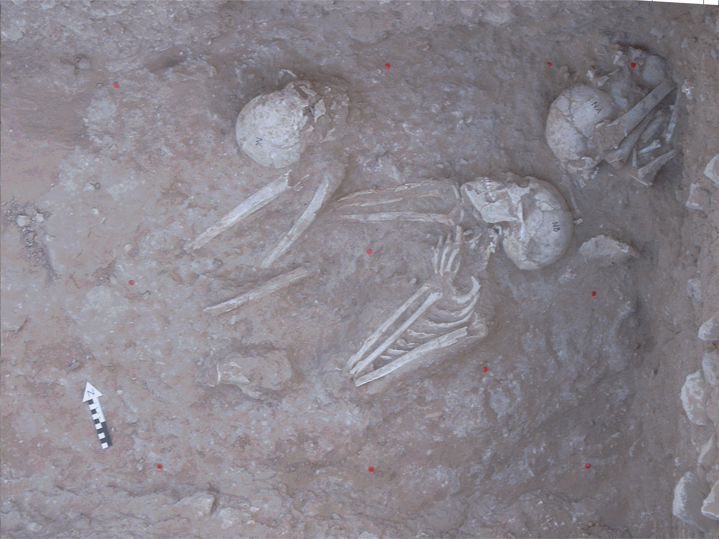
Individuals NA, NB, NC and ND in situ. Abtrag 9
Full Resolution
Full Resolution
Inidvidual NA:
Na, NB and NC in situ in W1.1, square 17/26. The pelvis fragment above NA is NH
Full Resolution
Full Resolution

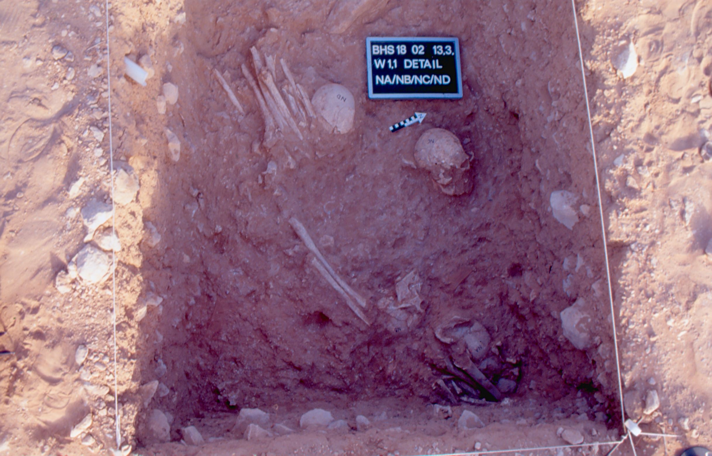
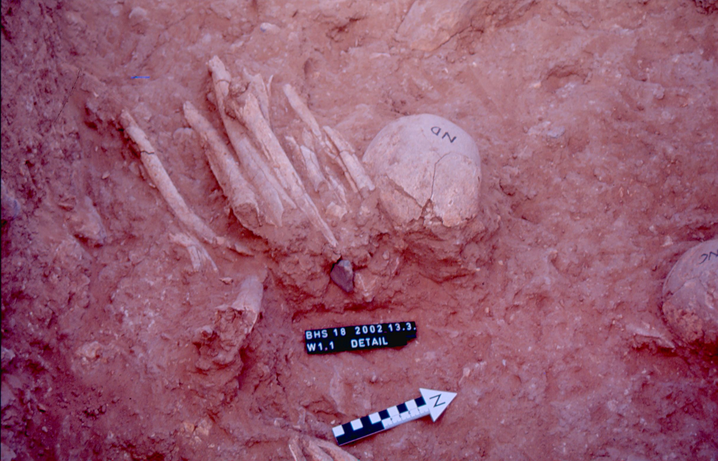
Inidvidual NA:
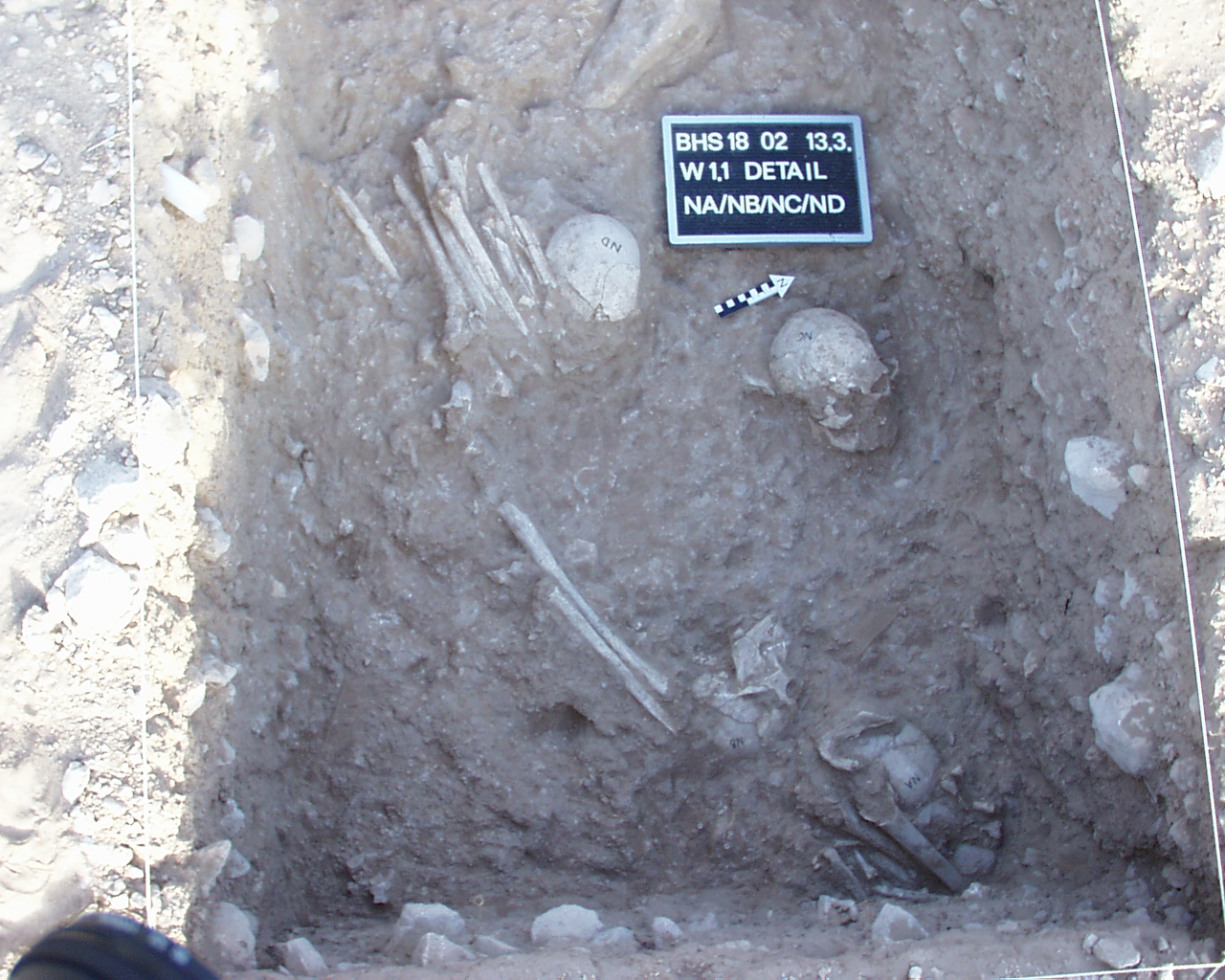
Detail NA/NB/NC/ND 13.03.2002
Full Resolution
Full Resolution

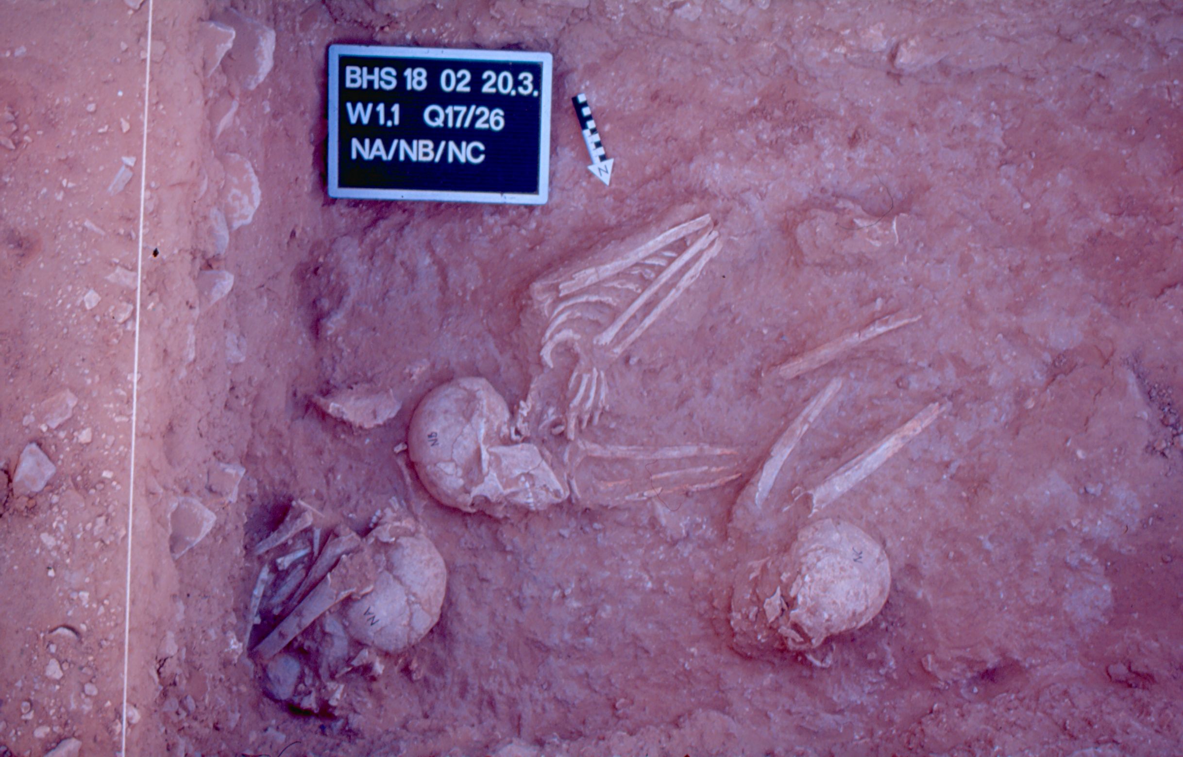
Inidvidual NA:
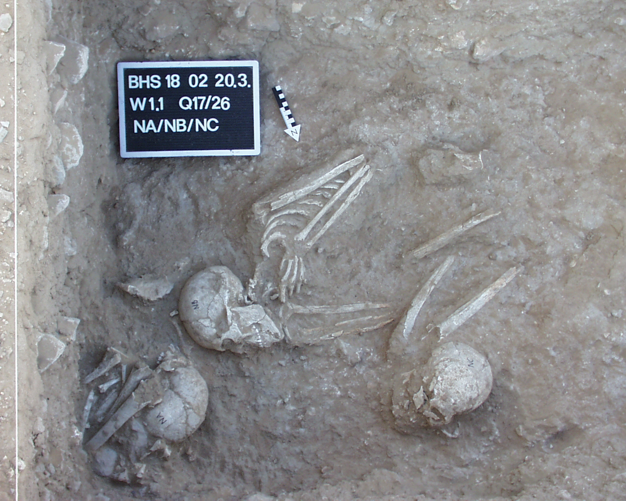
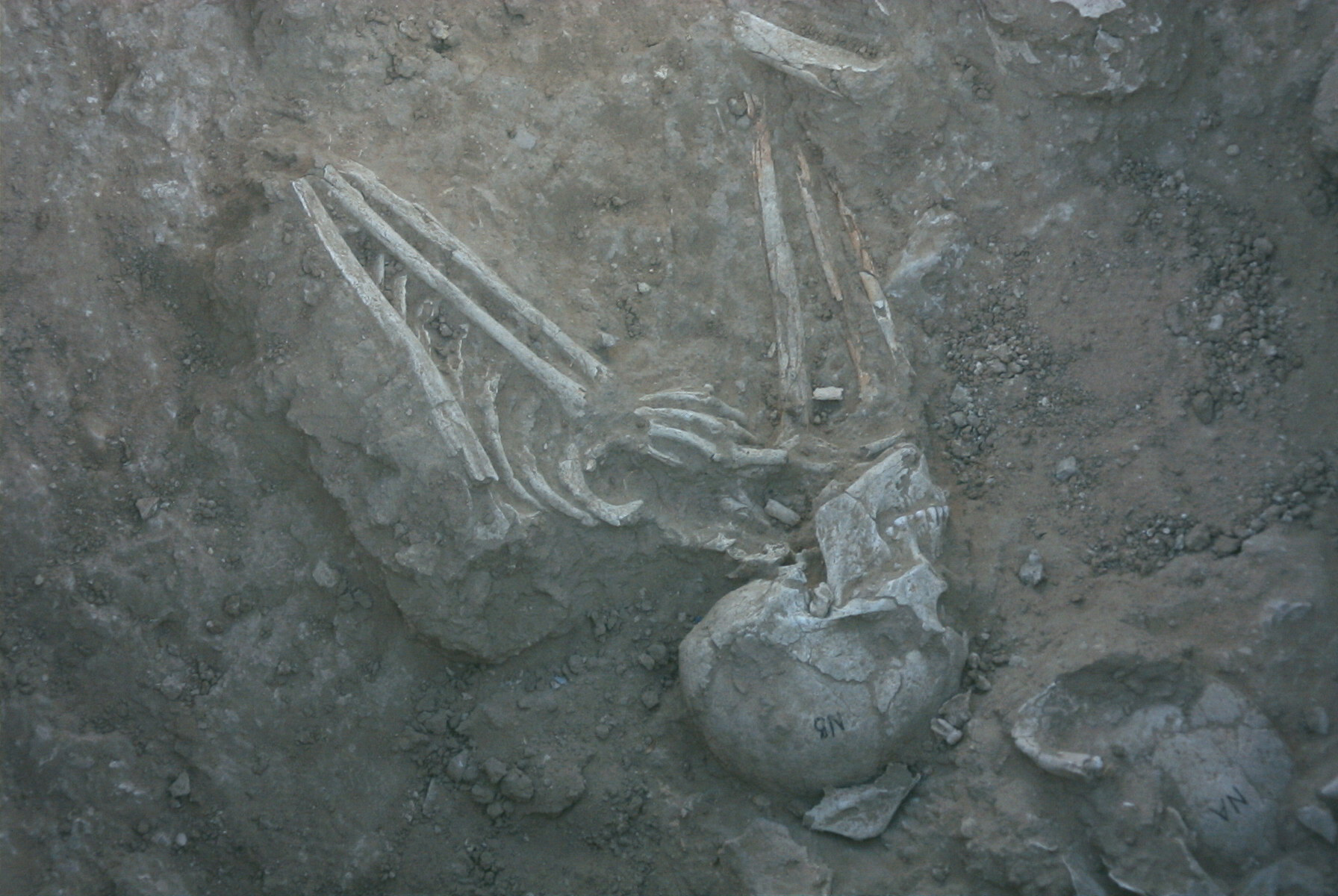
NA/NB/NC 20.03.2002
Full Resolution
Full Resolution
Inidvidual NB:
In situ burial NB
Full Resolution
Full Resolution
Inidvidual NB:
Burial NB, NC and NA in situ
Full Resolution
Full Resolution
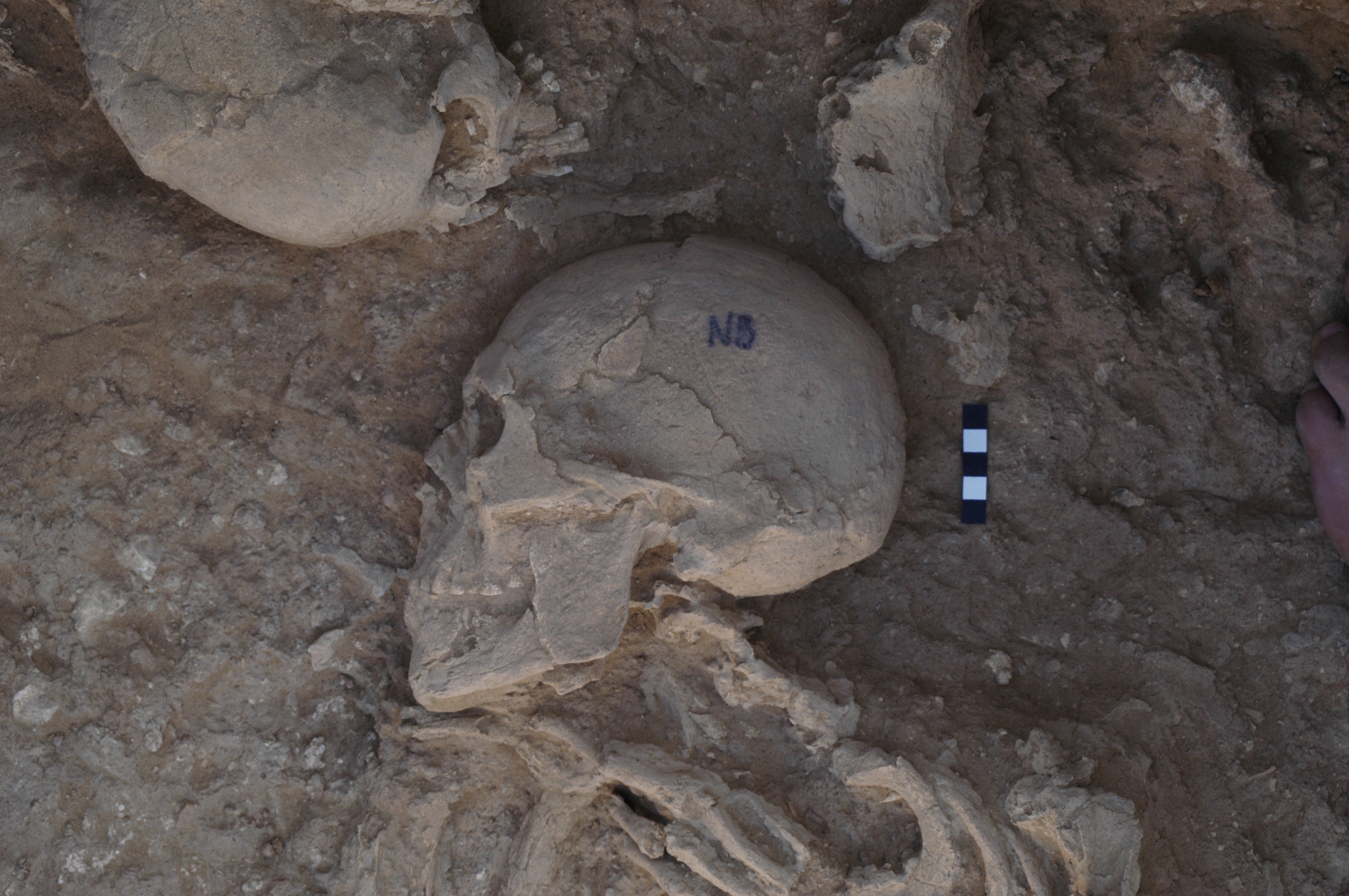
Inidvidual NB:
Detail of skull in situ
Full Resolution
Full Resolution

Inidvidual NB:
Individuals NA, NB, NC and ND in situ. Abtrag 9
Full Resolution
Full Resolution

Inidvidual NB:
Detail NA/NB/NC/ND 13.03.2002
Full Resolution
Full Resolution

Inidvidual NB:
NA/NB/NC 20.03.2002
Full Resolution
Full Resolution

Inidvidual NB:
NN, NB and NC in situ in W1.1, square 17/26. The pelvis fragment above NA is NH
Full Resolution
Full Resolution

Inidvidual NC:
Individuals NA, NB, NC and ND in situ. Abtrag 9
Full Resolution
Full Resolution

Inidvidual NC:
Na, NB and NC in situ in W1.1, square 17/26. The pelvis fragment above NA is NH
Full Resolution
Full Resolution
Inidvidual NC:
Detail NA/NB/NC/ND 13.03.2002
Full Resolution
Full Resolution
Inidvidual NC:
NA/NB/NC 20.03.2002
Full Resolution
Full Resolution

Inidvidual ND:
Individuals NA, NB, NC and ND in situ. Abtrag 9
Full Resolution
Full Resolution
Inidvidual ND:
Secondary burial ND W1.1 with stone artefact
Full Resolution
Full Resolution
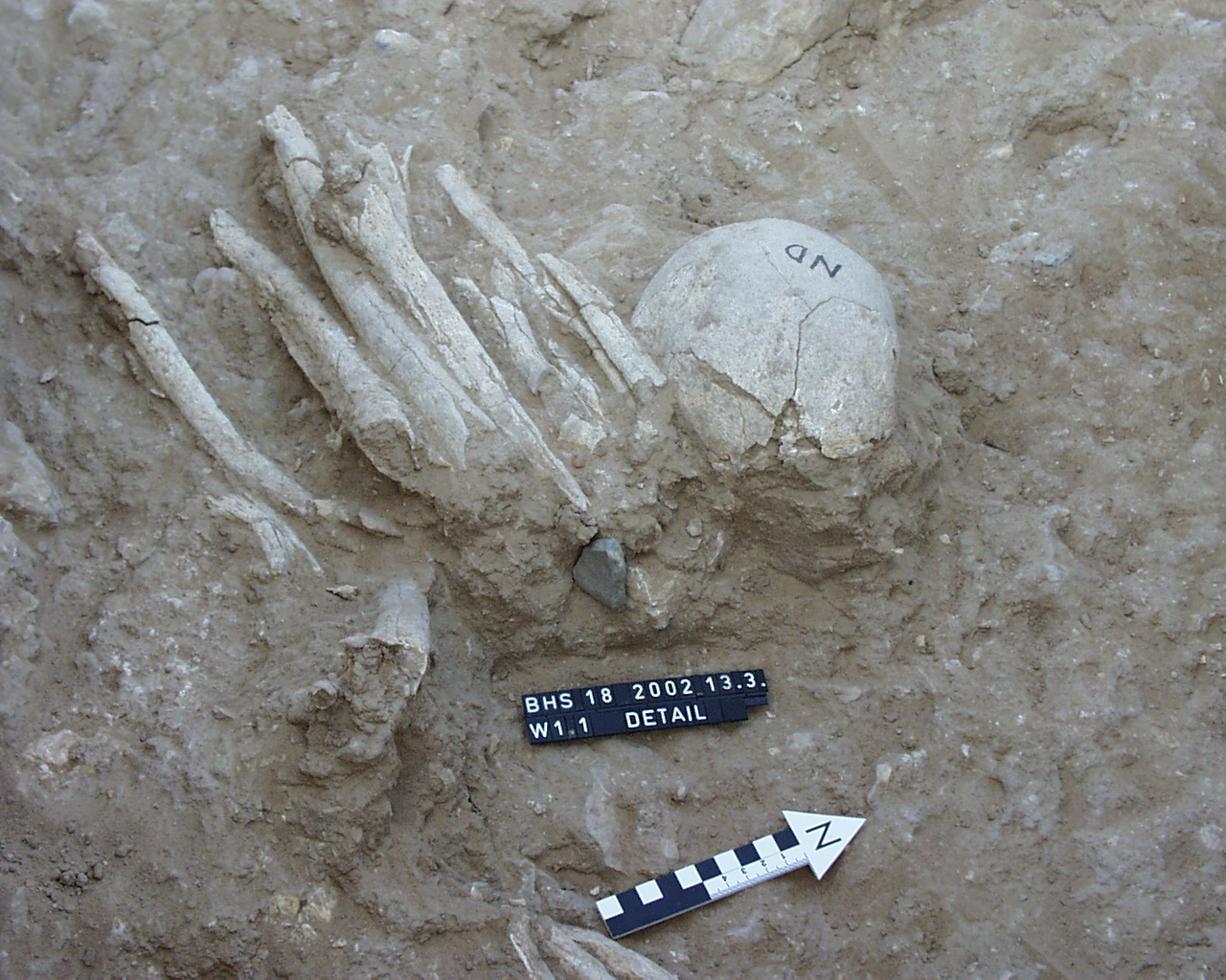
Inidvidual ND:
Detail ND 13.03.2002
Full Resolution
Full Resolution

Inidvidual ND:
Detail NA/NB/NC/ND 13.03.2002
Full Resolution
Full Resolution
Inidvidual NE:
Arm and hand in situ
Full Resolution
Full Resolution
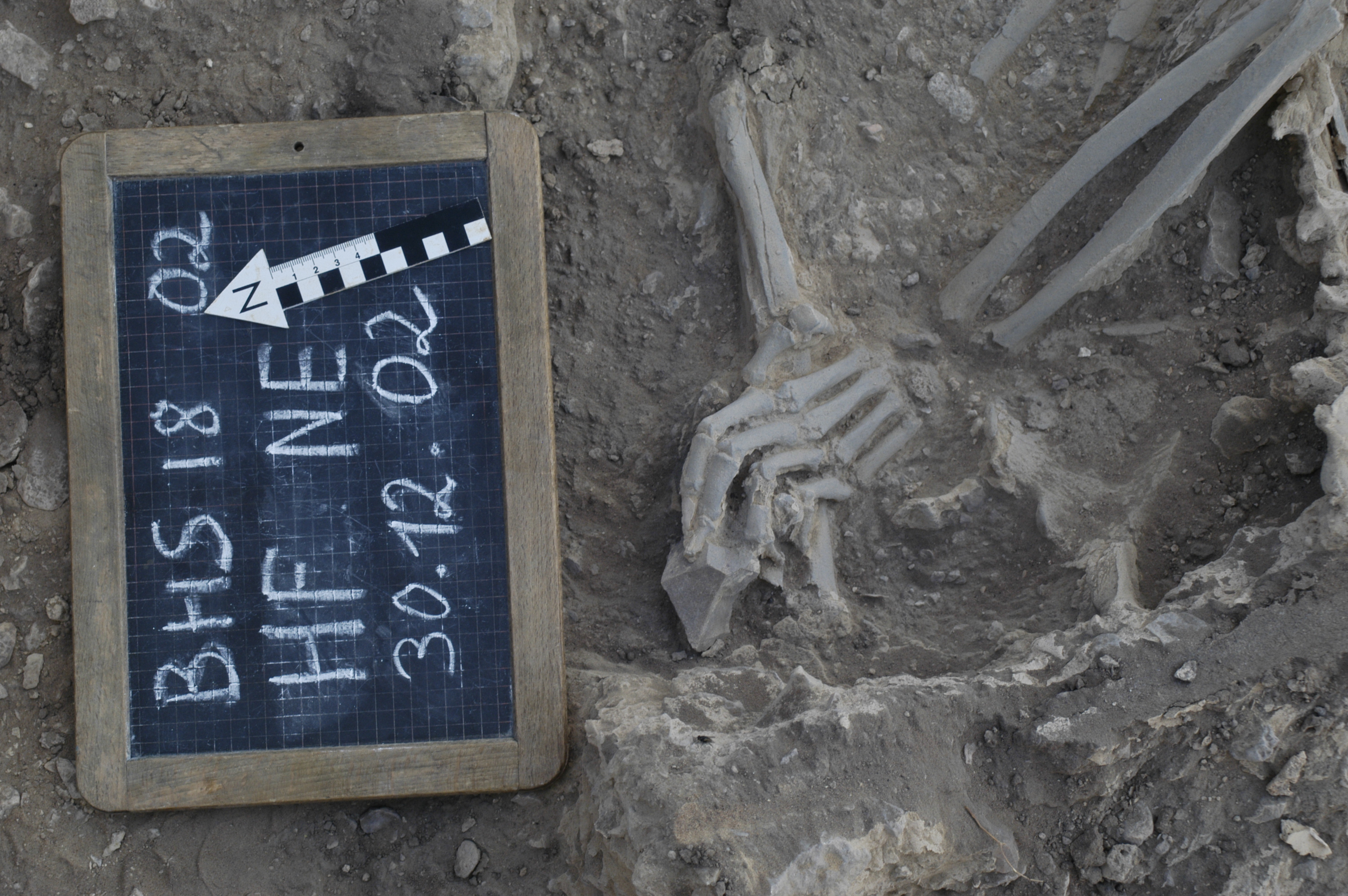
Inidvidual NE:
Individual NE (Hauptfläche HF)
Full Resolution
Full Resolution
Inidvidual NE:
Detail NE 26.03.2002
Full Resolution
Full Resolution
Inidvidual NE:
NE
Full Resolution
Full Resolution

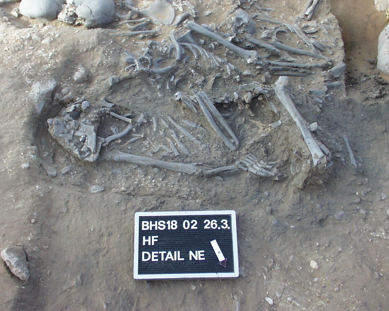
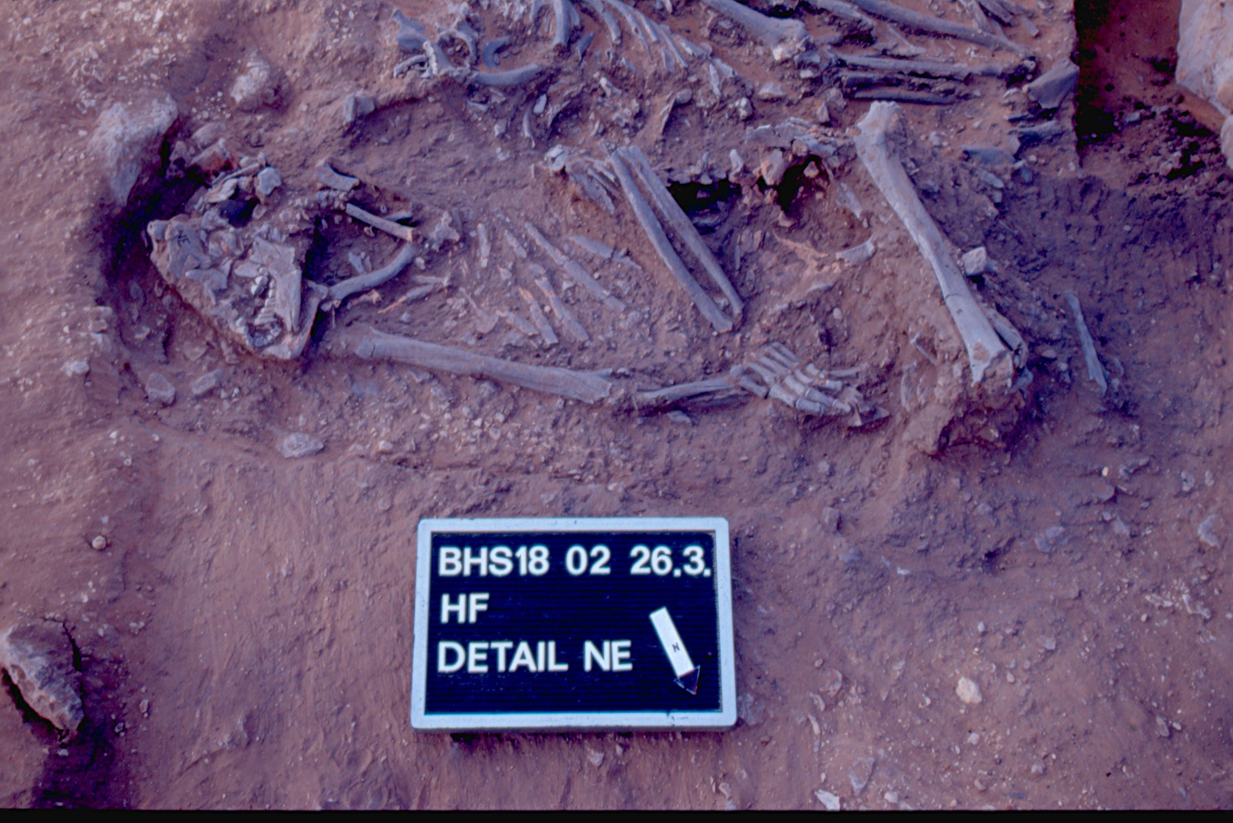
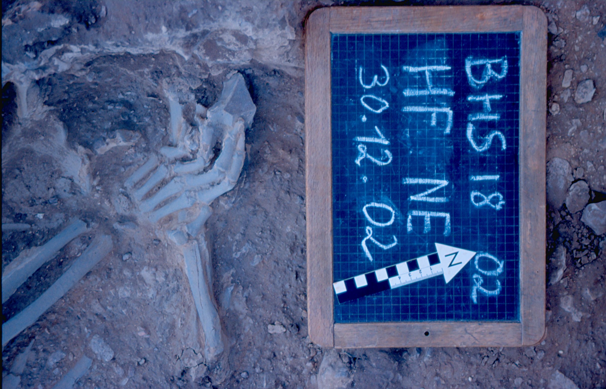
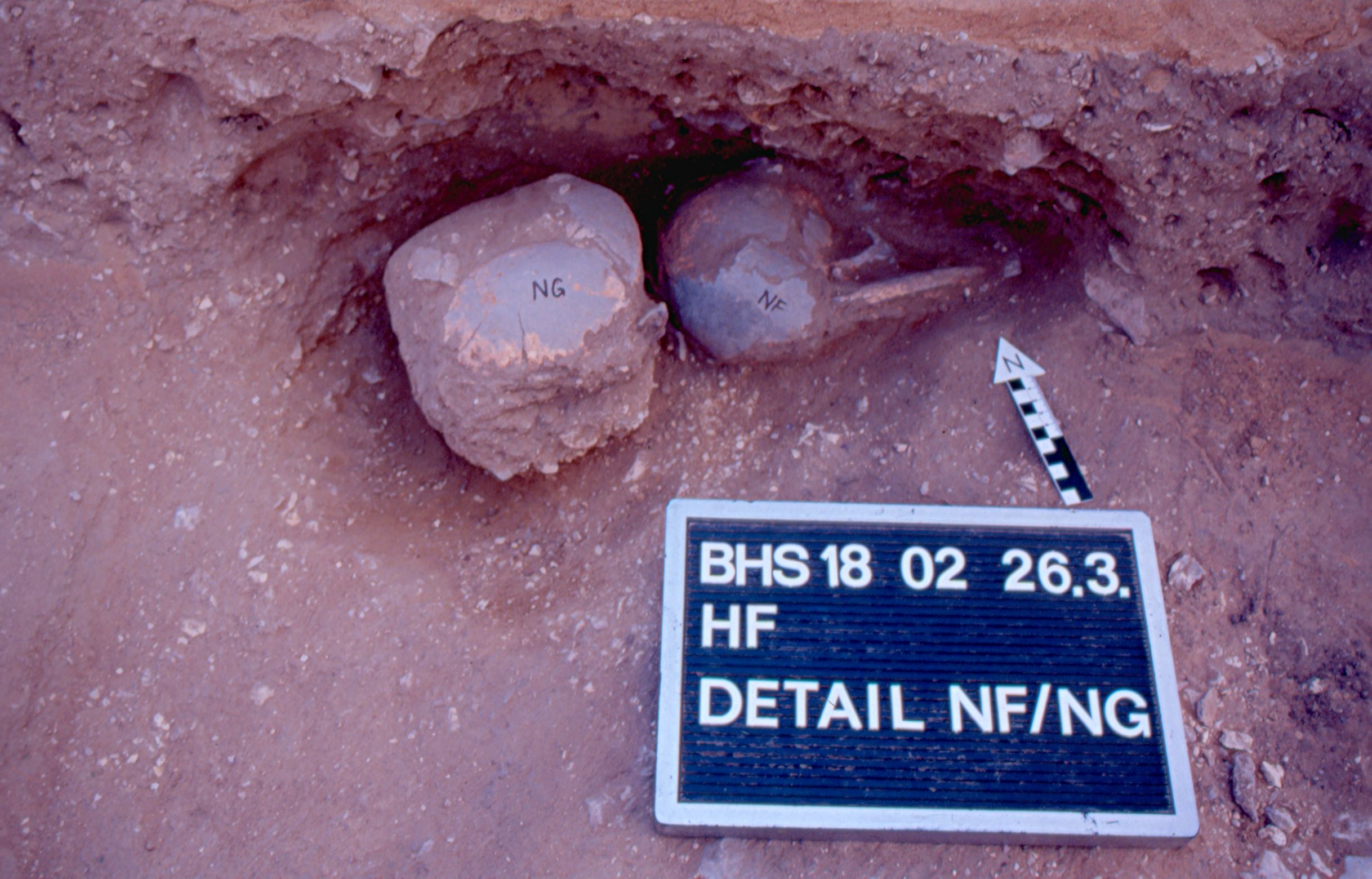
Inidvidual NF:
Skulls NF and NG in situ (Hauptfläche HF)
Full Resolution
Full Resolution
Inidvidual NF:
Detail NG/NF 26.03.2002
Full Resolution
Full Resolution
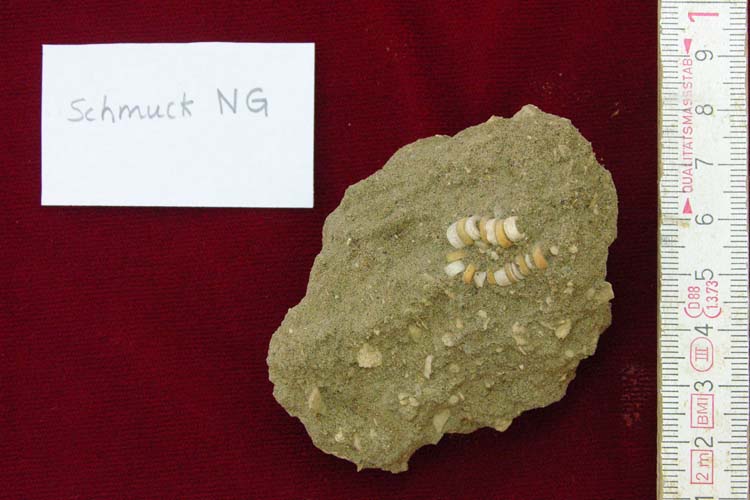
Inidvidual NG:
Adornments of NG
Full Resolution
Full Resolution
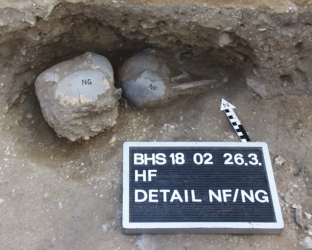
Inidvidual NG:
Skulls NF and NG in situ (Hauptfläche HF)
Full Resolution
Full Resolution

Inidvidual NG:
Detail NG/NF 26.03.2002
Full Resolution
Full Resolution
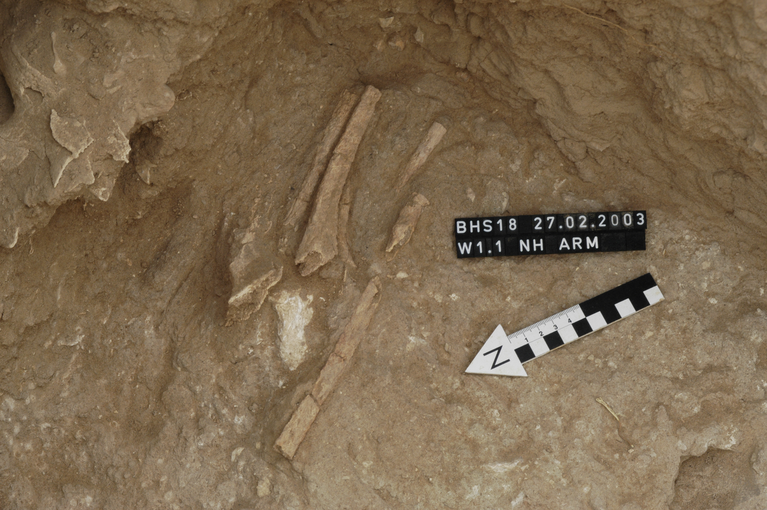
Inidvidual NH:
NH arm in situ
Full Resolution
Full Resolution

Inidvidual NH:
NA, NB and NC in situ in W1.1, square 17/26. The pelvis fragment above NA is NH
Full Resolution
Full Resolution
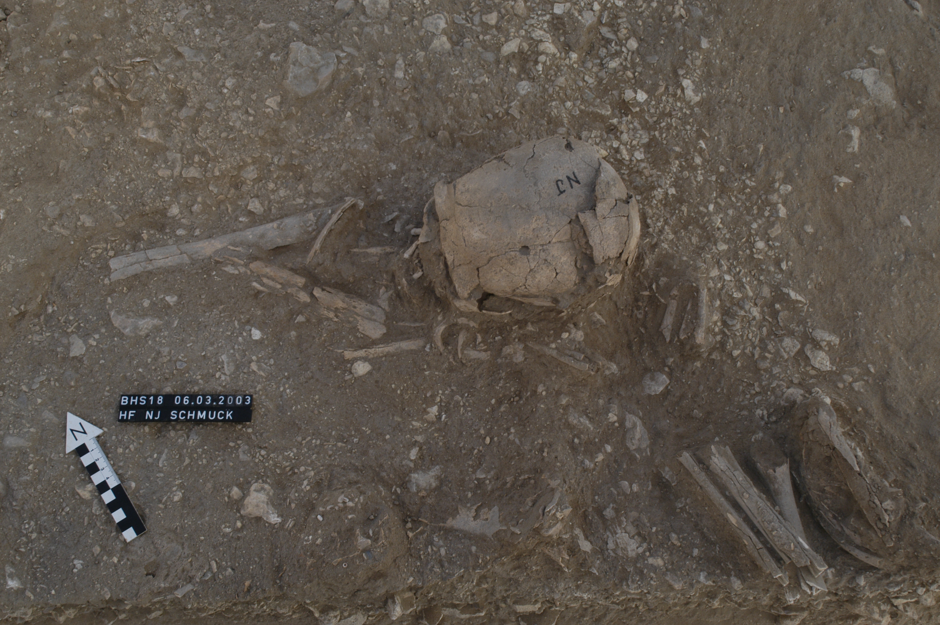
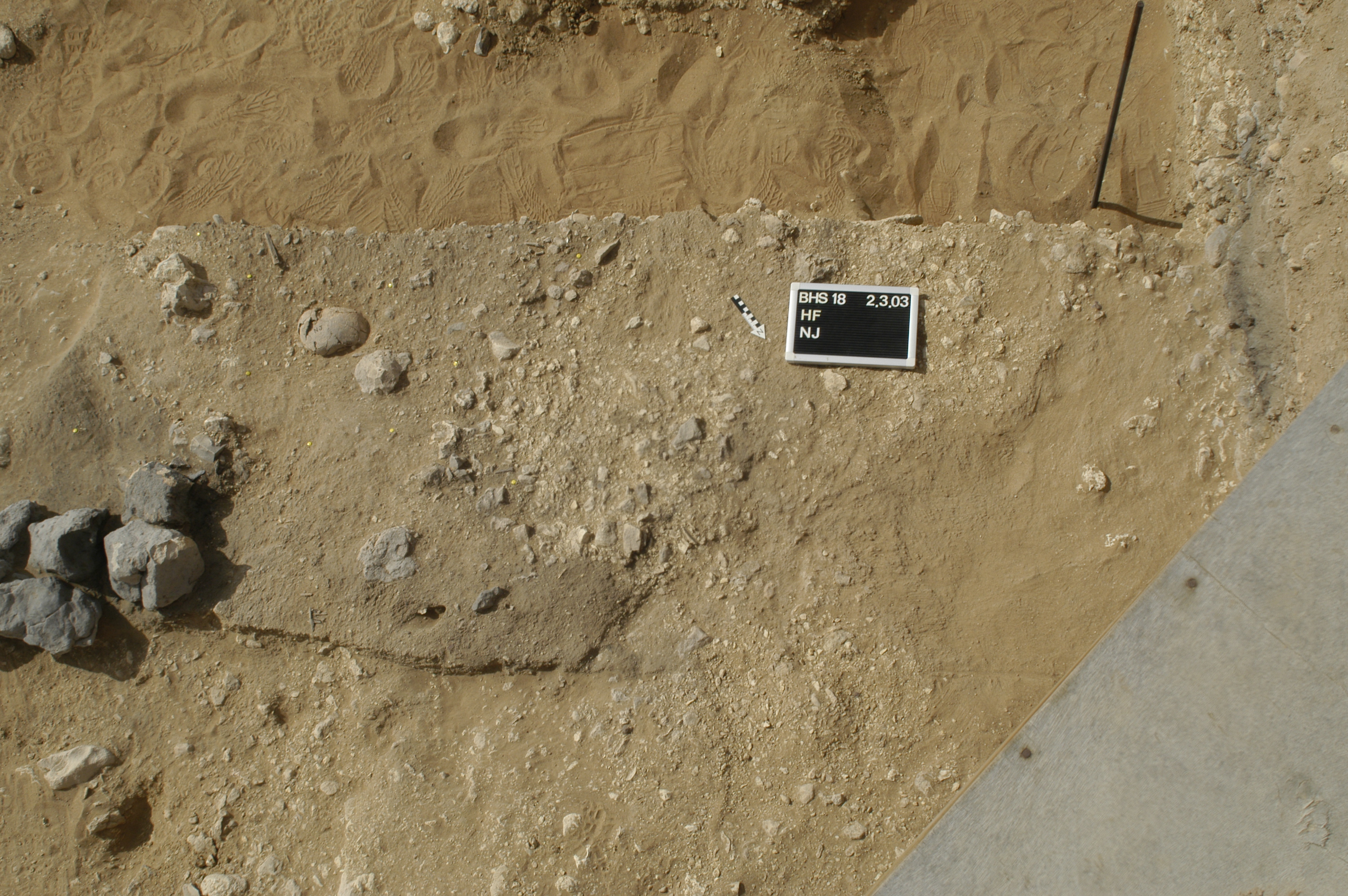
Inidvidual NJ:
In situ
Full Resolution
Full Resolution
Inidvidual NJ:
Skull NJ in situ, a pit is visible
Full Resolution
Full Resolution
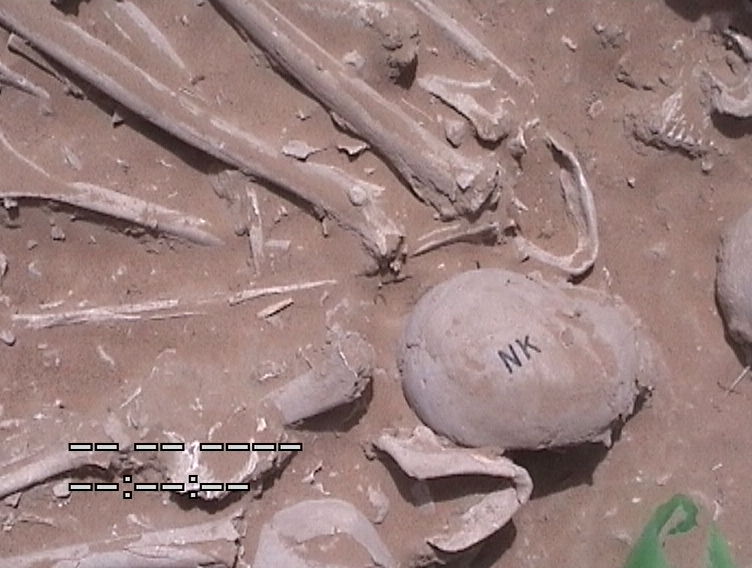
Inidvidual NK:
Skull in situ
Full Resolution
Full Resolution
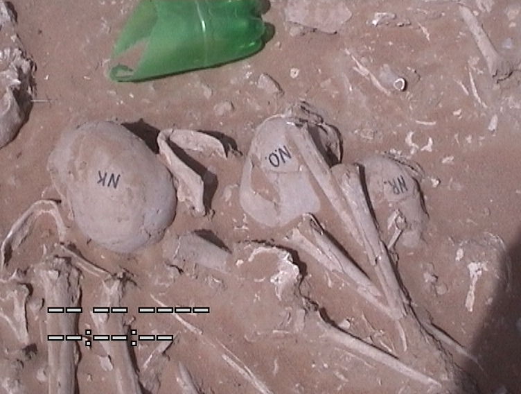
Inidvidual NK:
NK, NO, NR in situ
Full Resolution
Full Resolution

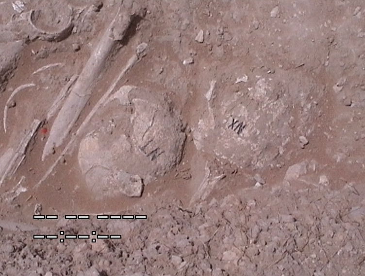
Inidvidual NM:
In situ picture of NM, NN, NK, NV, NY, NQ, NT, NU, NR, NO (orthorectified picture).
Full Resolution
Full Resolution
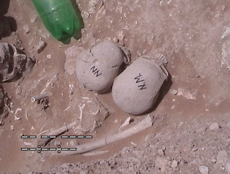
Inidvidual NN:
NN and NM in situ
Full Resolution
Full Resolution
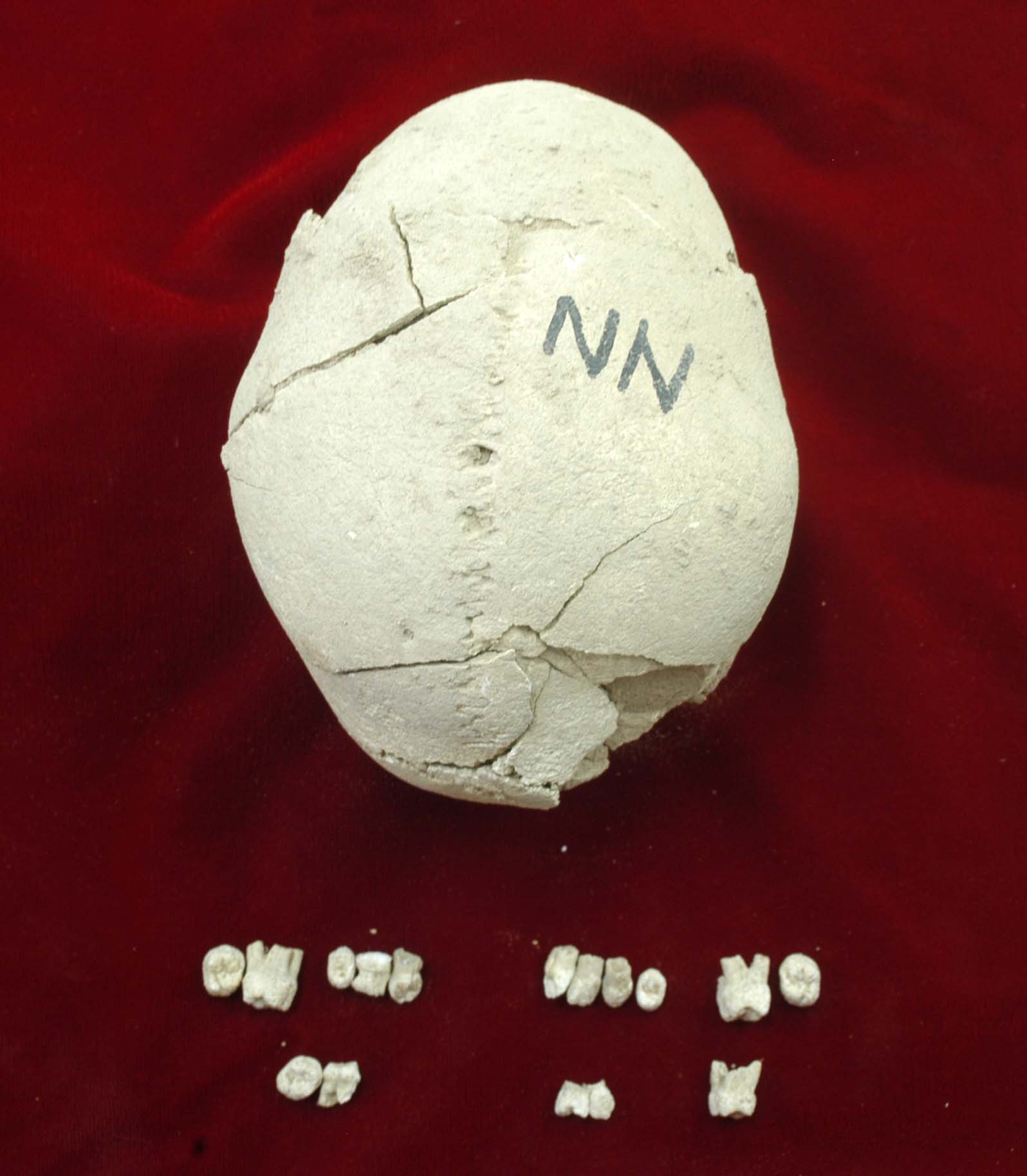
Inidvidual NN:
Premature suture closure of individual NN, teeth
Full Resolution
Full Resolution
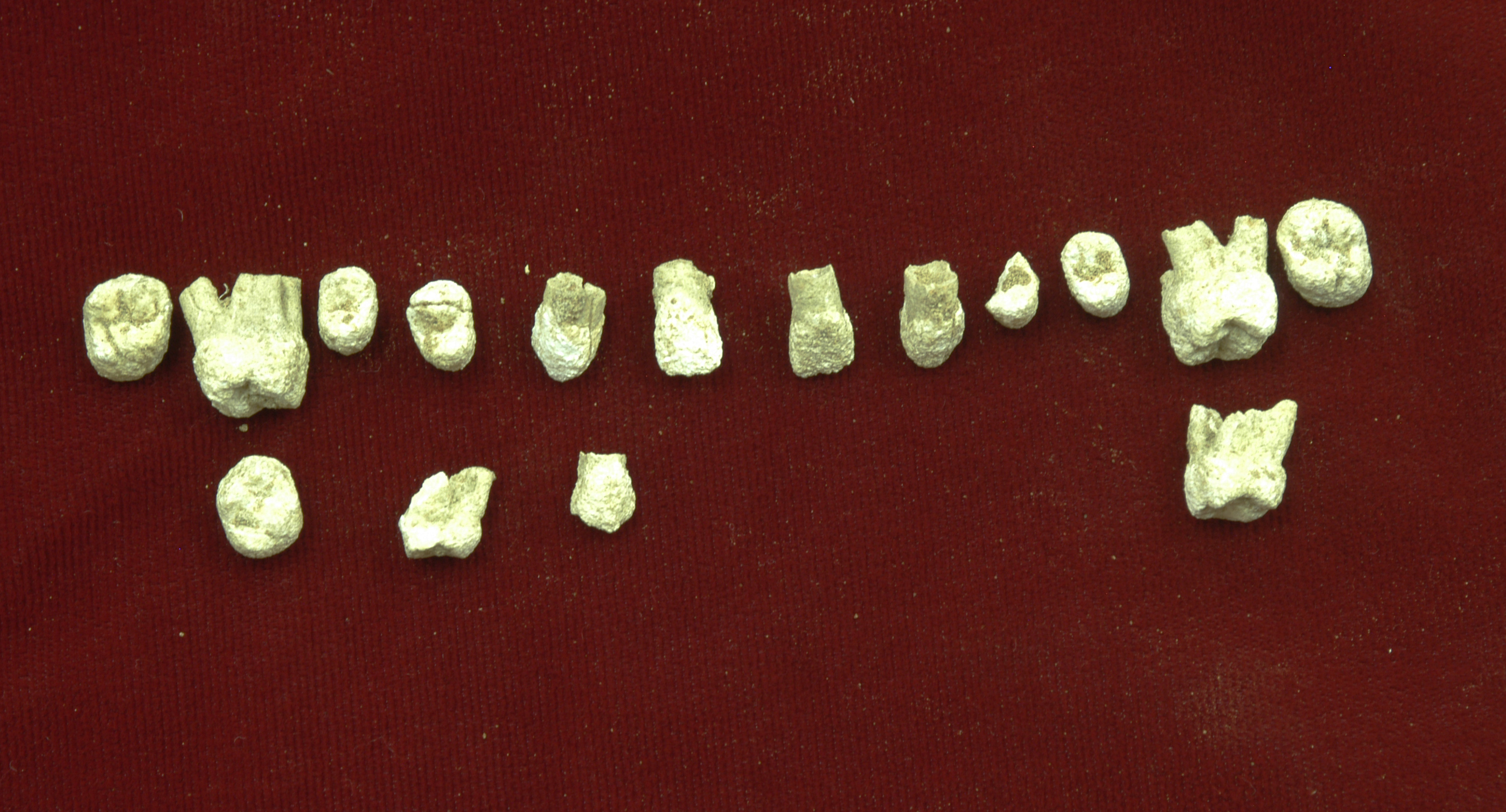
Inidvidual NN:
Teeth of individual NN
Full Resolution
Full Resolution

Inidvidual NN:
NN in situ
Full Resolution
Full Resolution

Inidvidual NO:
NO, NR, NK in situ
Full Resolution
Full Resolution

Inidvidual NQ:
Skulls in situ
Full Resolution
Full Resolution
Inidvidual NT:
In situ with NX
Full Resolution
Full Resolution

Inidvidual NT:
Skulls in situ
Full Resolution
Full Resolution

Inidvidual NU1:
NU1 (on photo NU) in situ
Full Resolution
Full Resolution

Inidvidual NV:
In situ
Full Resolution
Full Resolution
Inidvidual NW:
Skull frontal, individual NW
Full Resolution
Full Resolution

Inidvidual NW:
NW in situ, the skull lies on its side
Full Resolution
Full Resolution
Inidvidual NX:
NX and other skulls in situ
Full Resolution
Full Resolution
Inidvidual NY:
In situ
Full Resolution
Full Resolution

Inidvidual NY:
NY in situ
Full Resolution
Full Resolution
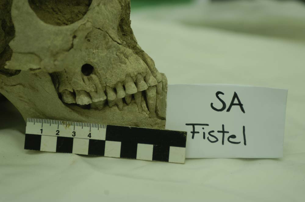
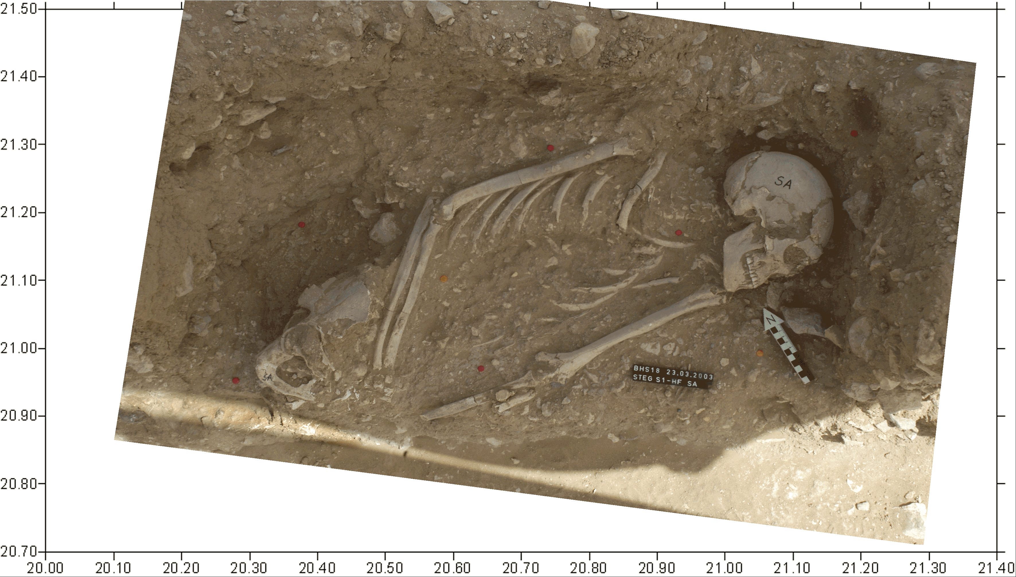
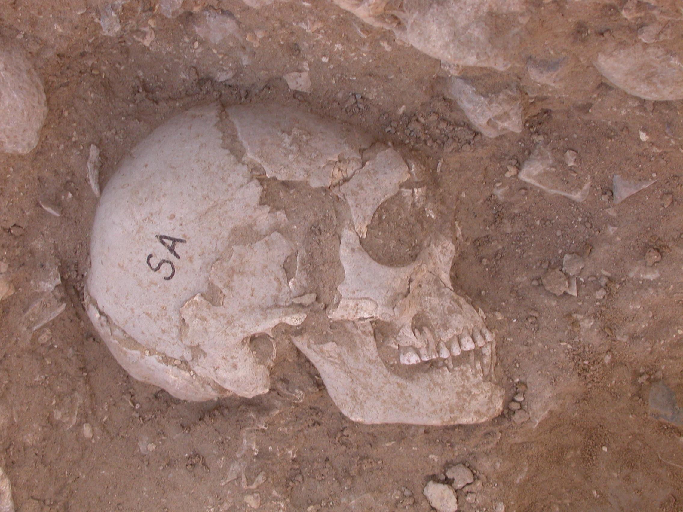
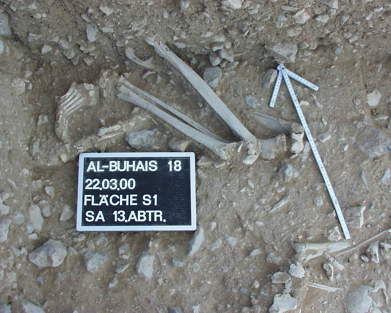
Inidvidual SA:
Tooth Fistula
Full Resolution
Full Resolution
Inidvidual SA:
Upper body and skull in situ
Full Resolution
Full Resolution
Inidvidual SA:
Detail of skull in situ
Full Resolution
Full Resolution

Inidvidual SA:
Pelvis individual SA
Full Resolution
Full Resolution
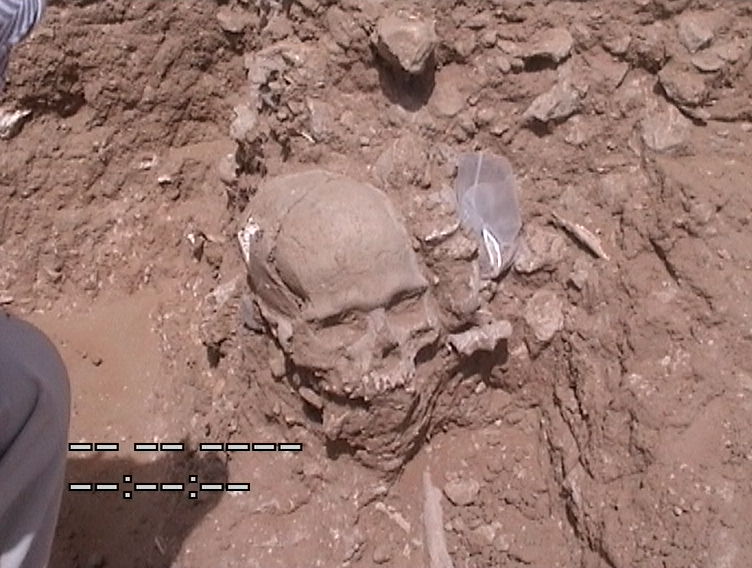
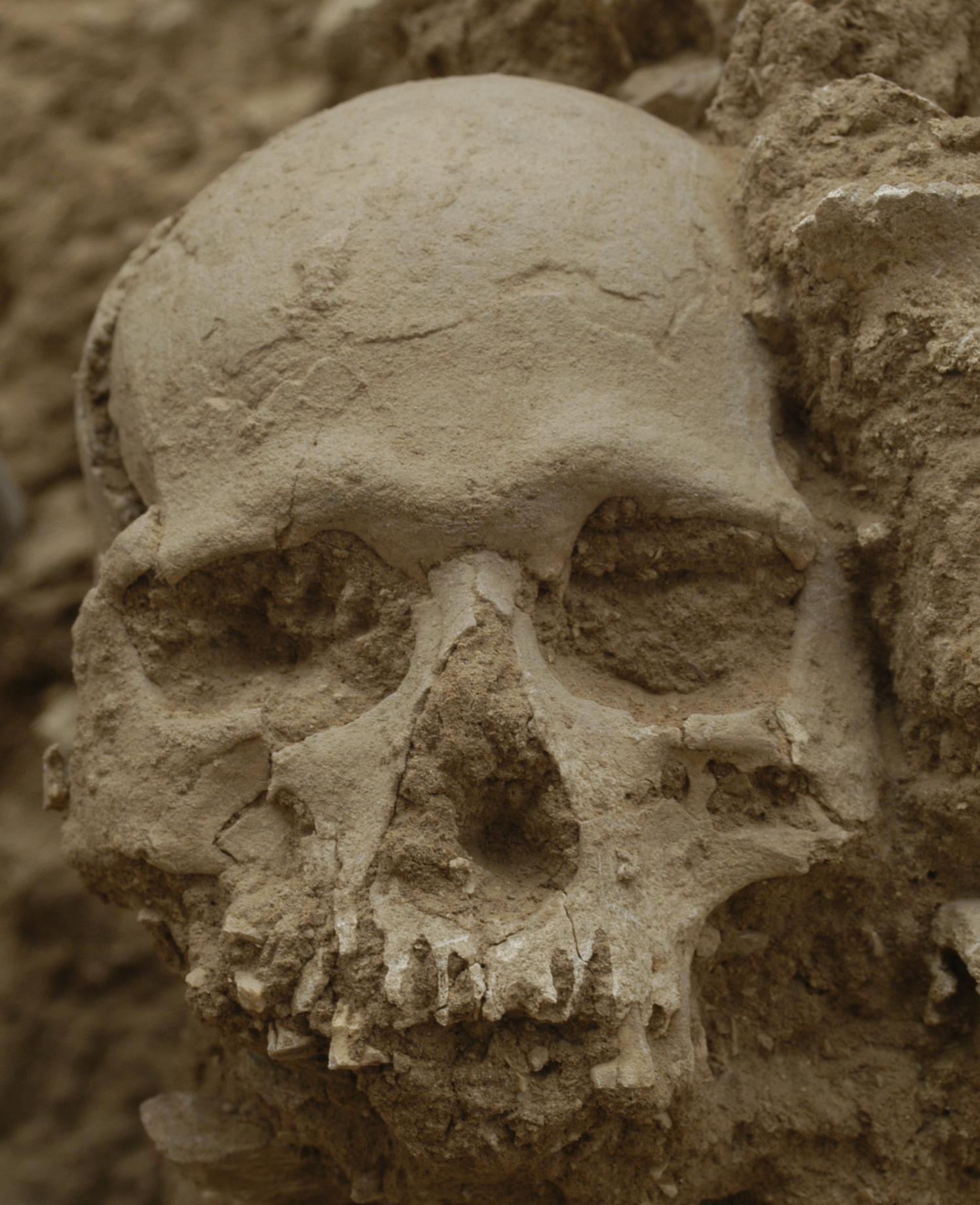
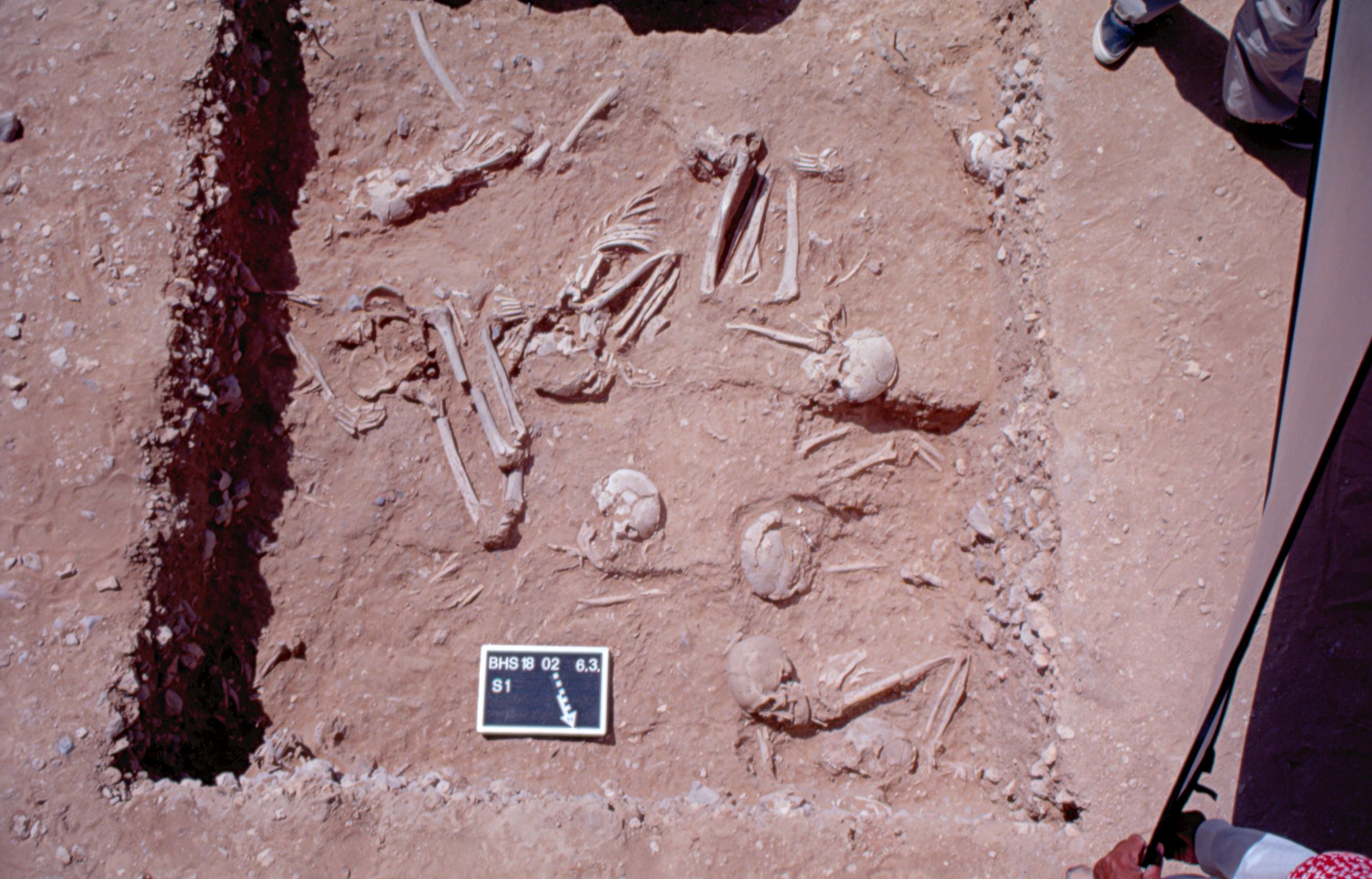
Inidvidual SA:
SA in S1
Full Resolution
Full Resolution
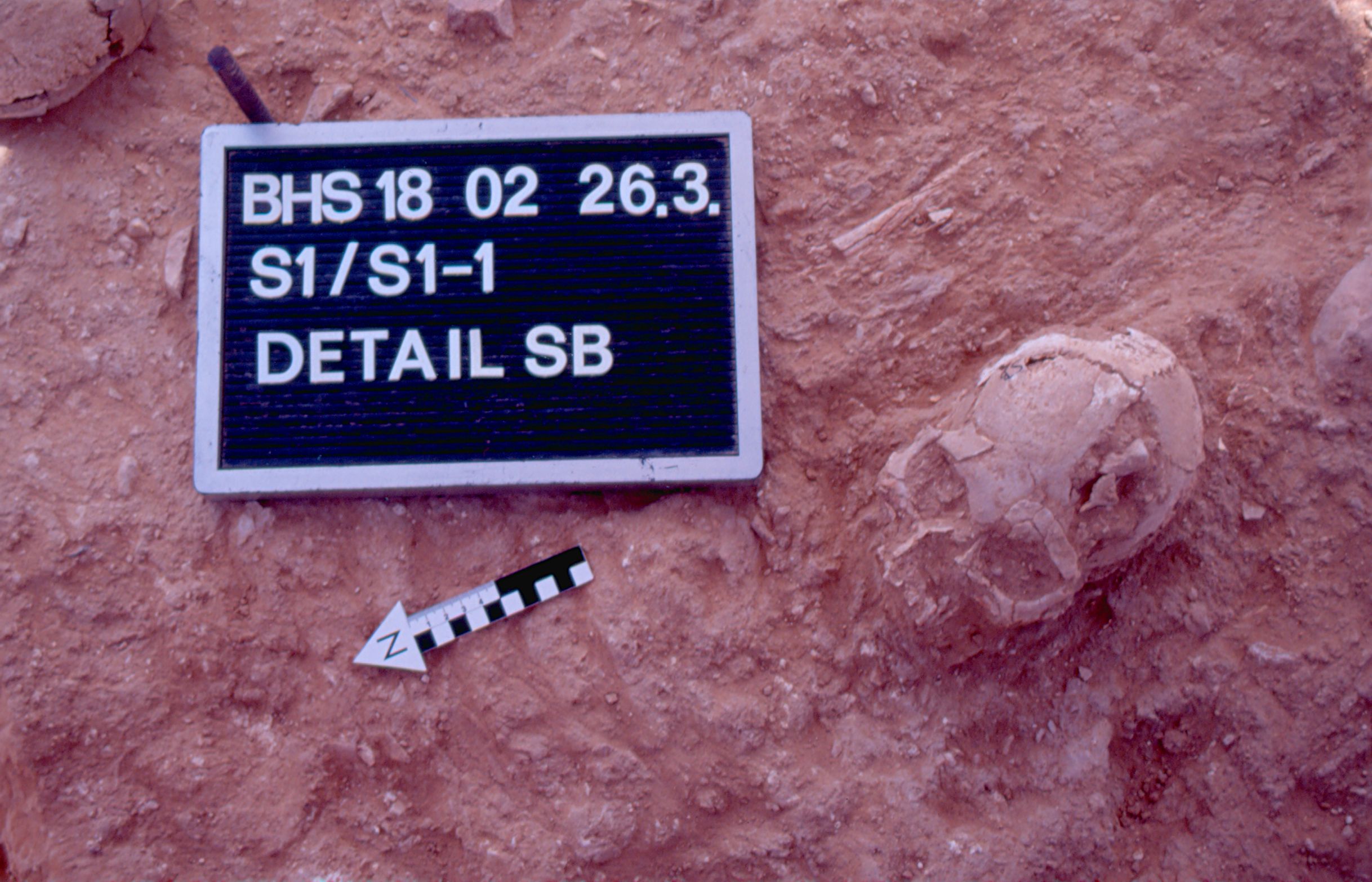
Inidvidual SB:
Secondary burial SB
Full Resolution
Full Resolution
Inidvidual SB:
Skull SB in situ in S1-1
Full Resolution
Full Resolution
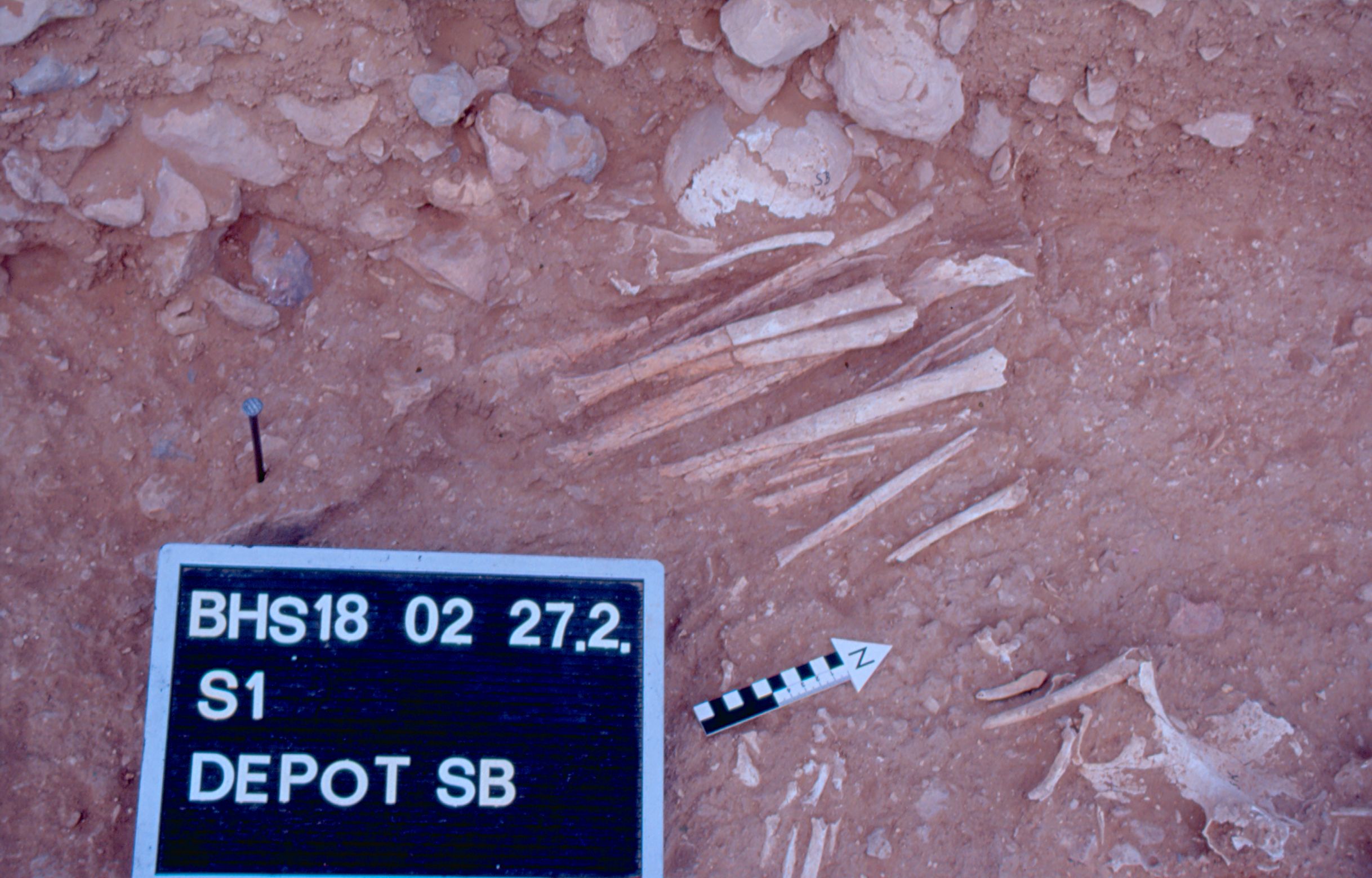
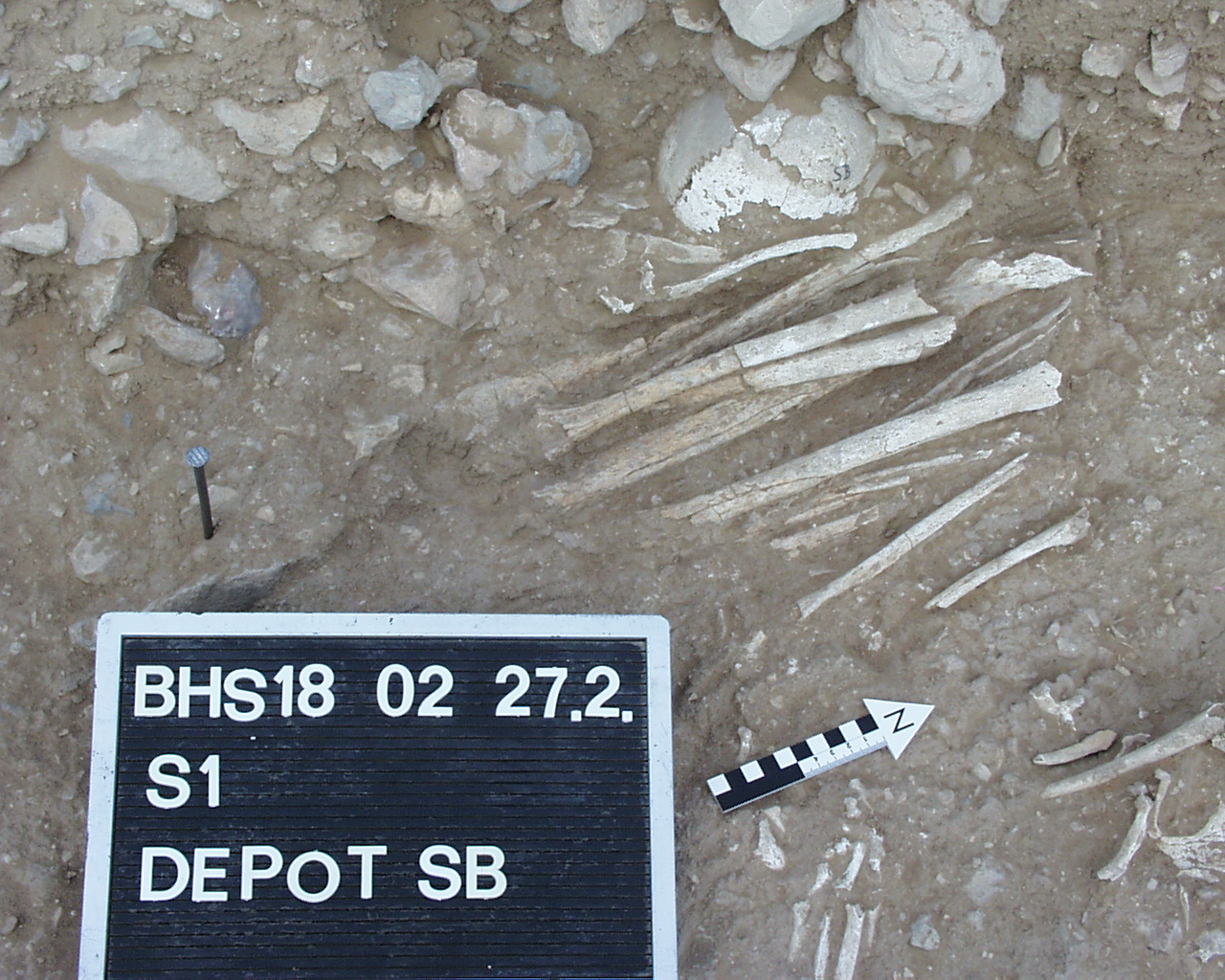
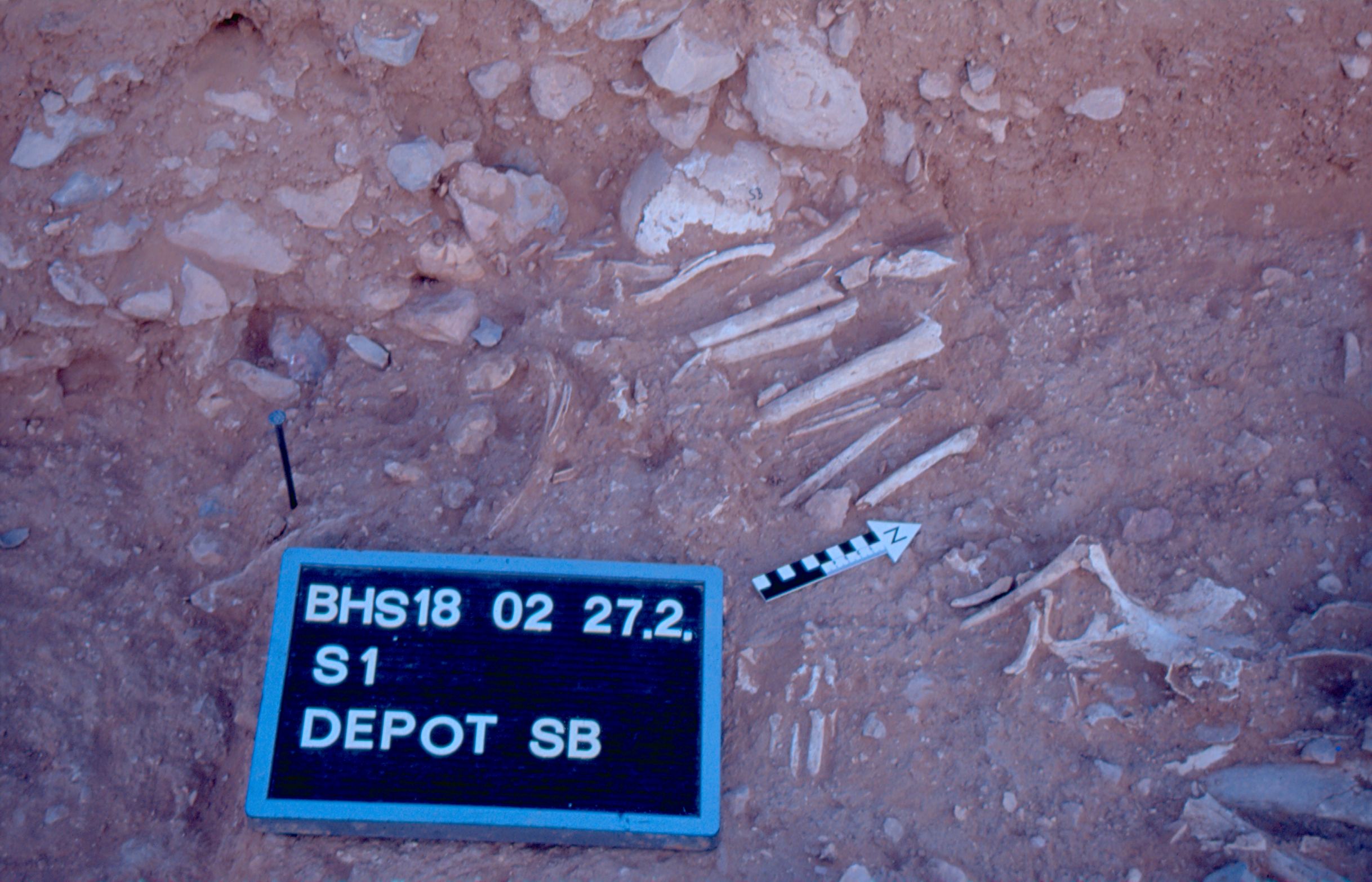
Inidvidual SB:
Depot SB 27.02.2002
Full Resolution
Full Resolution
Inidvidual SB:
Depot SB 27.02.2002
Full Resolution
Full Resolution
Inidvidual SB:
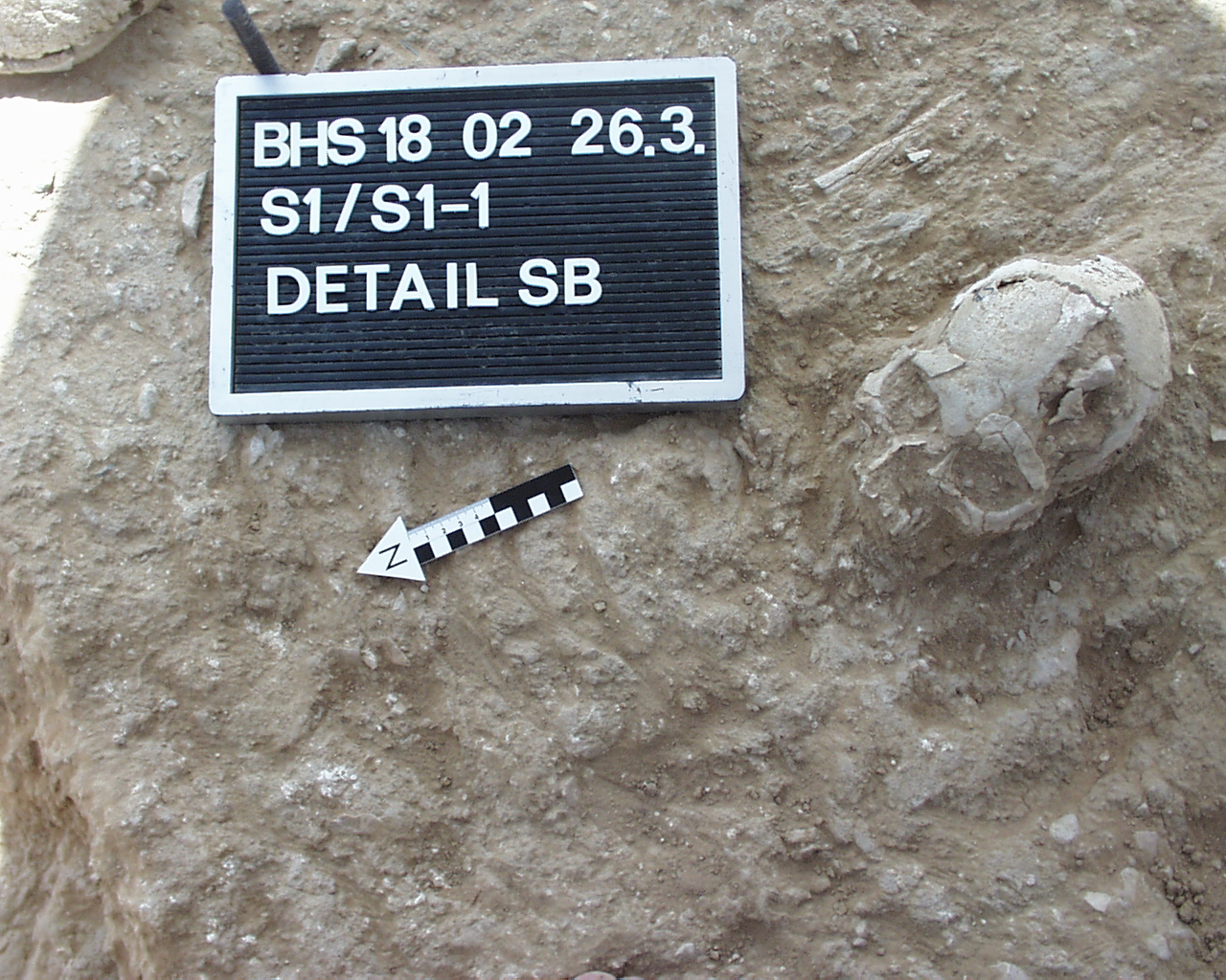
Detail SB 26.03.2002
Full Resolution
Full Resolution
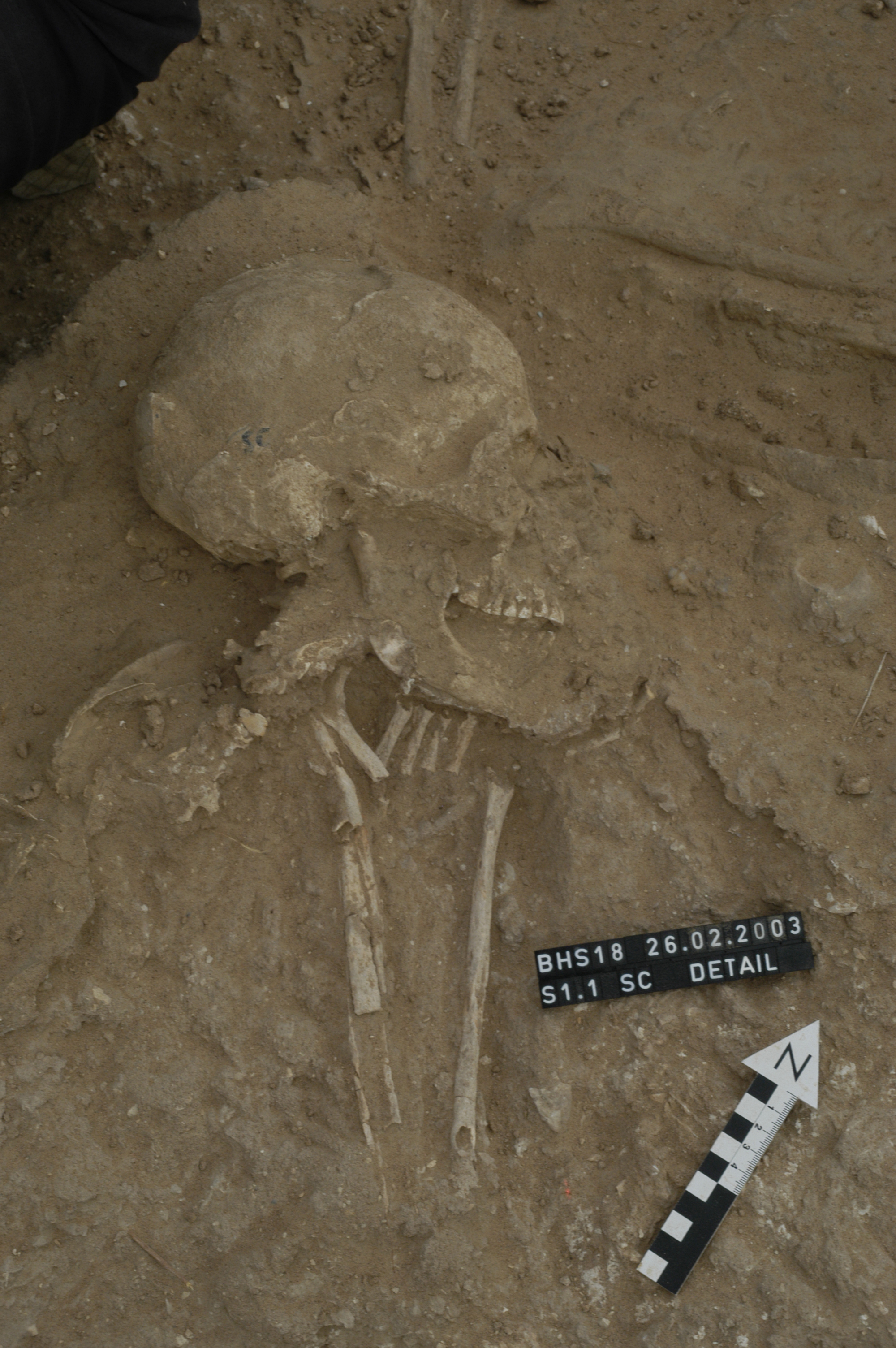
Inidvidual SC:
Skull and upper body in situ
Full Resolution
Full Resolution
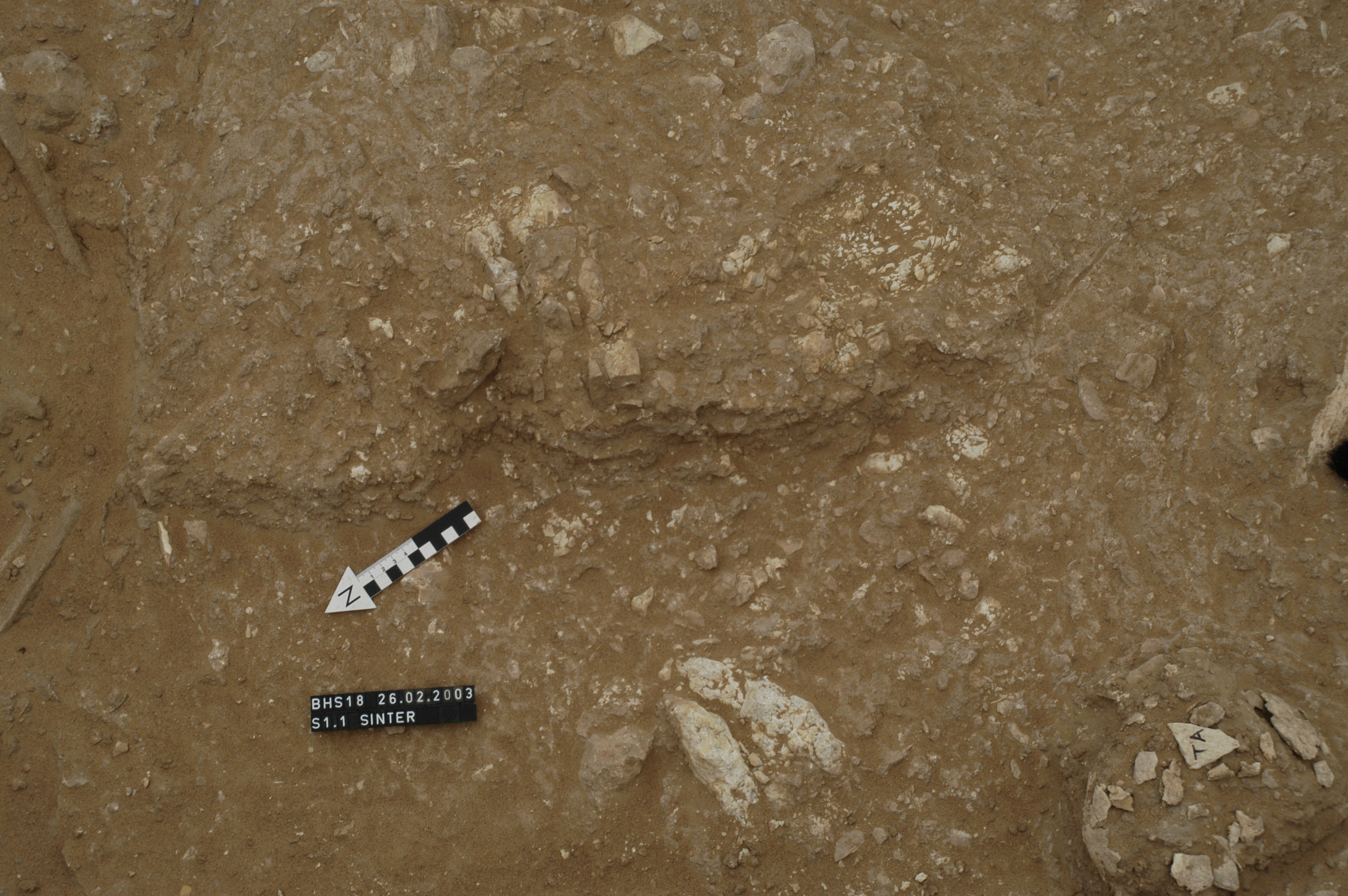
Inidvidual SC:
Sinter below burial SC in S1
Full Resolution
Full Resolution
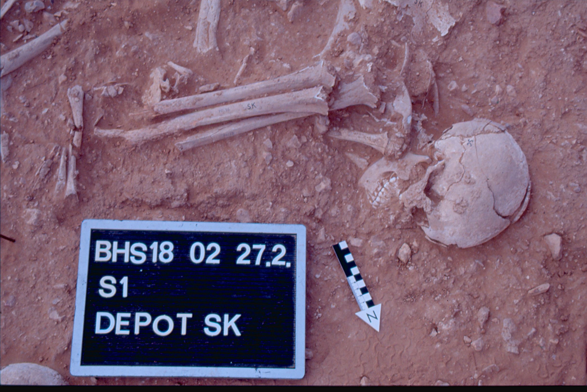
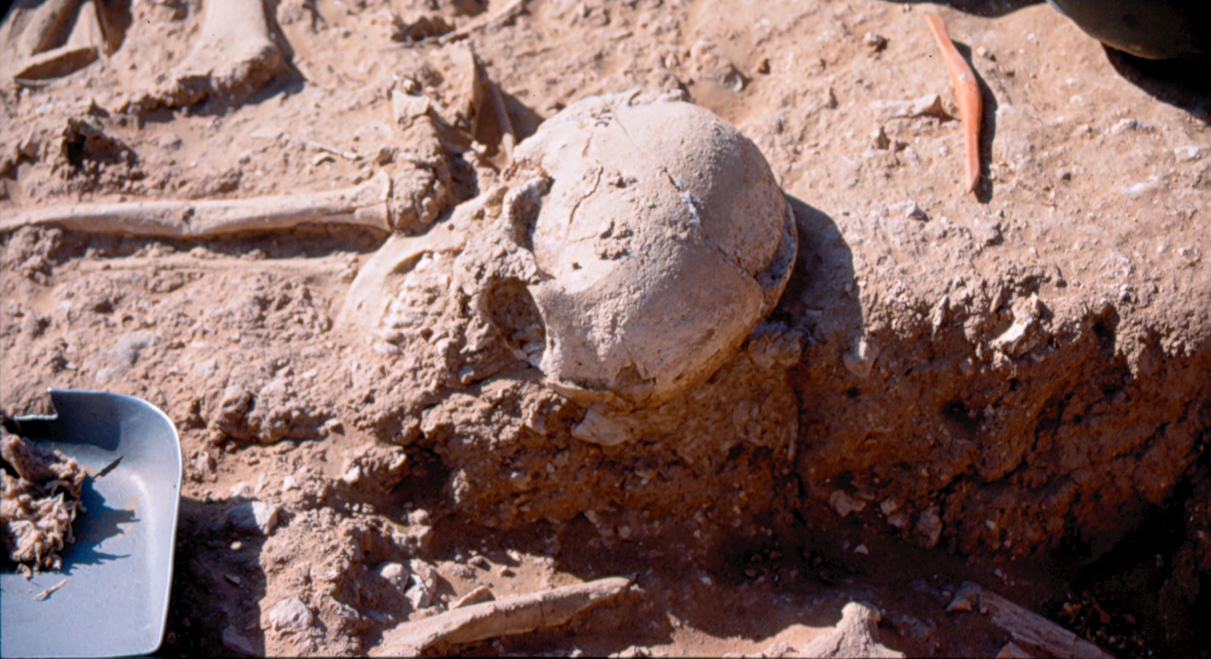
Inidvidual SC:
Depot SK including SK/SC 27.02.2002
Full Resolution
Full Resolution
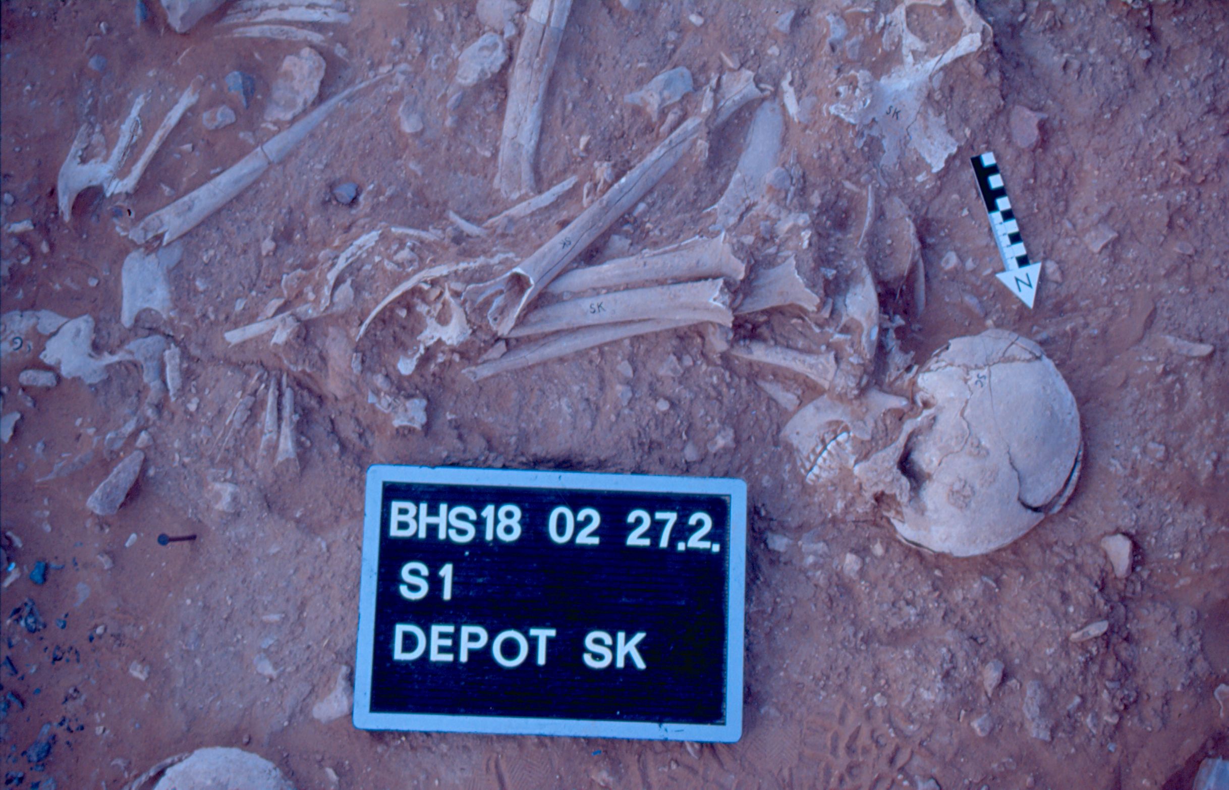
Inidvidual SC:
Depot SK including SK/SC 27.02.2002
Full Resolution
Full Resolution
Inidvidual SC:
SC
Full Resolution
Full Resolution
Inidvidual SC:
SC
Full Resolution
Full Resolution

Inidvidual SC:
SG and SC (left side of picture) in situ
Full Resolution
Full Resolution

Inidvidual SD:
In situ in S1, SD (upper left corner), other burials are SE, SG. Orthorectified.
Full Resolution
Full Resolution
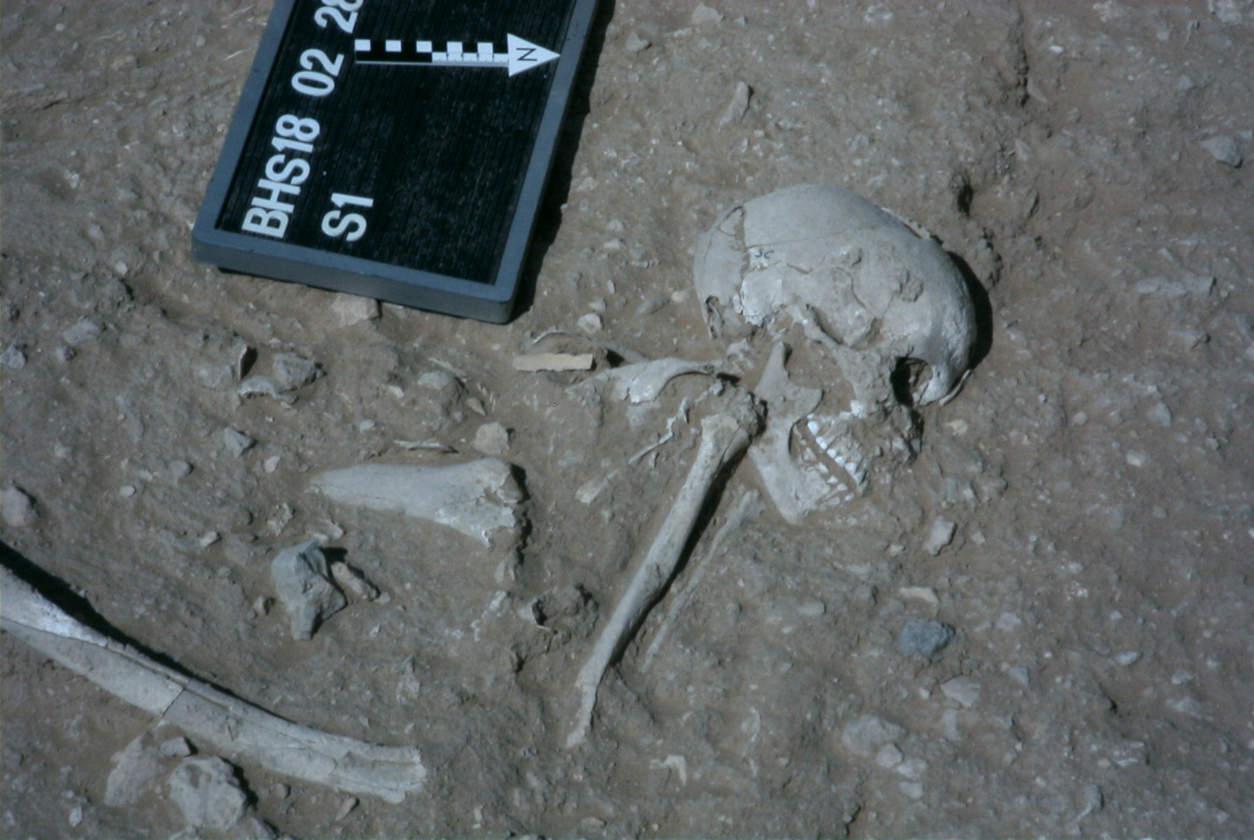
Inidvidual SD:
SD and SO, detail
Full Resolution
Full Resolution

Inidvidual SD:
SE, SG and SD in situ.
Full Resolution
Full Resolution

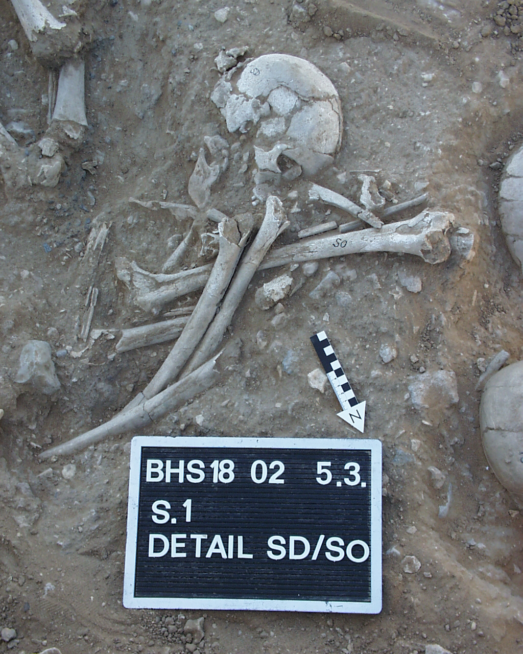
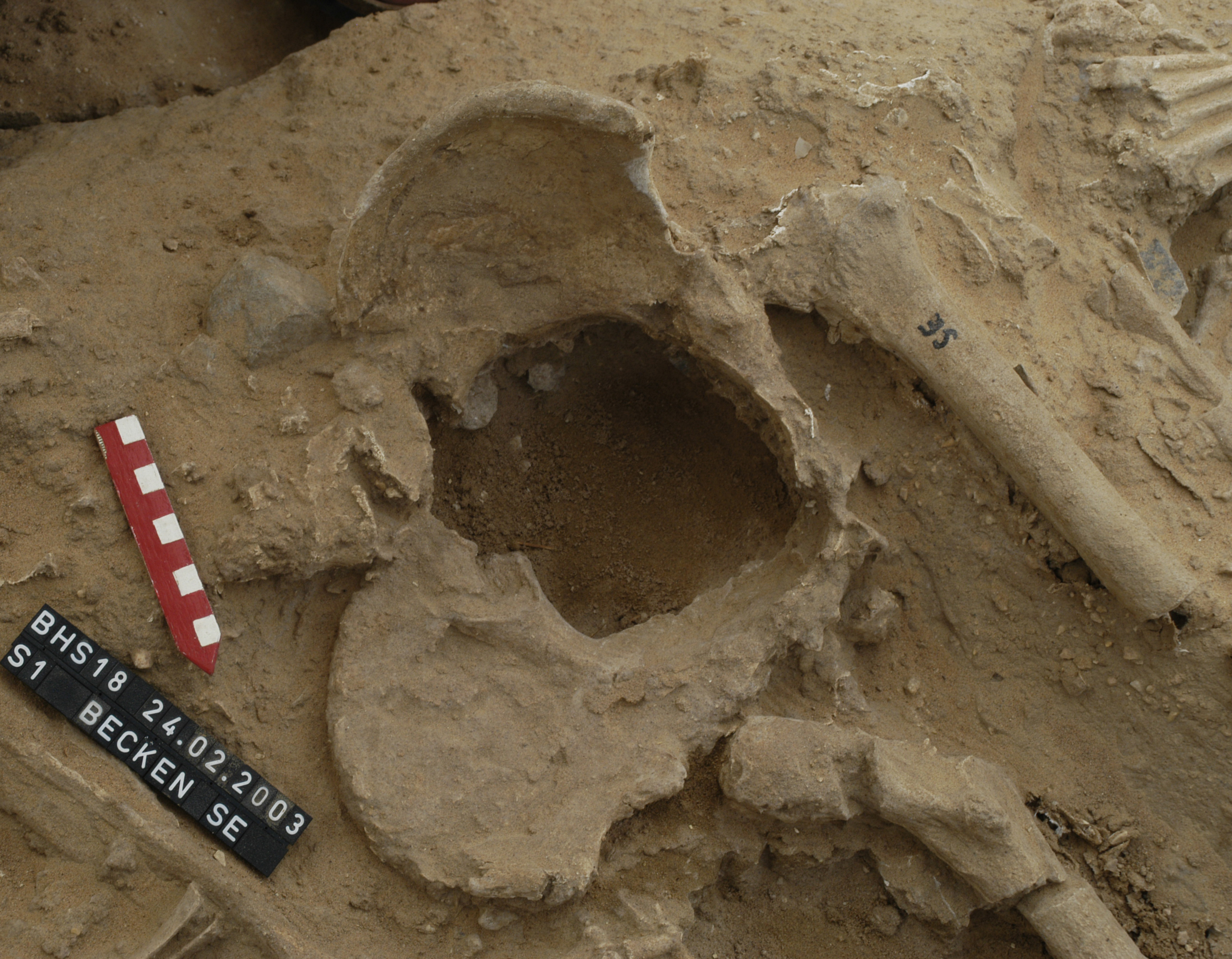
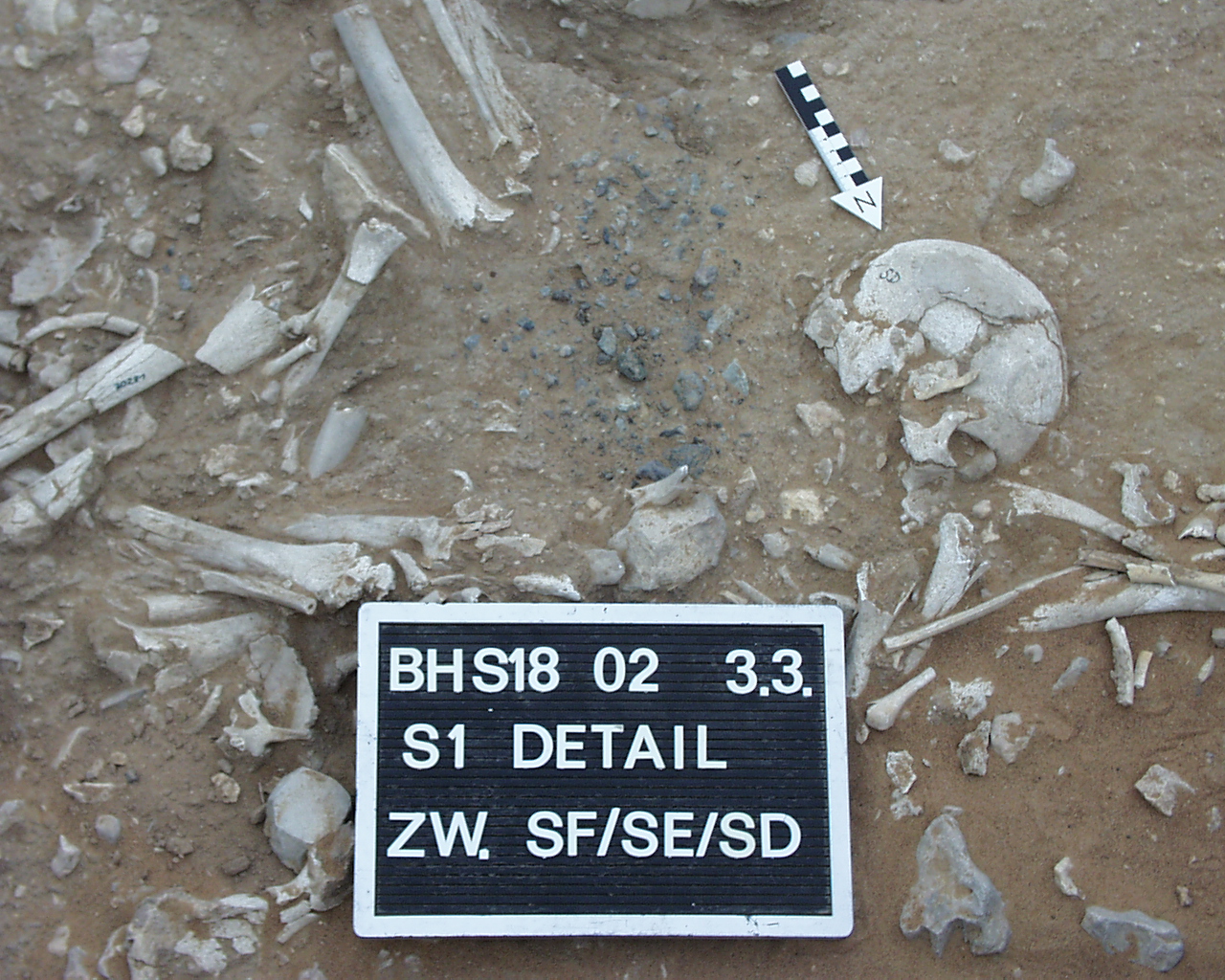
Inidvidual SD:
Detail of non-local ophiolite concentration between individuals SF, SE and SD.
Full Resolution
Full Resolution

Inidvidual SD:
SD/SO 05.03.2002
Full Resolution
Full Resolution
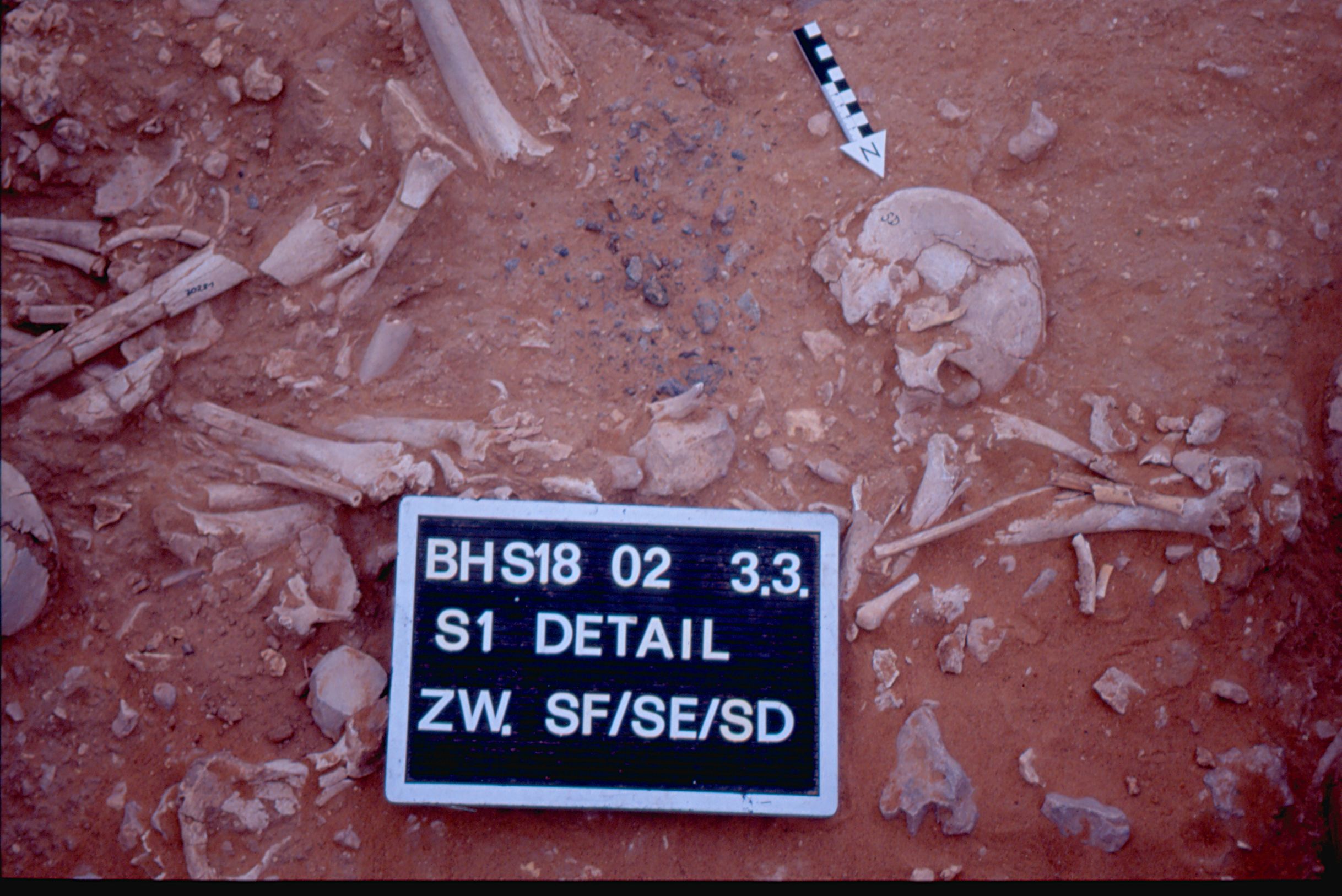
Inidvidual SD:
SF/SE/SD 03.03.2002
Full Resolution
Full Resolution
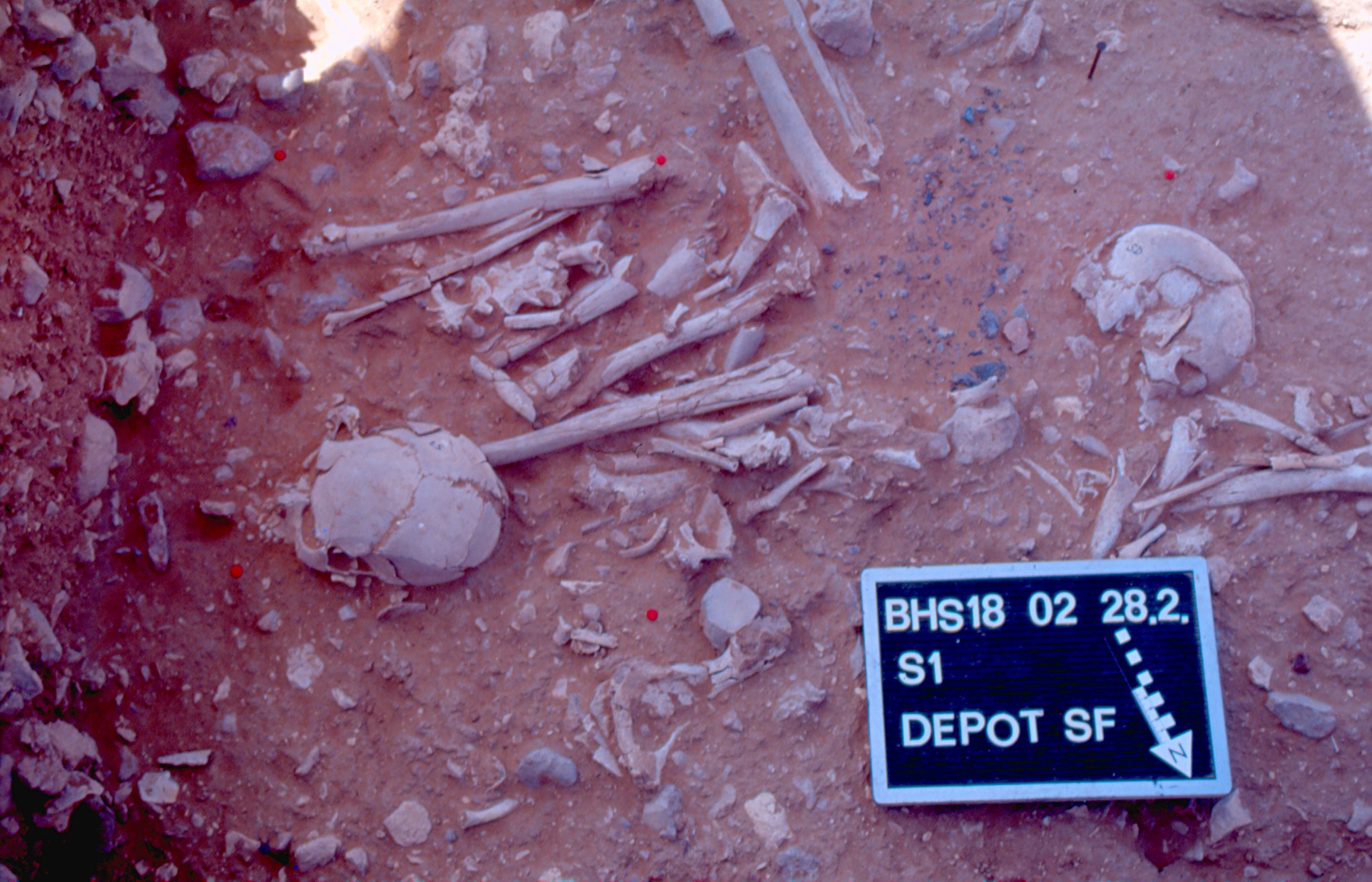
Inidvidual SD:
Depot SF including SF/SD. Non-local Ophiolite is visible near SF
Full Resolution
Full Resolution
Inidvidual SE:
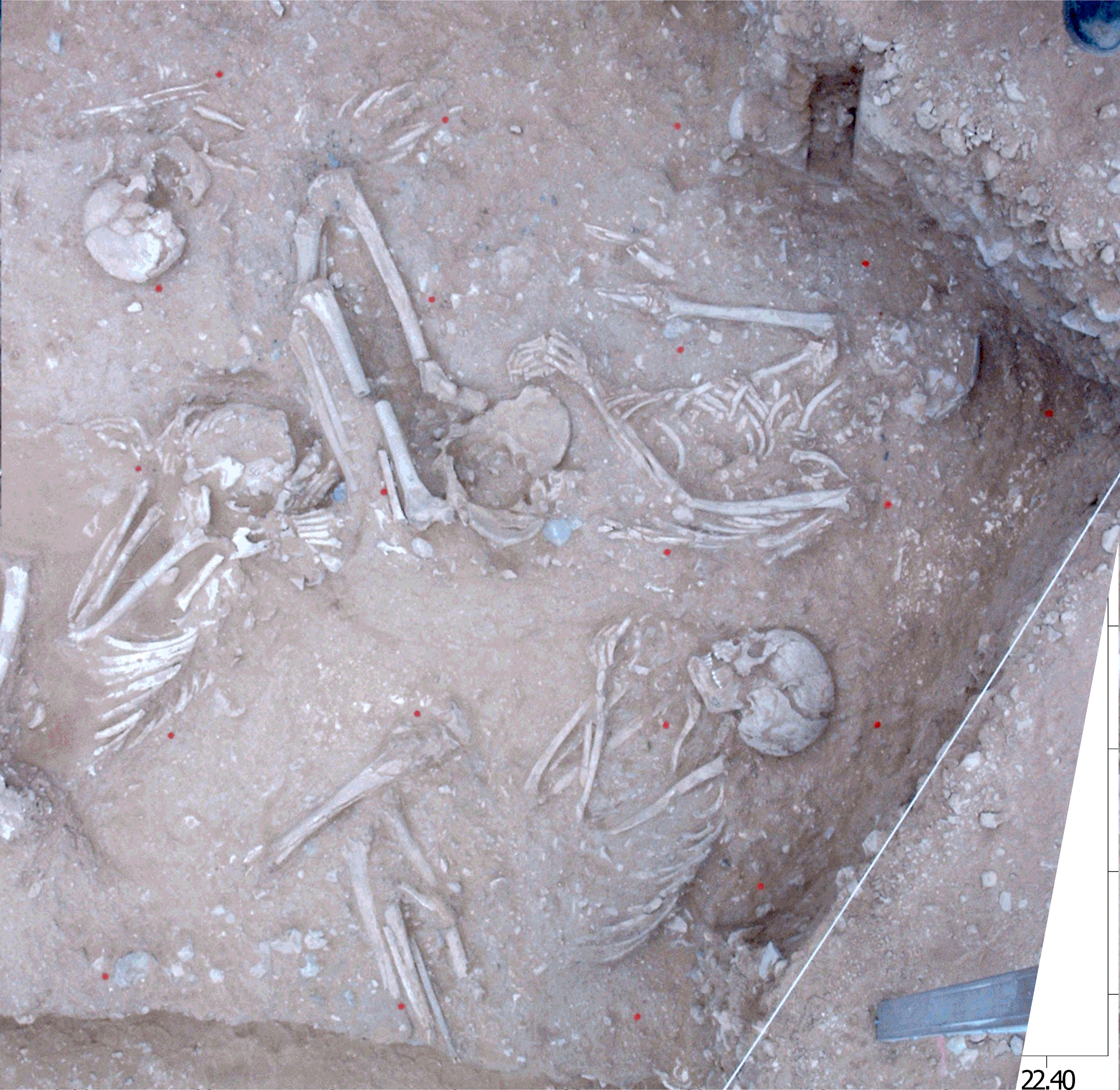
Orthorectified picture of SE, SG, SD in situ in S1
Full Resolution
Full Resolution

Inidvidual SE:
Orthorectified picture of primary burials in situ in S1: SE, SG, SD, SI, SL and secondary burial of SS and SR in the right upper corner
Full Resolution
Full Resolution
Inidvidual SE:
Pelvis in situ
Full Resolution
Full Resolution

Inidvidual SE:
Skull SE, detail with adornments in situ. Carnelian bead
Full Resolution
Full Resolution
Inidvidual SE:
SE, SG and SD in situ.
Full Resolution
Full Resolution
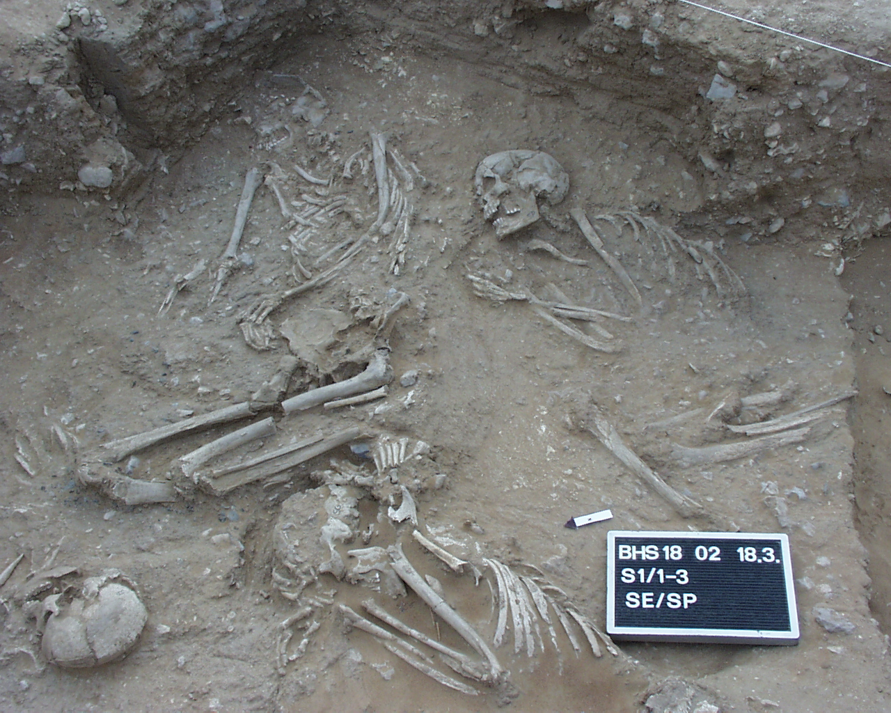
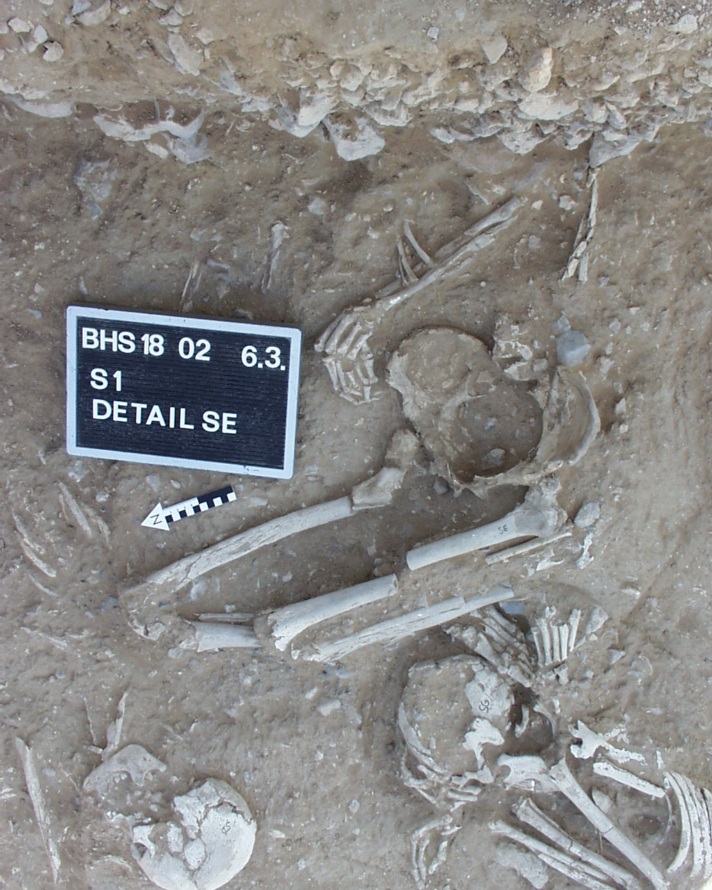
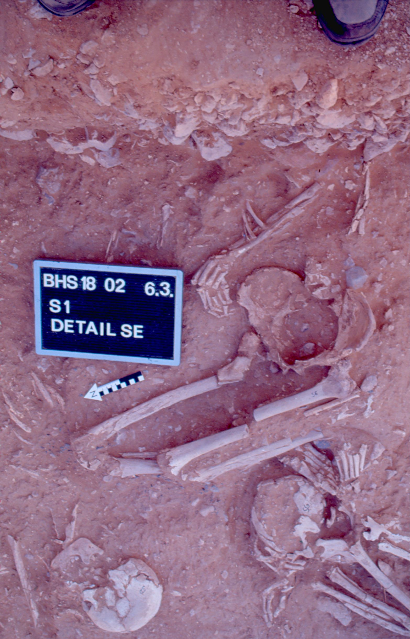
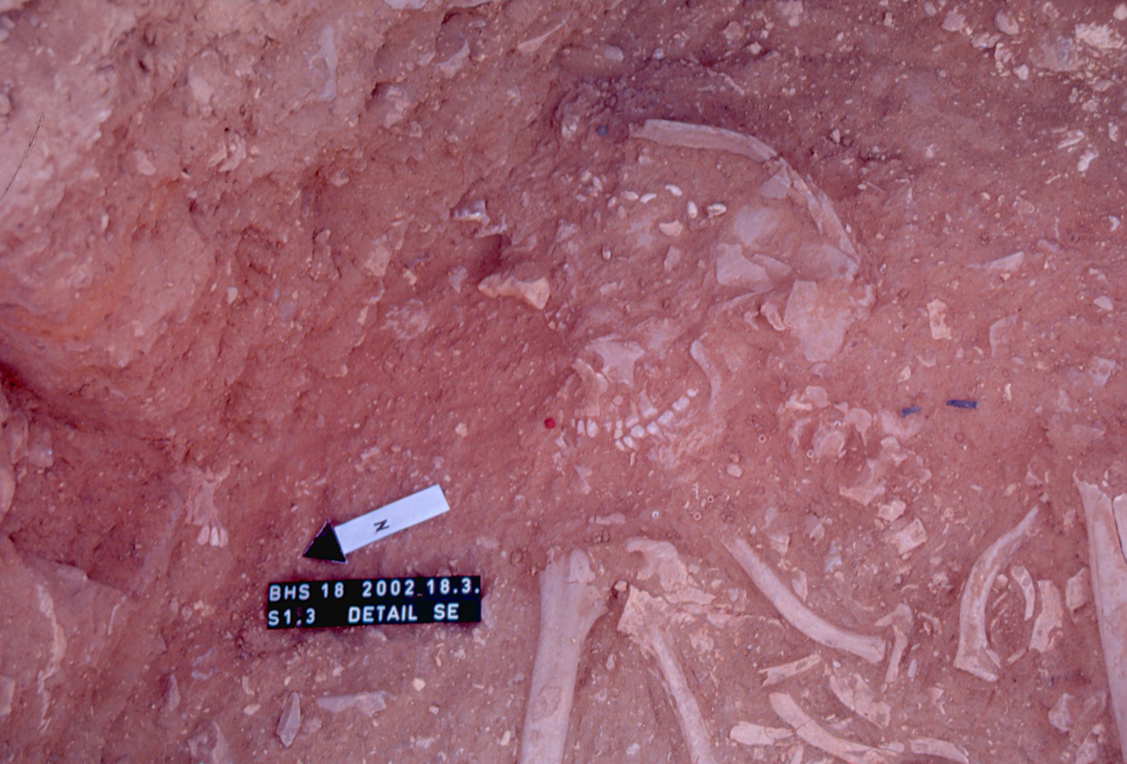
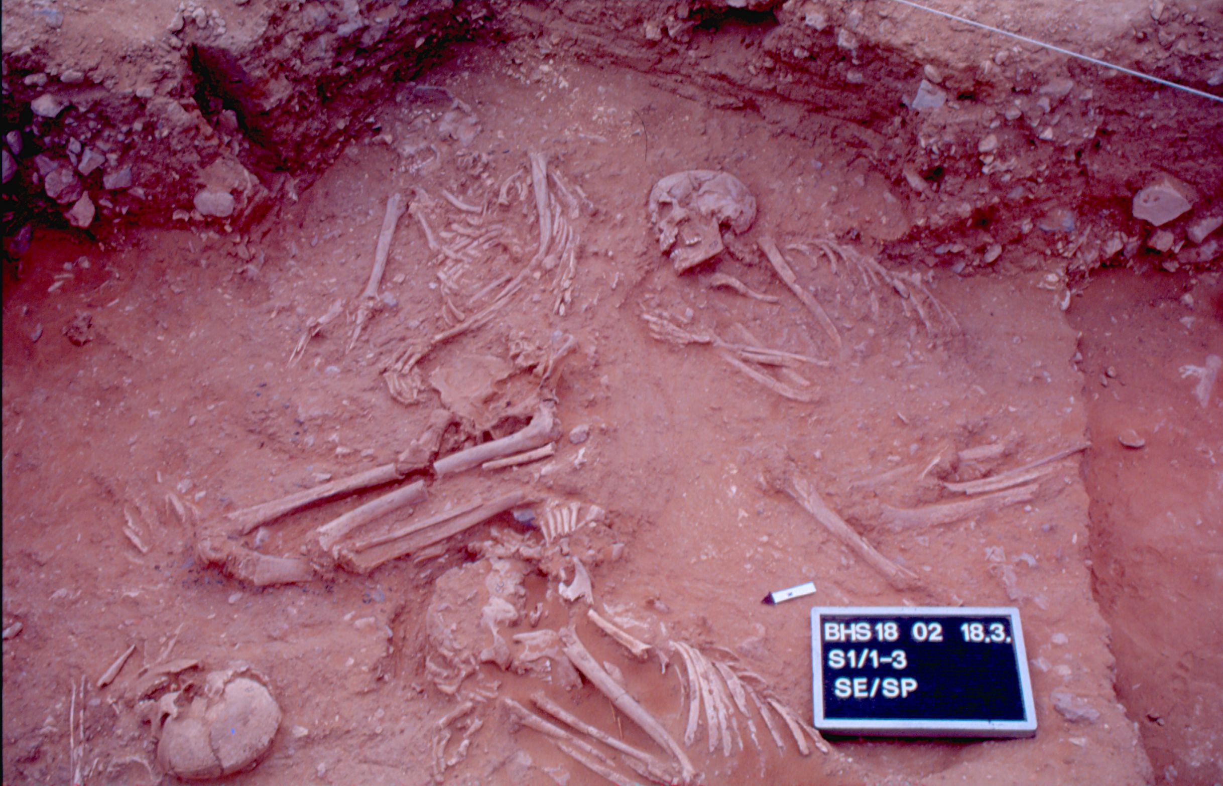
Inidvidual SE:
SE and SP in situ in S1/1-3
Full Resolution
Full Resolution

Inidvidual SE:
Detail of ophiolite concentration between individuals SF, SE and SD. Ophiolite.
Full Resolution
Full Resolution

Inidvidual SE:
SF/SE/SD 03.03.2002
Full Resolution
Full Resolution
Inidvidual SE:
Detail SE 06.03.2002
Full Resolution
Full Resolution
Inidvidual SE:
Detail SE 18.03.2002
Full Resolution
Full Resolution

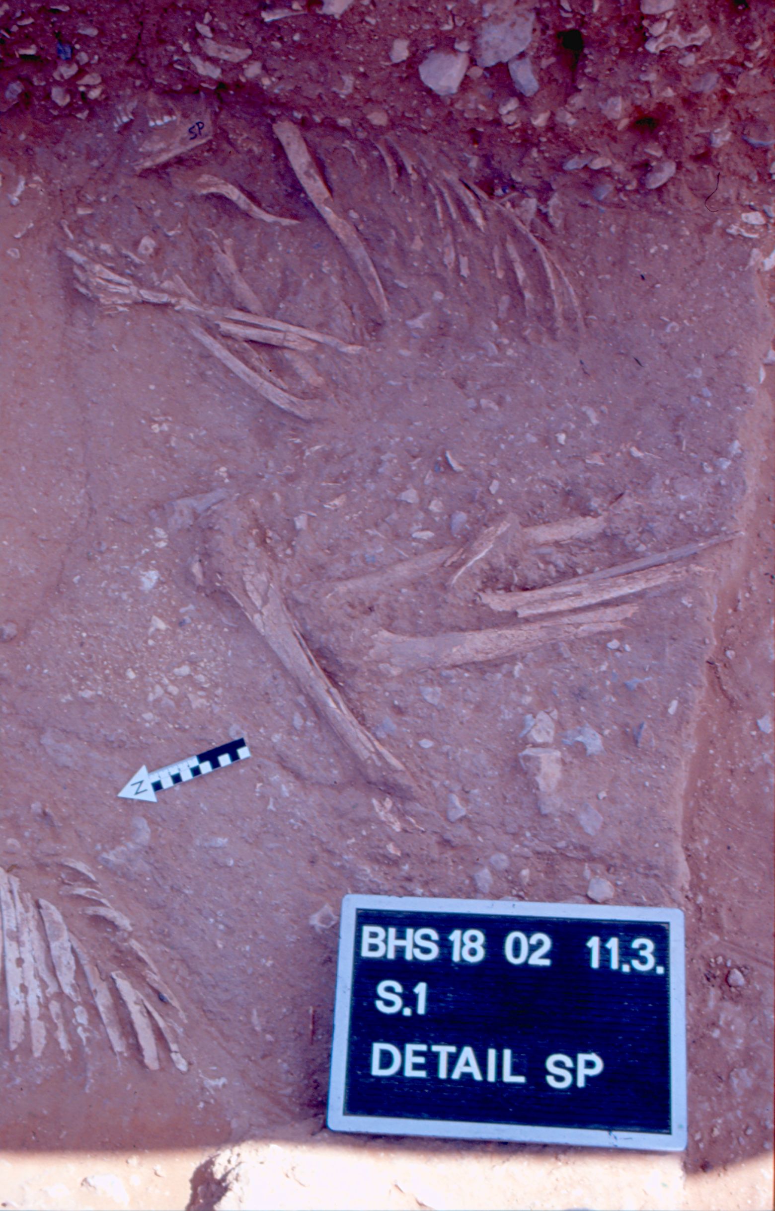
Inidvidual SE:
SE/SP 18.03.2002
Full Resolution
Full Resolution
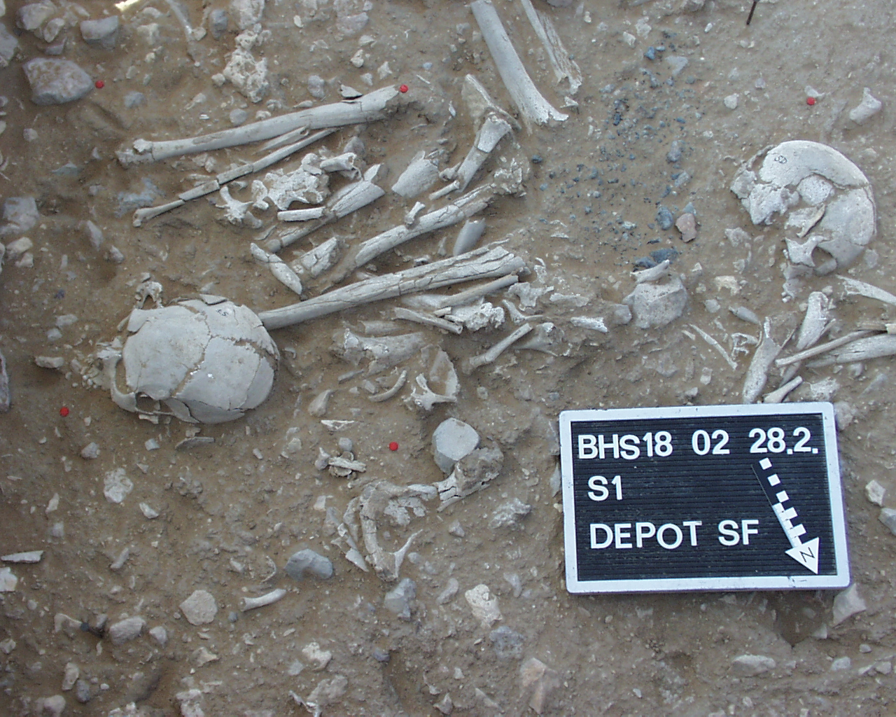
Inidvidual SF:
Burial SD and Depot SF
Full Resolution
Full Resolution
Inidvidual SF:
Detail depot SF with adornments
Full Resolution
Full Resolution
Inidvidual SF:
S1, Depot SF with adornments and skull
Full Resolution
Full Resolution
Inidvidual SF:
Detail of ophiolite concentration between individuals SF, SE and SD. Ophiolite supposedly from primary burial in a wadi.
Full Resolution
Full Resolution

Inidvidual SF:
SF/SE/SD 03.03.2002
Full Resolution
Full Resolution
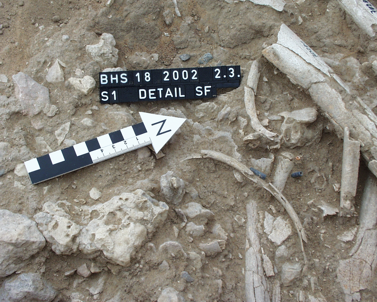
Inidvidual SF:
Detail SF 02.03.2002
Full Resolution
Full Resolution

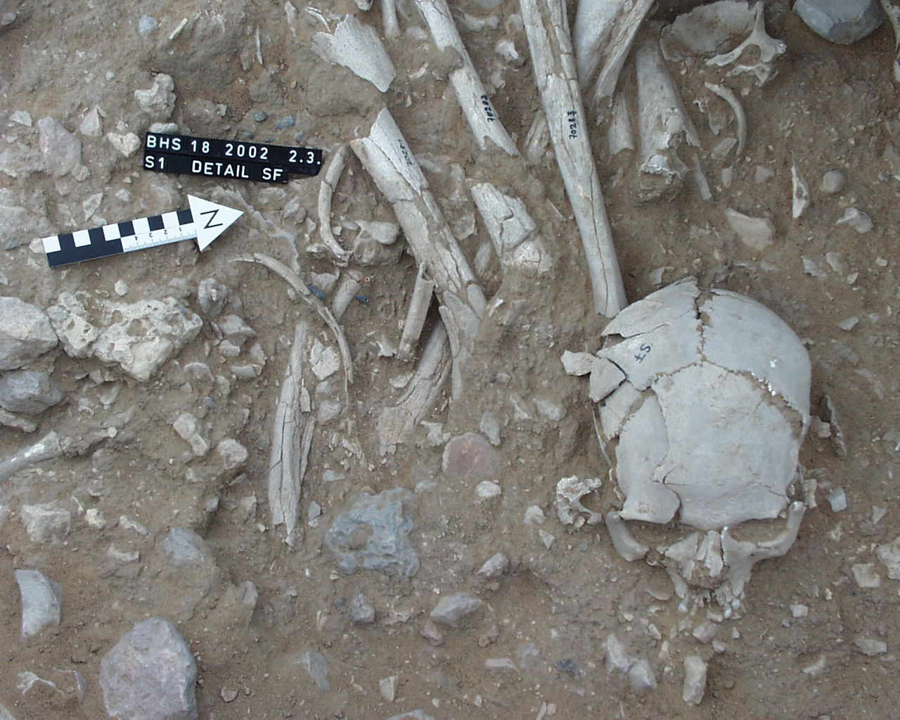
Inidvidual SF:
close Detail SF 02.03.2002
Full Resolution
Full Resolution

Inidvidual SF:
Depot SF 28.02.2002
Full Resolution
Full Resolution
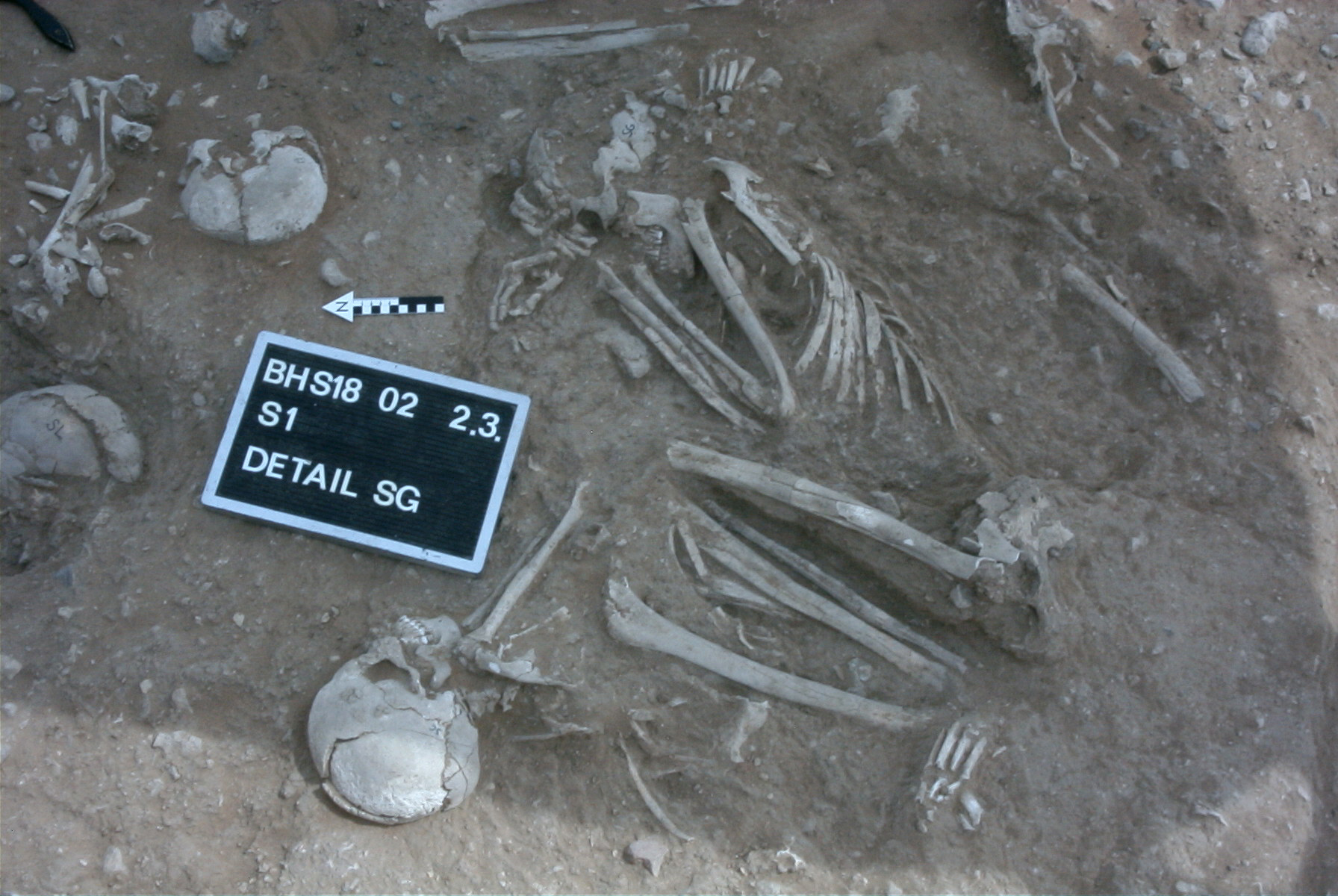
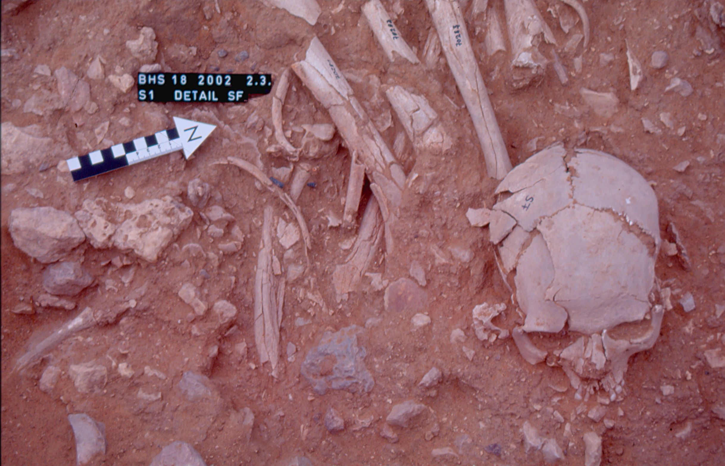
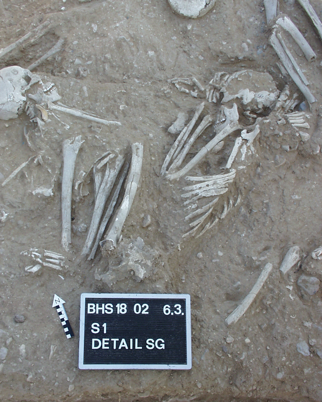
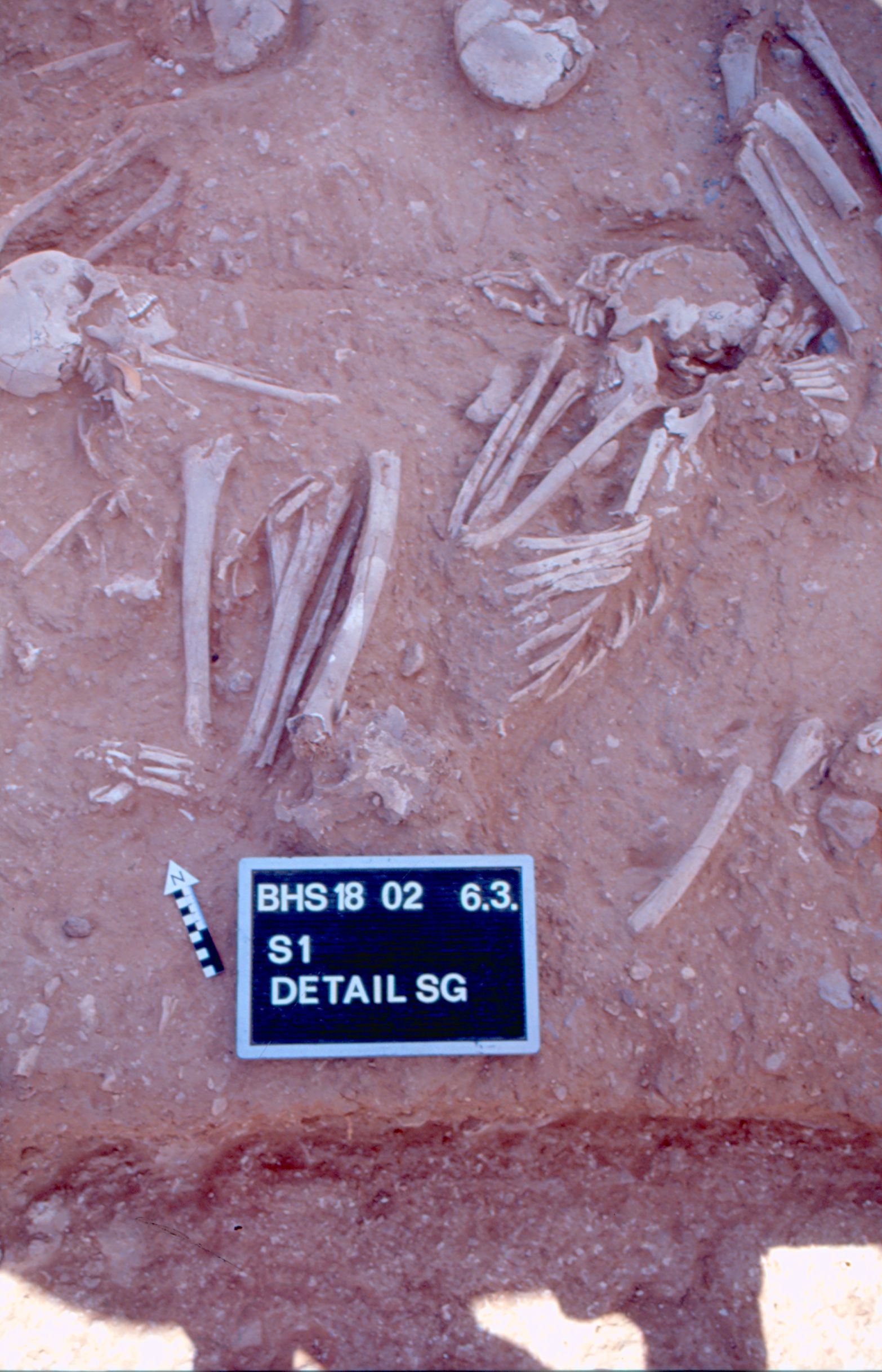
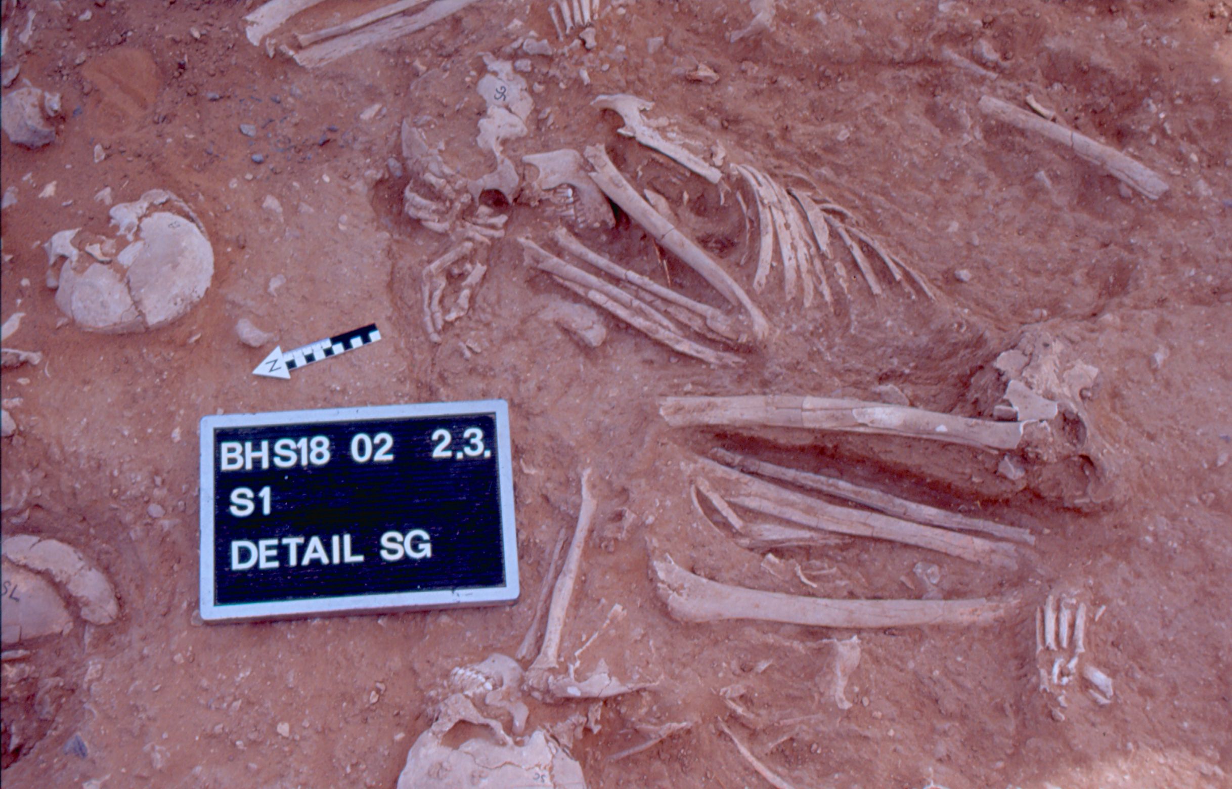
Inidvidual SG:
SG in situ
Full Resolution
Full Resolution
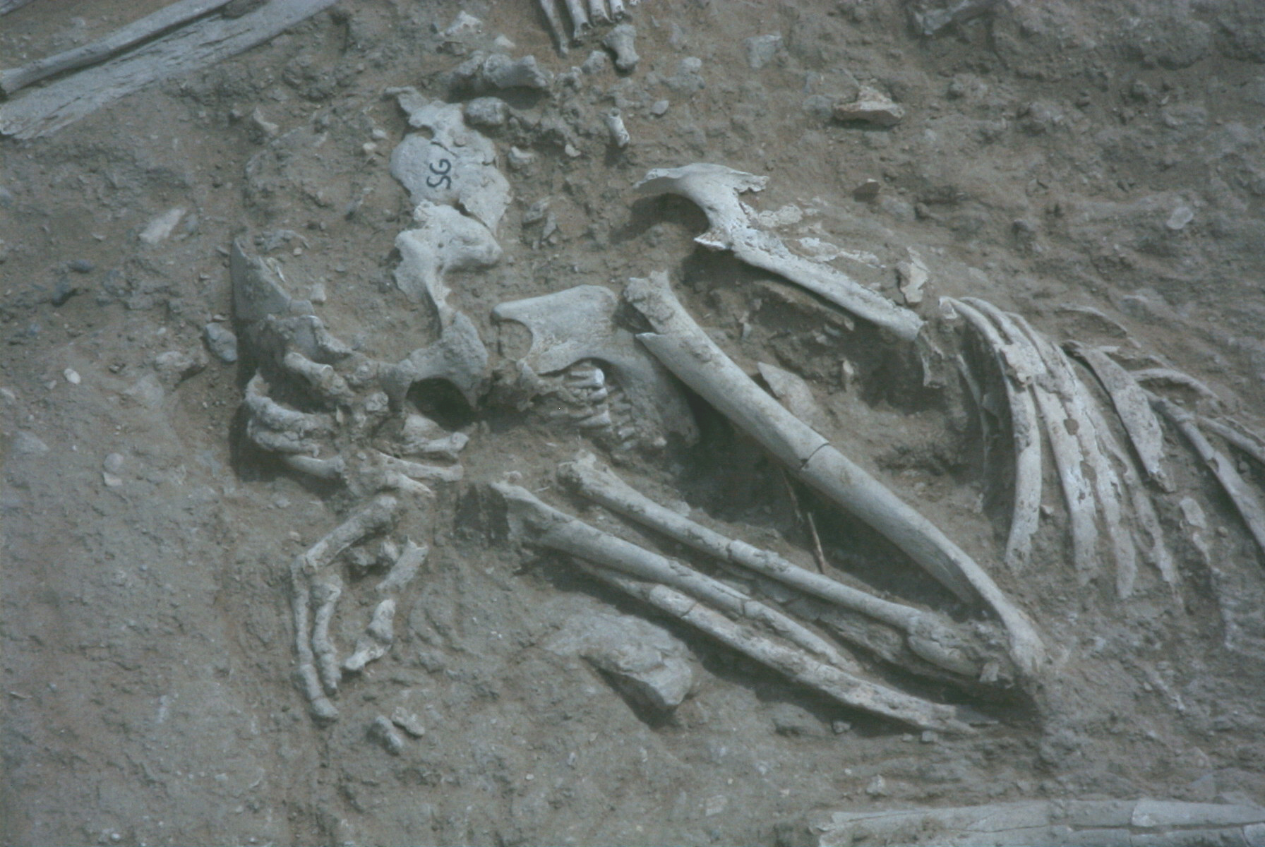
Inidvidual SG:
SG in situ detail upper body part
Full Resolution
Full Resolution
Inidvidual SG:
SG and SC in situ
Full Resolution
Full Resolution

Inidvidual SG:
SE, SG and SD in situ.
Full Resolution
Full Resolution
Inidvidual SG:
SG (scanned slide)
Full Resolution
Full Resolution
Inidvidual SG:
Detail SG 02.03.2002
Full Resolution
Full Resolution
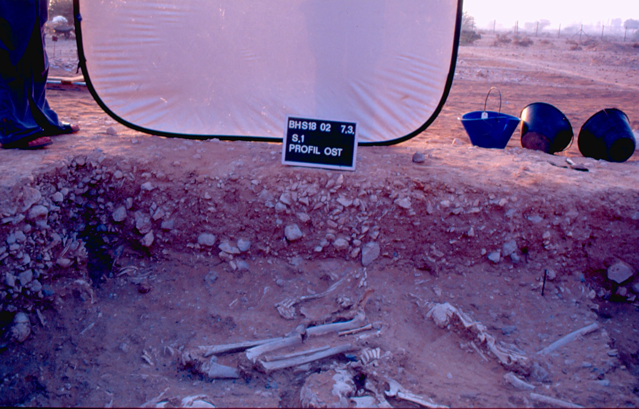
Inidvidual SG:
Profil OST S.1 with SG 07.03.2002
Full Resolution
Full Resolution

Inidvidual SG:
Detail SE with SG 06.03.2002
Full Resolution
Full Resolution
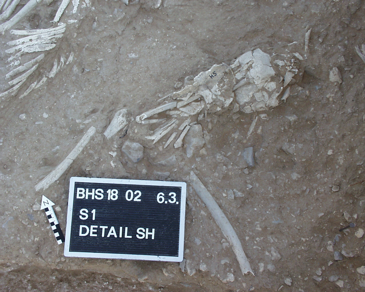
Inidvidual SH:
Individual SH in S1
Full Resolution
Full Resolution
Inidvidual SH:
Individual SH in S1 06.03.2002
Full Resolution
Full Resolution
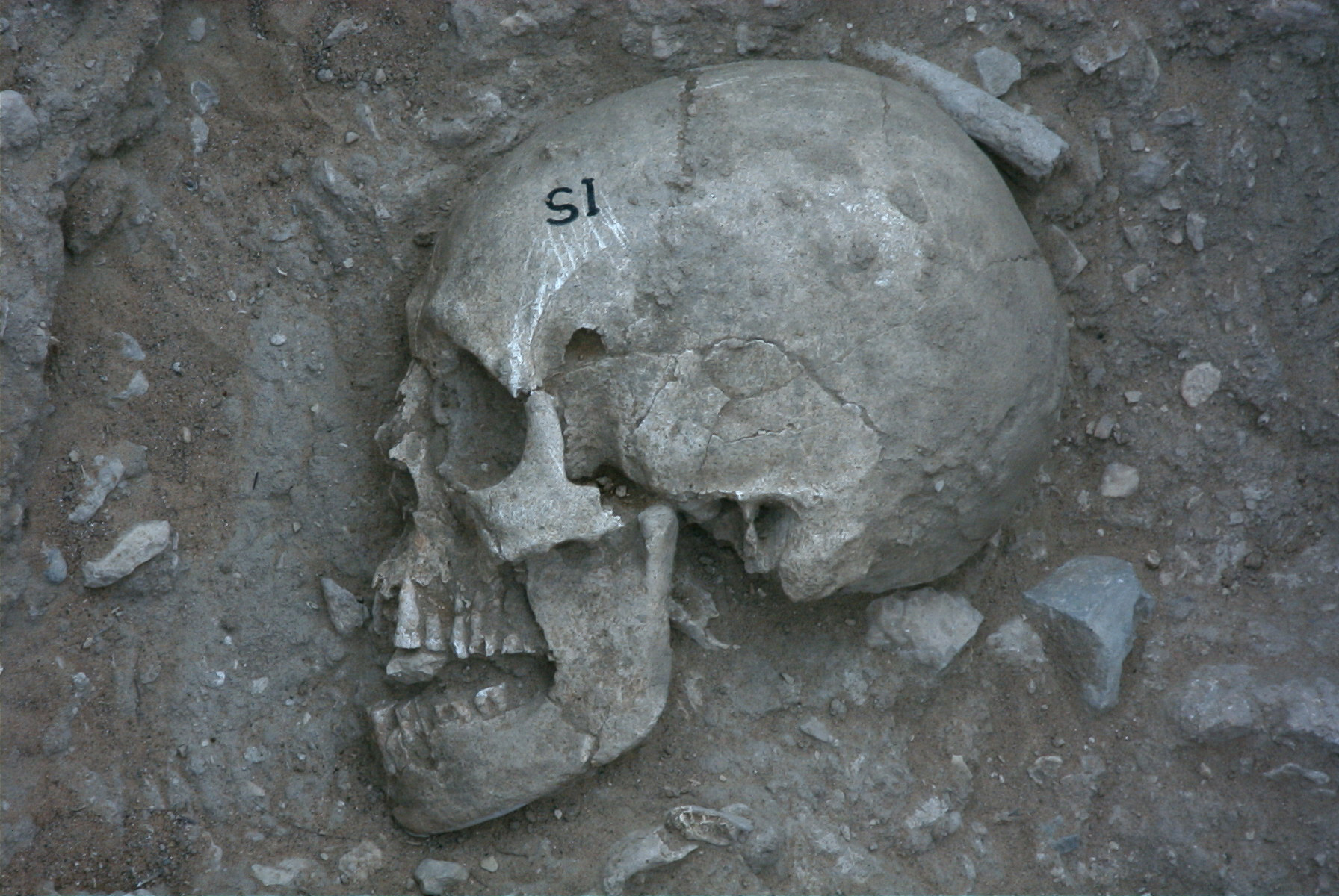
Inidvidual SI:
Skull SI in situ
Full Resolution
Full Resolution

Inidvidual SI:
SI in situ, orthorectified
Full Resolution
Full Resolution
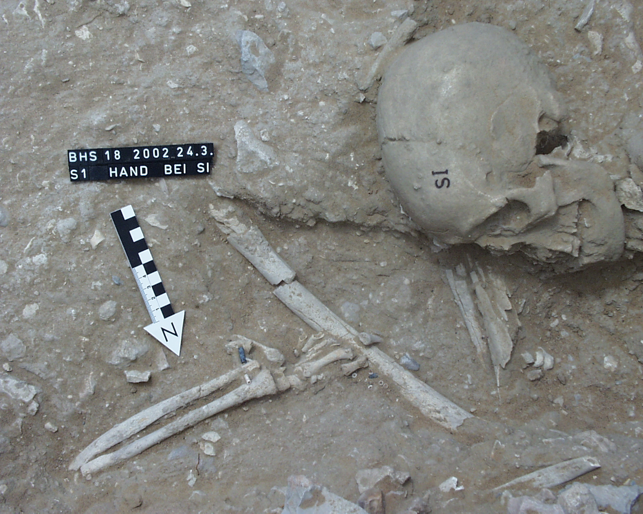
Inidvidual SI:
hand and arm with adornemnets near individual SI
Full Resolution
Full Resolution

Inidvidual SI:
SI/SL/SM 06.03.2002
Full Resolution
Full Resolution

Inidvidual SI:
S1 with SI/SL and others
Full Resolution
Full Resolution
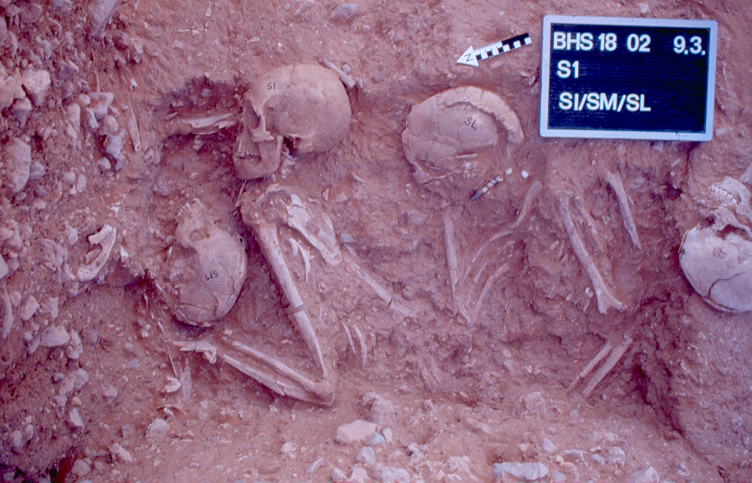
Inidvidual SI:
S1 with SI and others
Full Resolution
Full Resolution
Inidvidual SI:
SI/SM/SL 09.03.2002
Full Resolution
Full Resolution
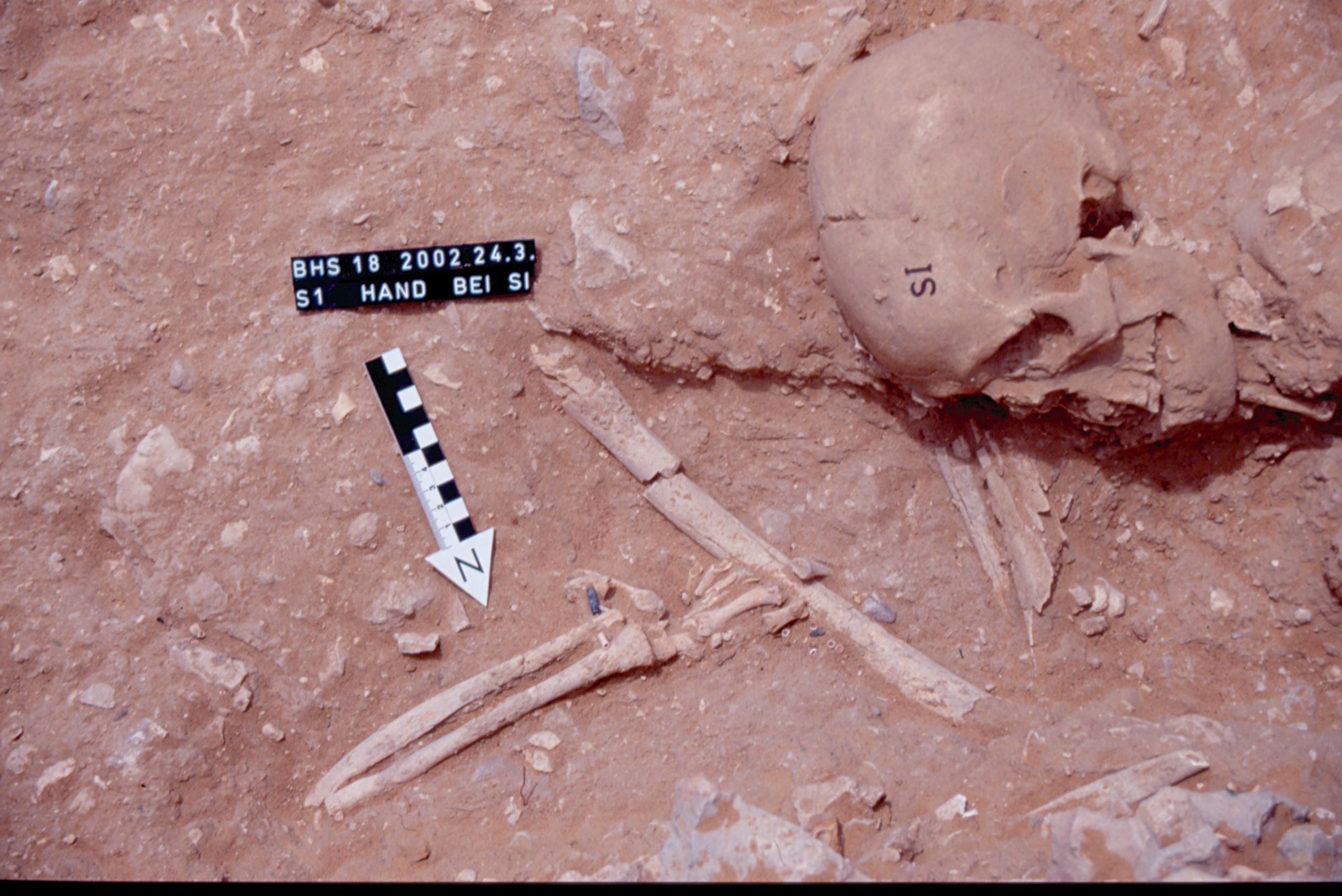
Inidvidual SI:
hand close to SI
Full Resolution
Full Resolution
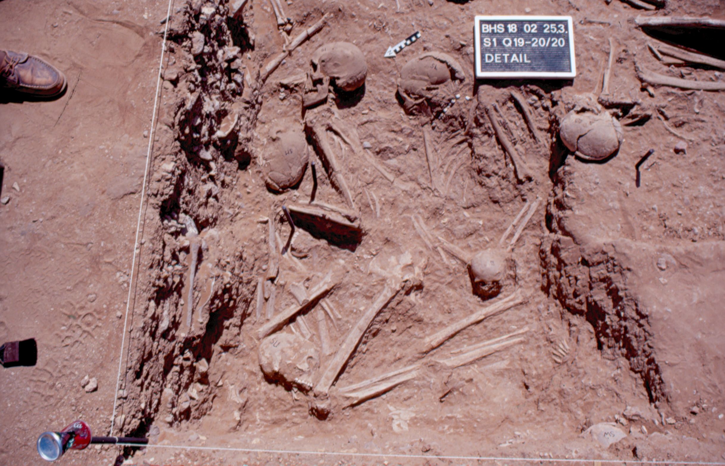
Inidvidual SI:
Detail SU/SM/SI/SL 25.03.2002
Full Resolution
Full Resolution
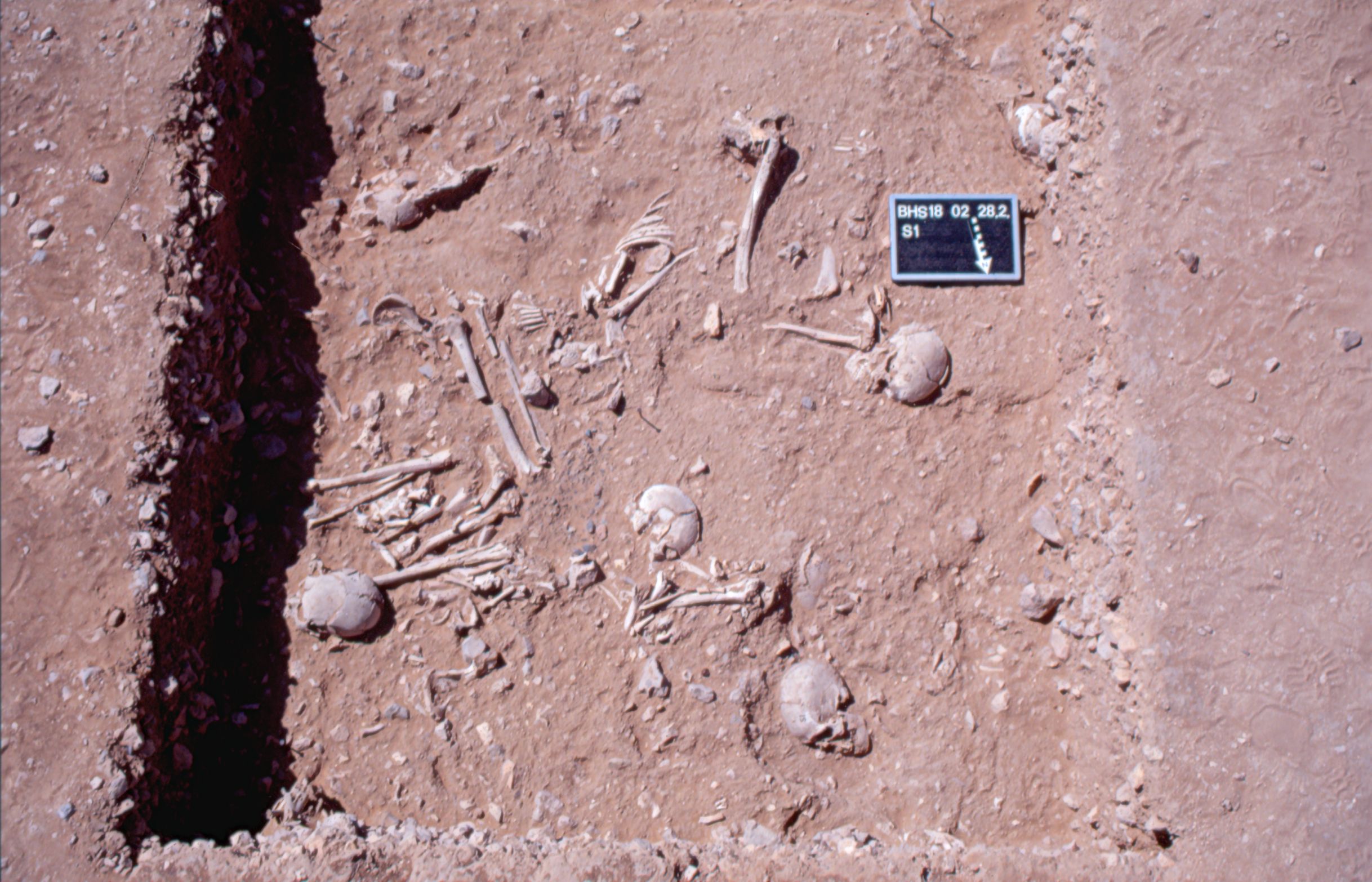
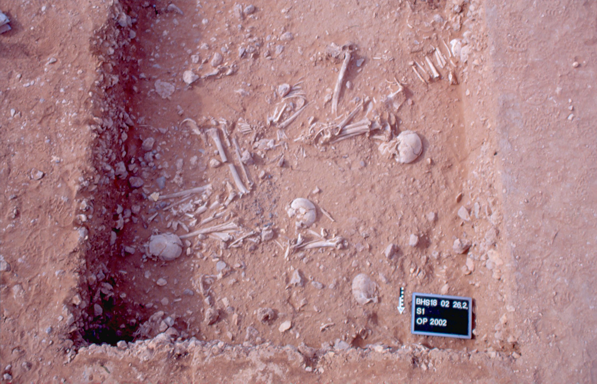
Inidvidual SI:
S1 with several individuals, 28.02.2002
Full Resolution
Full Resolution
Inidvidual SJ:
Skull SJ in situ
Full Resolution
Full Resolution
Inidvidual SJ:
Skull SJ in situ
Full Resolution
Full Resolution
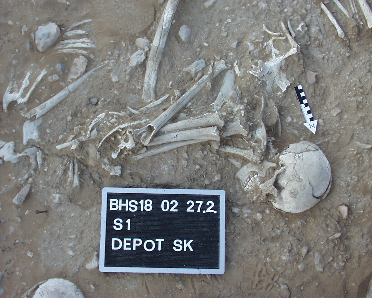
Inidvidual SK:
Secondary burial SK in situ
Full Resolution
Full Resolution

Inidvidual SK:
Depot SK including SK/SC 27.02.2002
Full Resolution
Full Resolution

Inidvidual SK:
Depot SK including SK/SC 27.02.2002
Full Resolution
Full Resolution
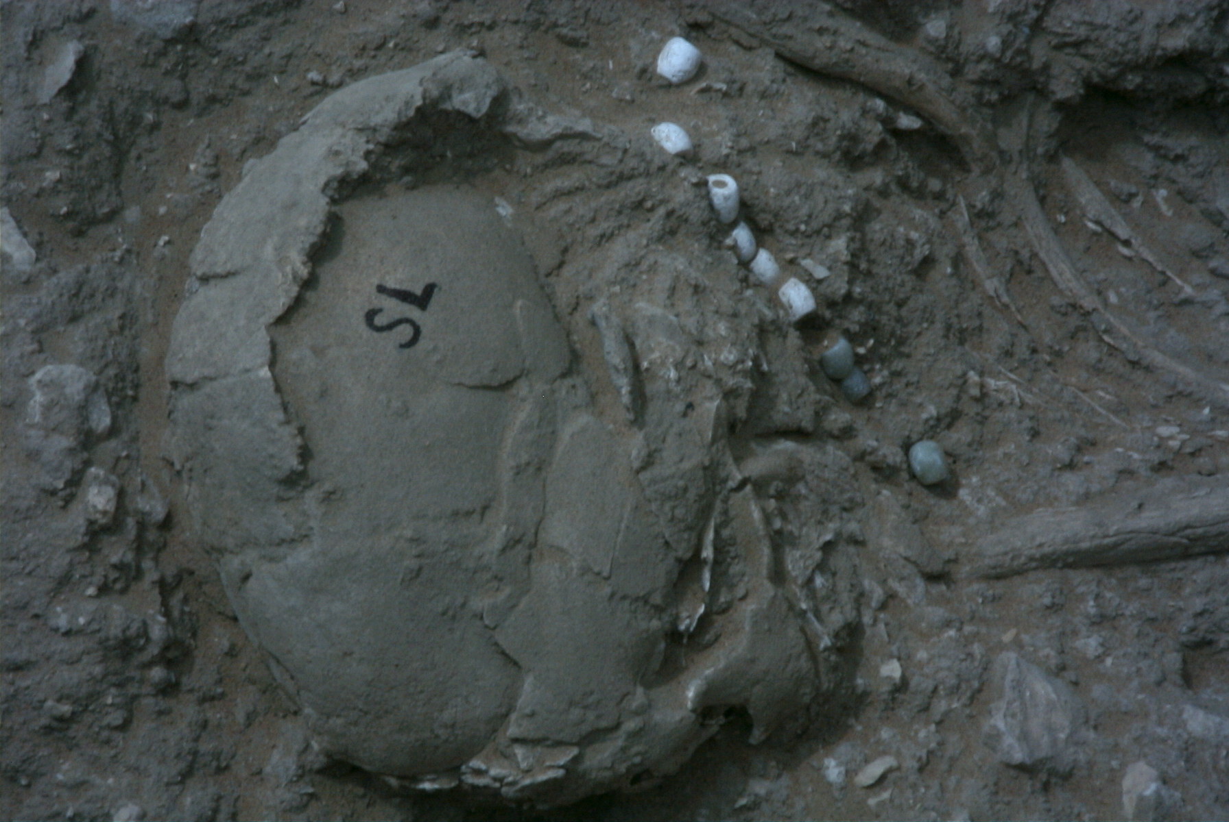
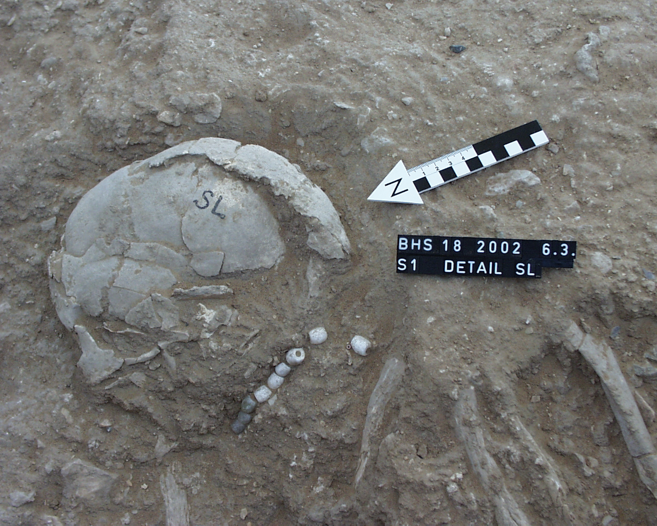
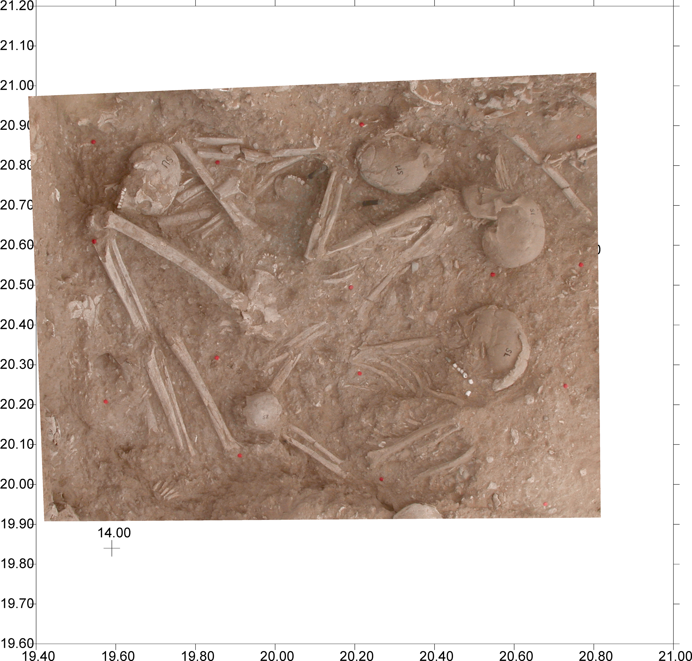
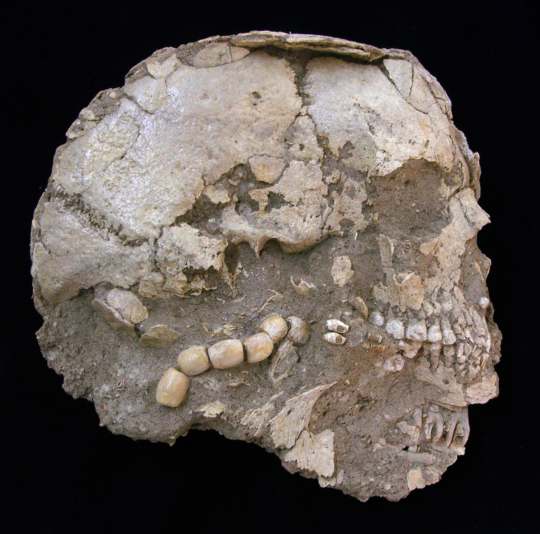
Inidvidual SL:
Skull SL in situ. Bracelet
Full Resolution
Full Resolution
Inidvidual SL:
Detail of adornments SL in situ
Full Resolution
Full Resolution
Inidvidual SL:
S1 with SI/SL and others 06.03.2002
Full Resolution
Full Resolution

Inidvidual SL:
SI/SM/SL 09.03.2002
Full Resolution
Full Resolution

Inidvidual SL:
Detail SU/SM/SI/SL 25.03.2002
Full Resolution
Full Resolution
Inidvidual SL:
SI and SL and SZ in situ, orthorectified picture
Full Resolution
Full Resolution
Inidvidual SL:
Skull SL, right side, cleaned with adornments. Exhibited in the Archaeological Museum Sharjah (UAE)
Full Resolution
Full Resolution
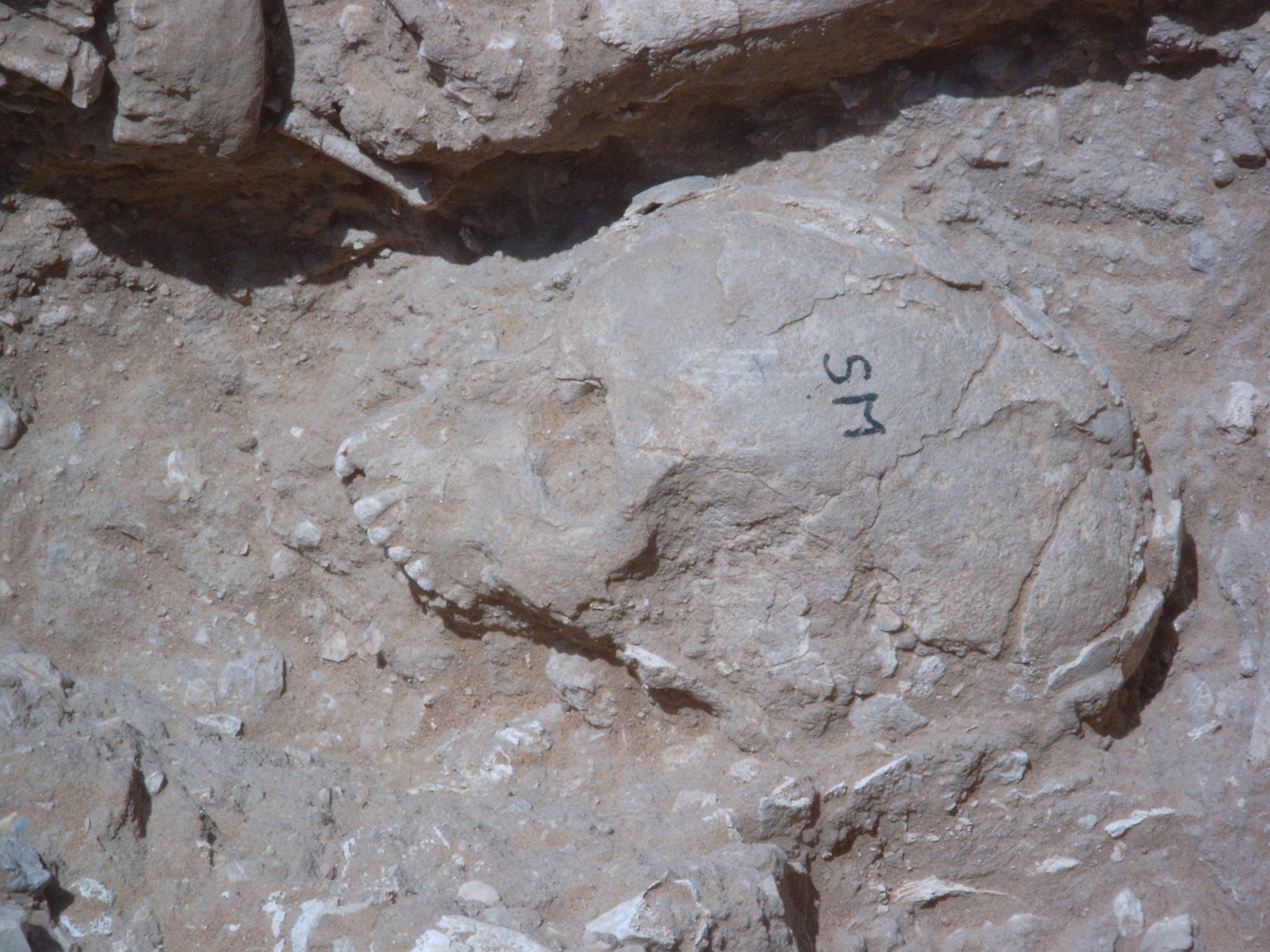
Inidvidual SM:
Skull SM in S1 in situ
Full Resolution
Full Resolution
Inidvidual SM:
SI/SM/SL 06.03.2002
Full Resolution
Full Resolution

Inidvidual SM:
SI/SM/SL 09.03.2002
Full Resolution
Full Resolution
Inidvidual SM:
S1/S1-1/S1-1 with differnent individuals
Full Resolution
Full Resolution

Inidvidual SM:
Detail SU/SM/SI/SL 25.03.2002
Full Resolution
Full Resolution
Inidvidual SP:
Burial in situ
Full Resolution
Full Resolution

Inidvidual SP:
SE and SP in situ in S1/1-3
Full Resolution
Full Resolution
Inidvidual SP:
Detail SP 11.03.2002
Full Resolution
Full Resolution
Inidvidual SP:
SE/SP 18.03.2002
Full Resolution
Full Resolution
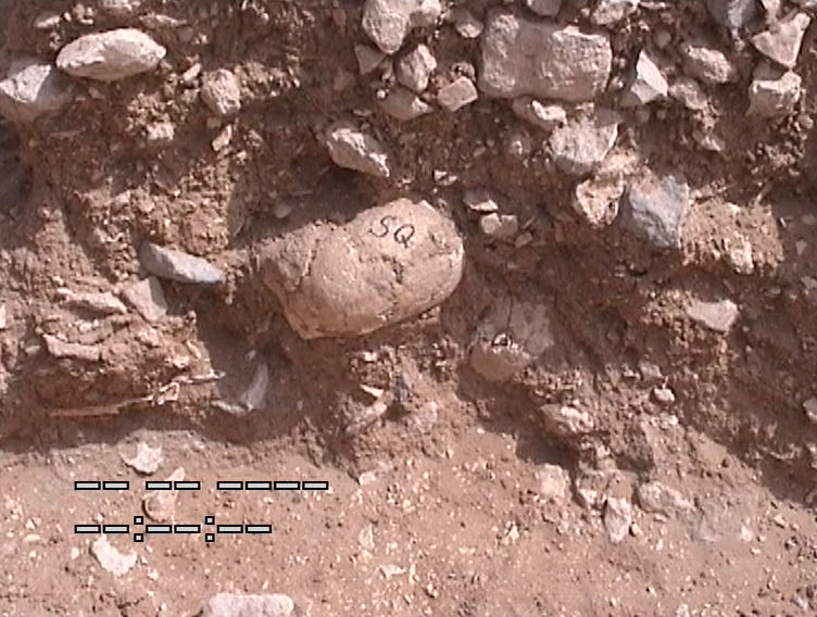
Inidvidual SQ:
Skull in situ situated in profile
Full Resolution
Full Resolution

Inidvidual SQ:
TU, TX, SQ and TR, orthorectified picture of Steg S1-Ax
Full Resolution
Full Resolution
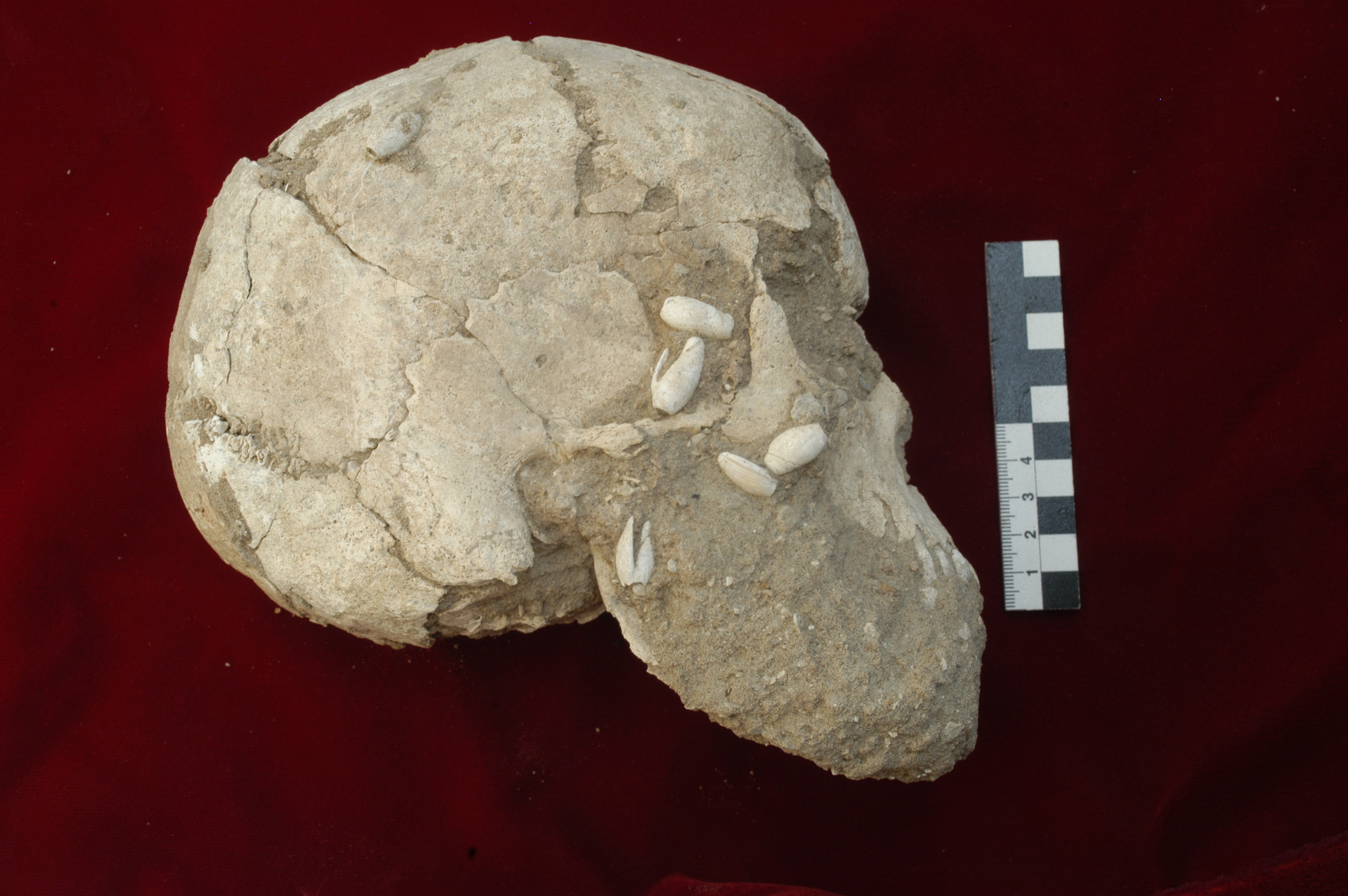
Inidvidual SQ:
SQ with adornments, bottom side after recovery
Full Resolution
Full Resolution
Inidvidual SQ:
Depot TR, TQ, TU, TX and skull of burial SQ
Full Resolution
Full Resolution

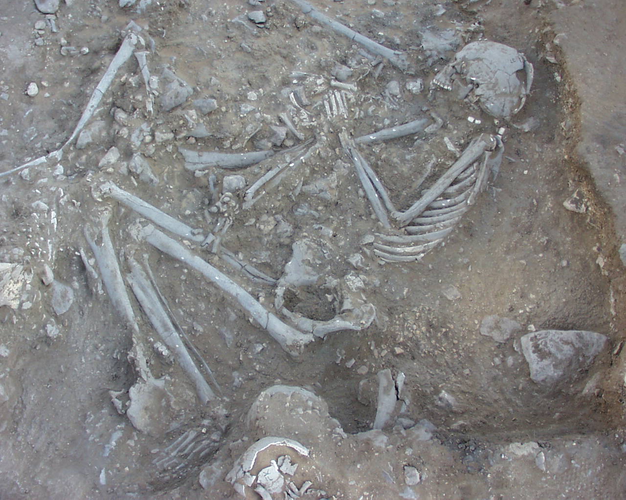
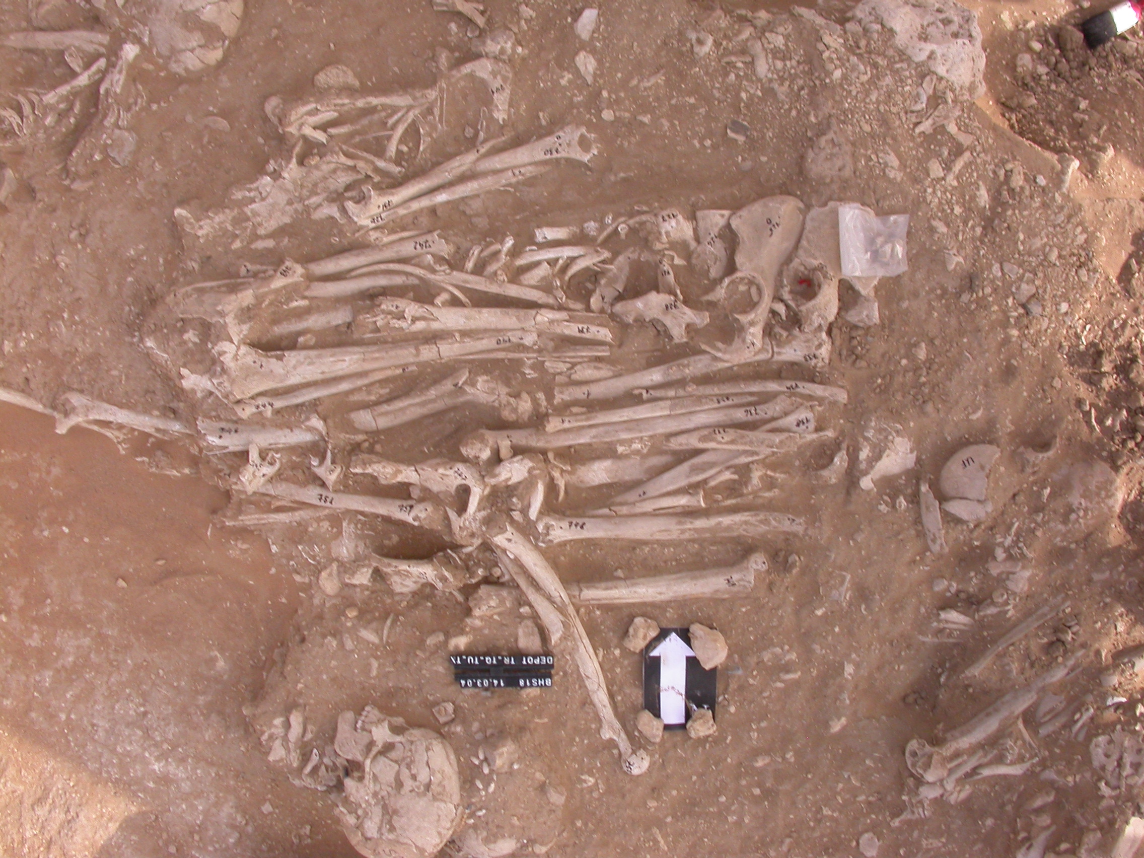
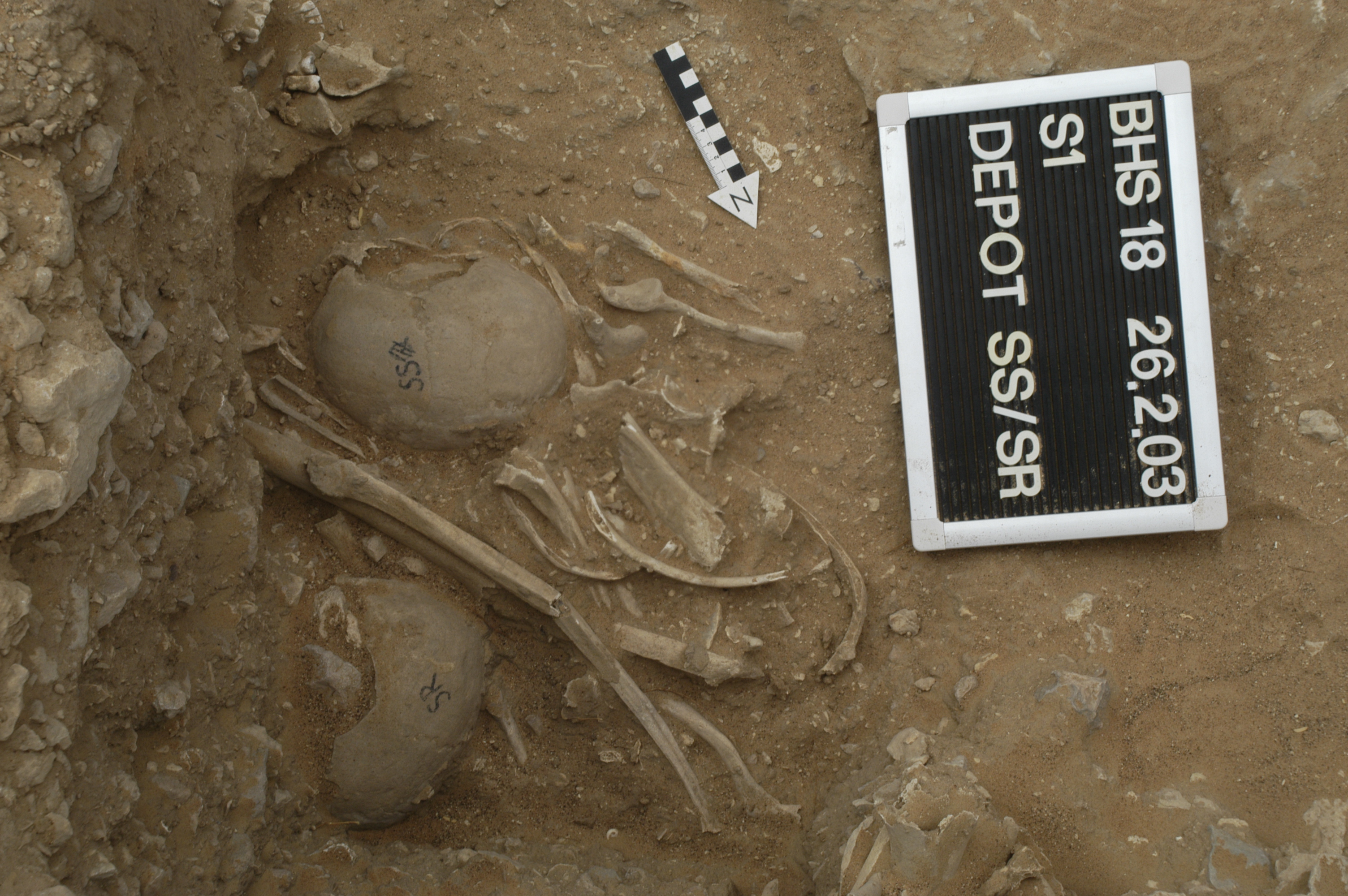
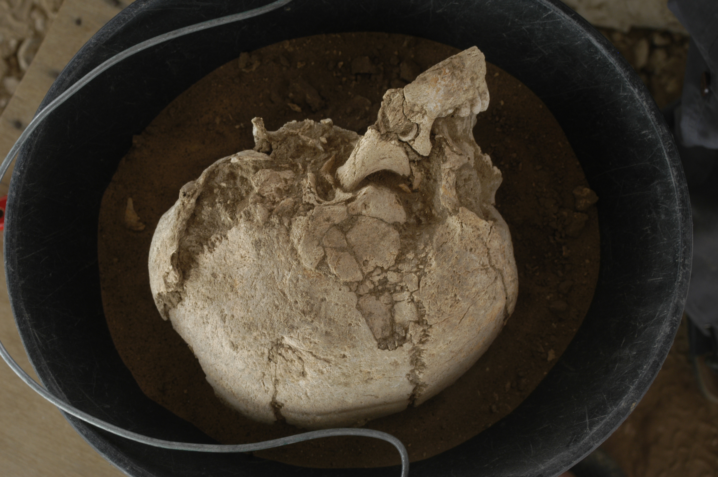
Inidvidual SR:
Individuals SS, ST and SR in S1
Full Resolution
Full Resolution
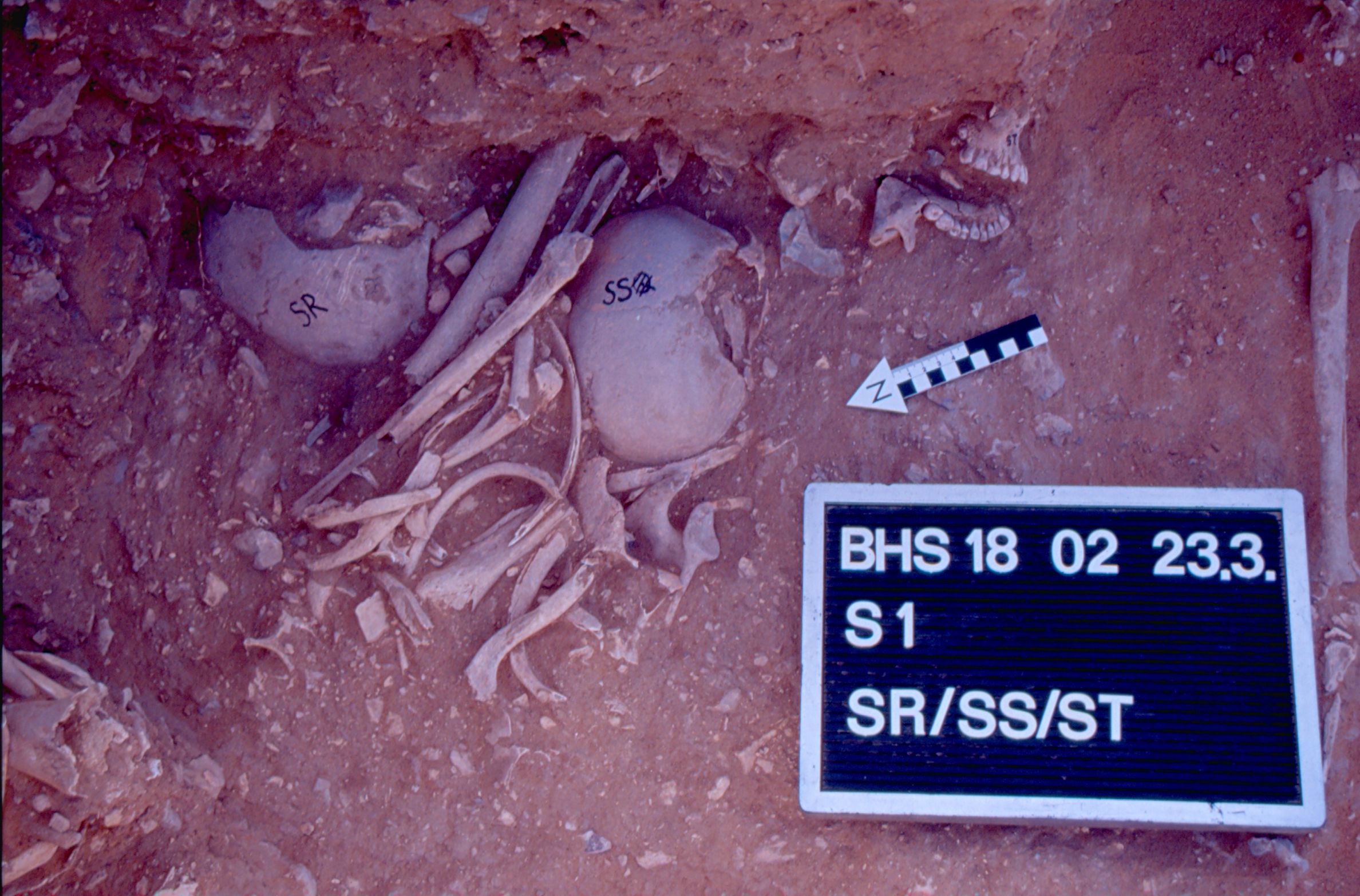
Inidvidual SR:
SR/SS/ST 23.03.2002
Full Resolution
Full Resolution
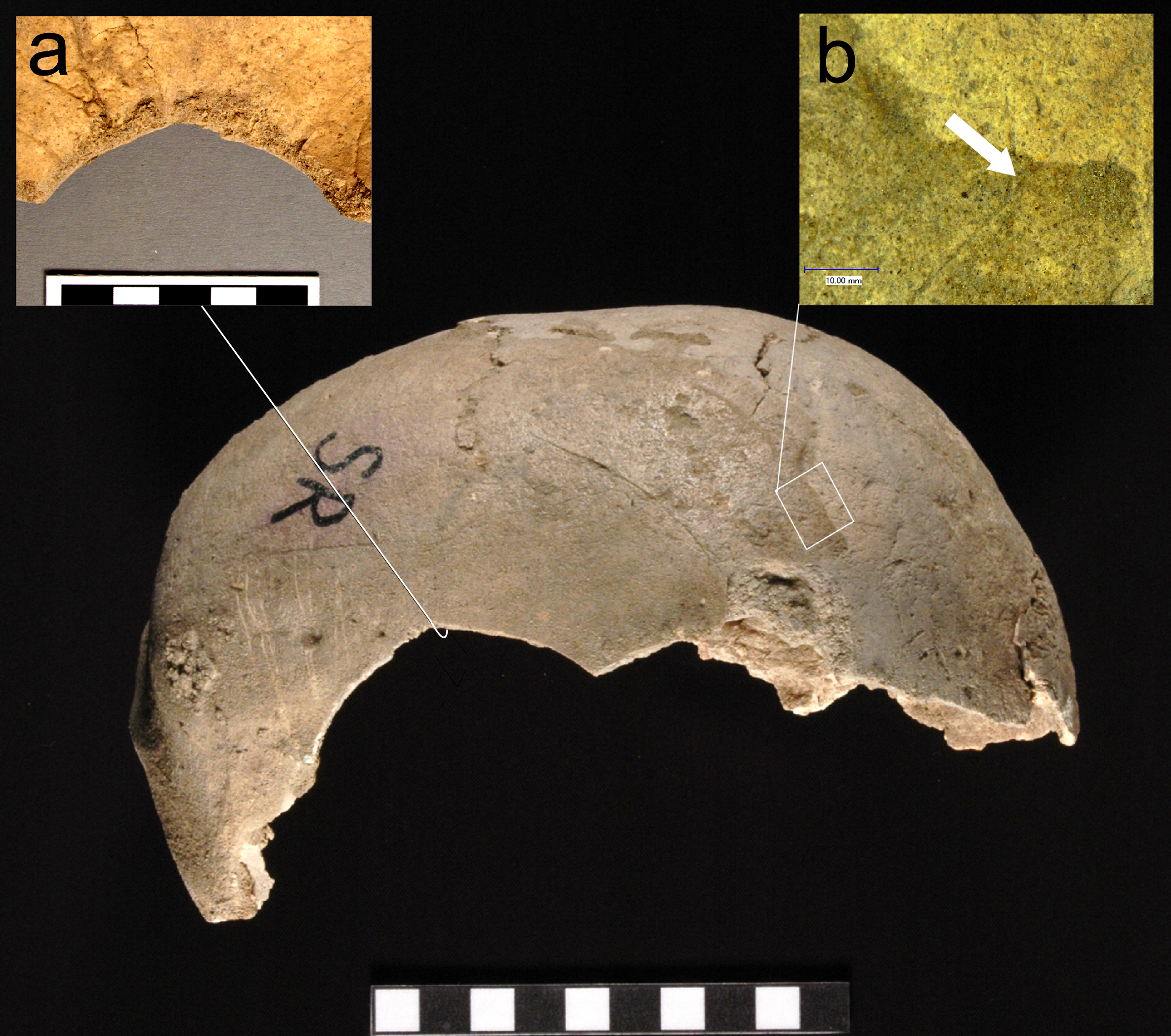
Inidvidual SR:
Peri-mortem blunt force trauma and traces of cutting (foto by Hilde Jensen). The microscopic image shows one of the cut-marks.
Full Resolution
Full Resolution
Inidvidual SS:
SS and SR in situ
Full Resolution
Full Resolution

Inidvidual SS:
Individuals SS, ST and SR in S1
Full Resolution
Full Resolution

Inidvidual SS:
SR/SS/ST 23.03.2002
Full Resolution
Full Resolution
Inidvidual ST:
Skull ST after recovery
Full Resolution
Full Resolution

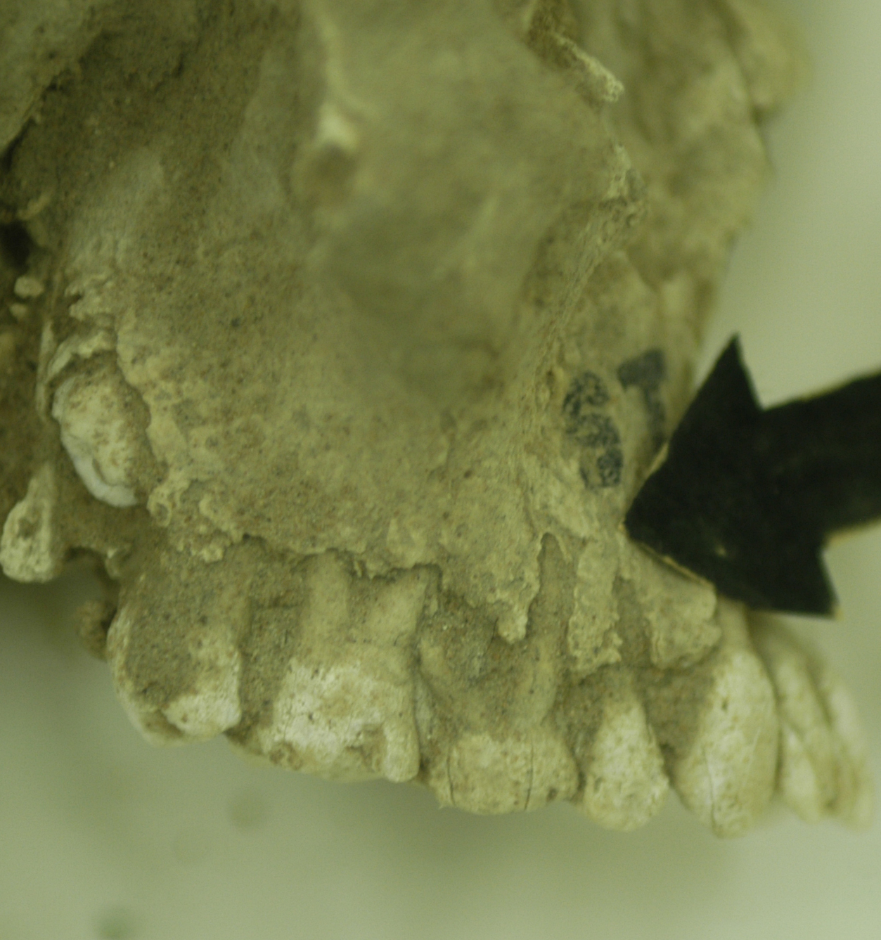
Inidvidual ST:
Mandible fragment ST
Full Resolution
Full Resolution
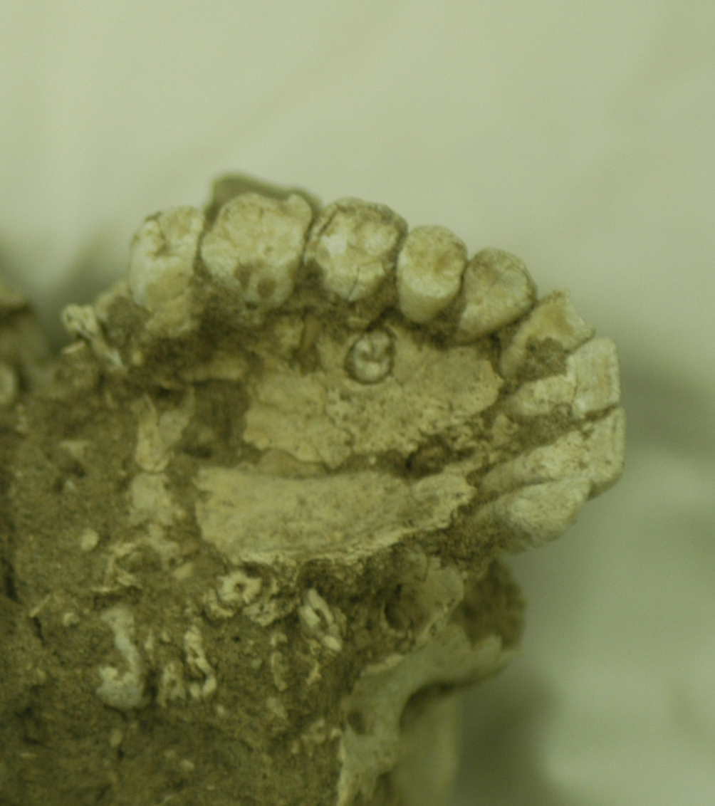
Inidvidual ST:
ST, persisting milk tooth, view from inferior
Full Resolution
Full Resolution
Inidvidual ST:
ST, persisting milk tooth. view from lateral
Full Resolution
Full Resolution
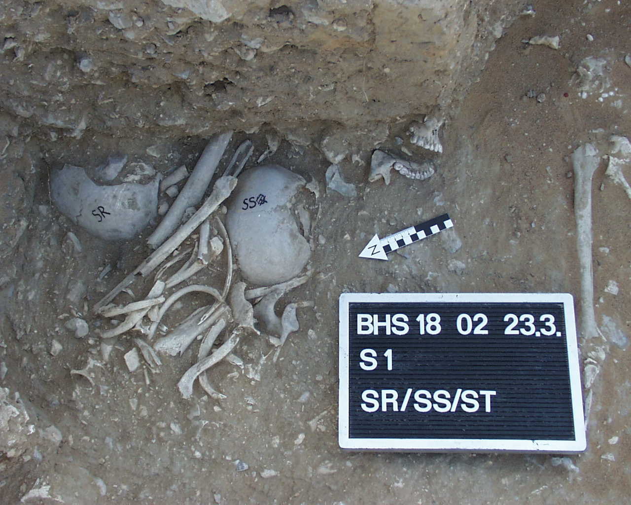
Inidvidual ST:
Individuals SS, ST and SR in S1
Full Resolution
Full Resolution

Inidvidual ST:
SR/SS/ST 23.03.2002
Full Resolution
Full Resolution

Inidvidual SU:
Detail SU/SM/SI/SL 25.03.2002
Full Resolution
Full Resolution

Inidvidual SU:
SU
Full Resolution
Full Resolution
Inidvidual SU:
SU
Full Resolution
Full Resolution
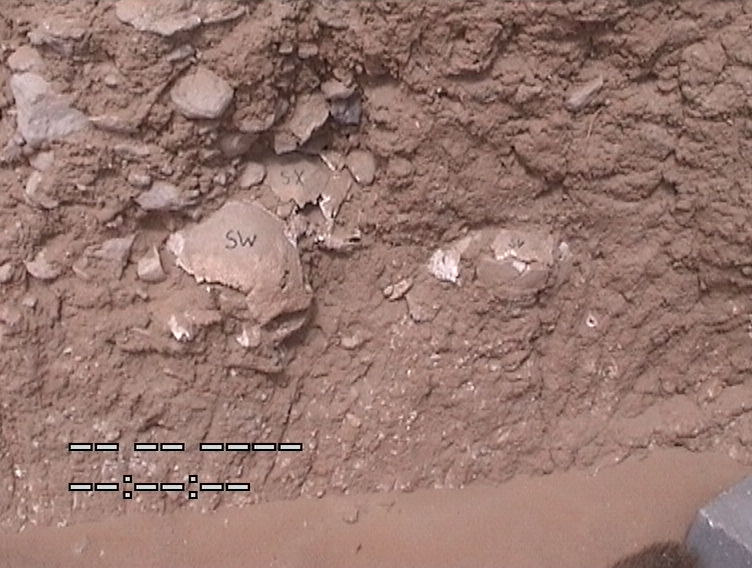
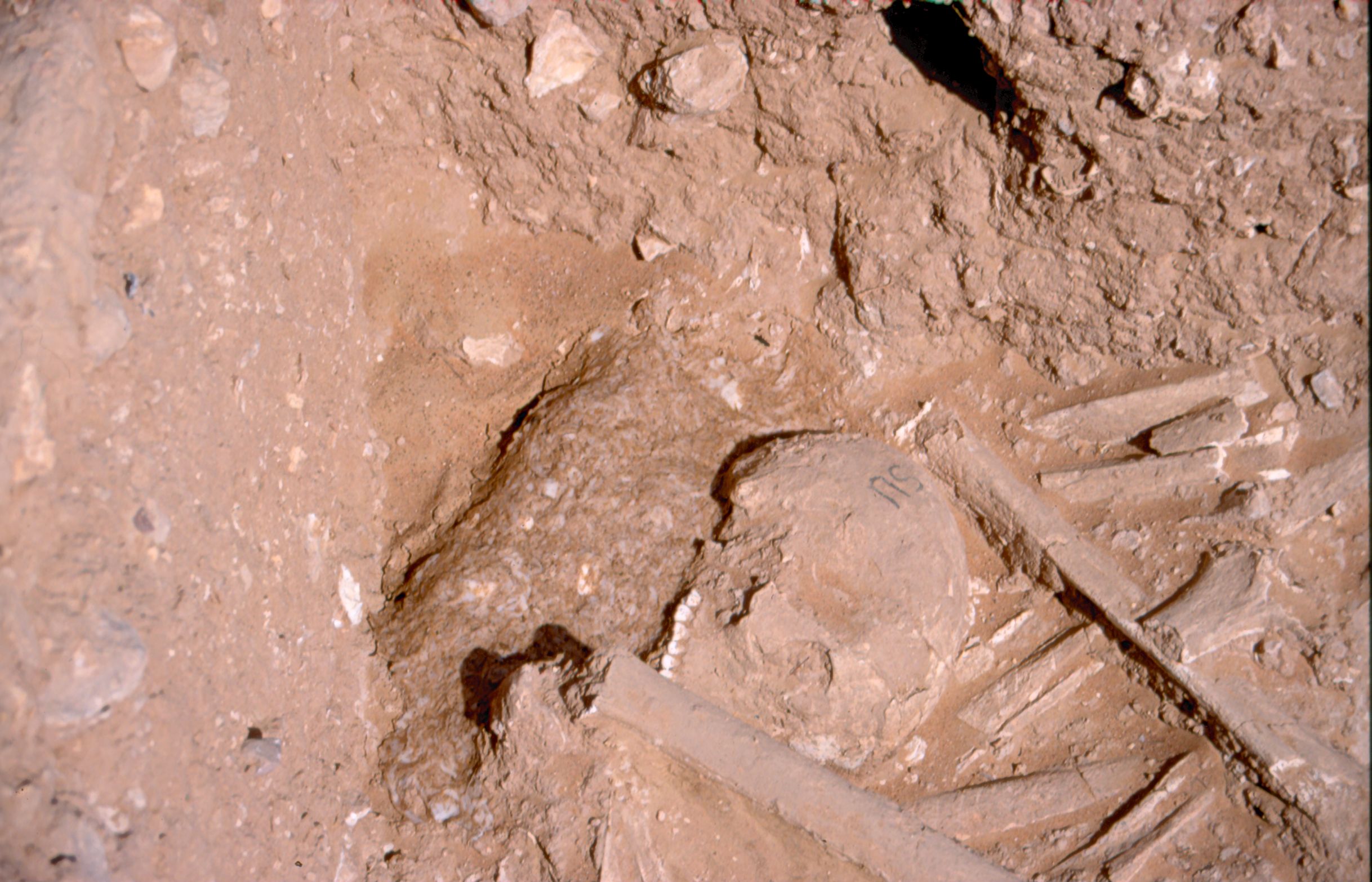
Inidvidual SW:
SW, SV and SX in situ
Full Resolution
Full Resolution
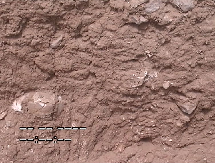
Inidvidual SY:
Skull in situ
Full Resolution
Full Resolution
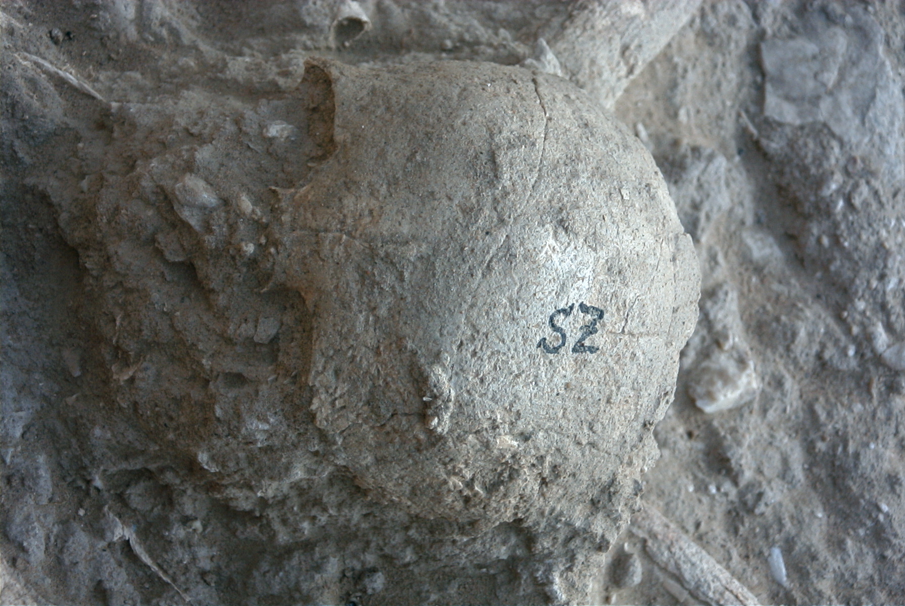
Inidvidual SZ:
Skull SZ in situ
Full Resolution
Full Resolution
Inidvidual SZ:
SZ 25.03.2002
Full Resolution
Full Resolution
Inidvidual TA:
In situ
Full Resolution
Full Resolution
Inidvidual TB:
TB in situ
Full Resolution
Full Resolution
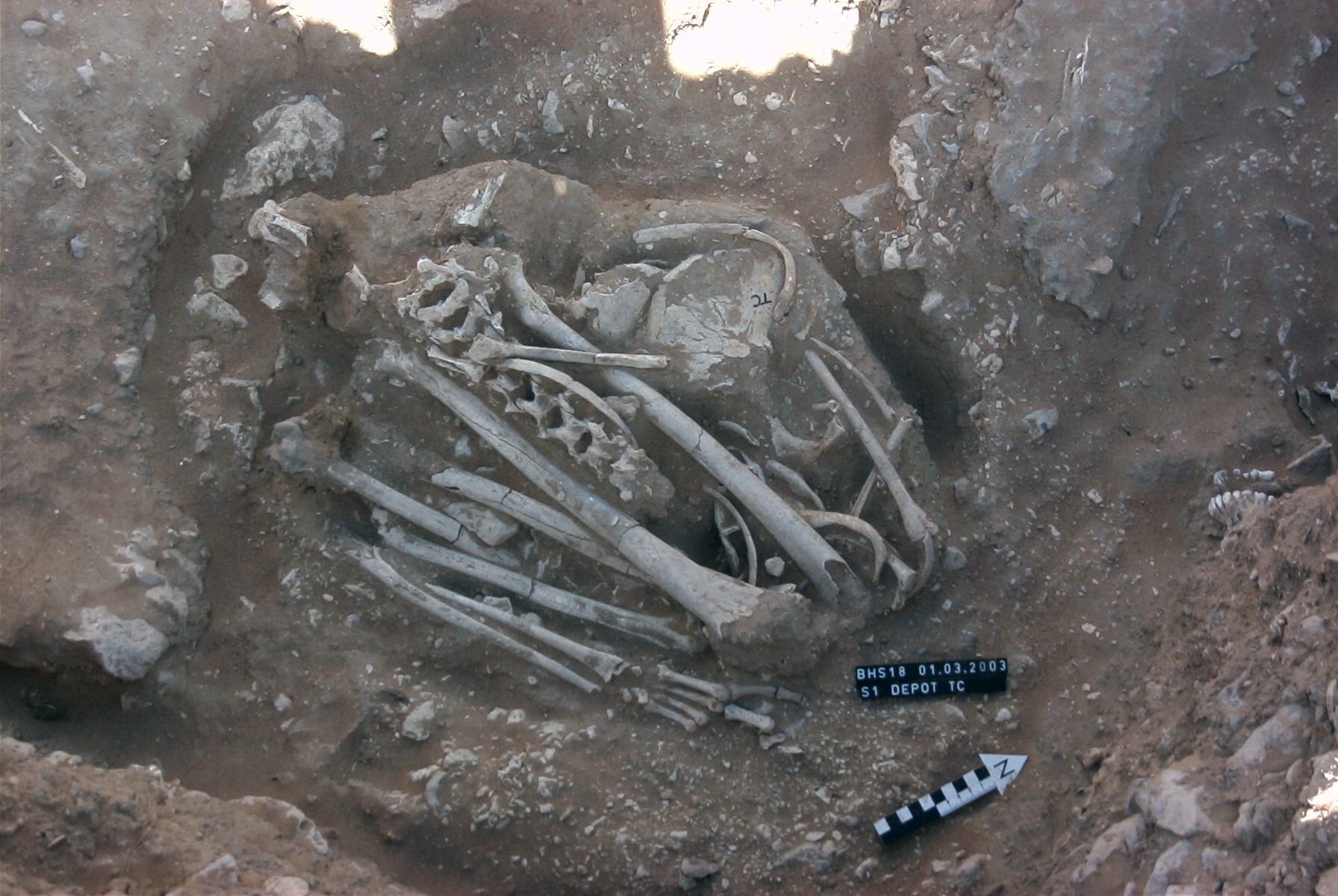
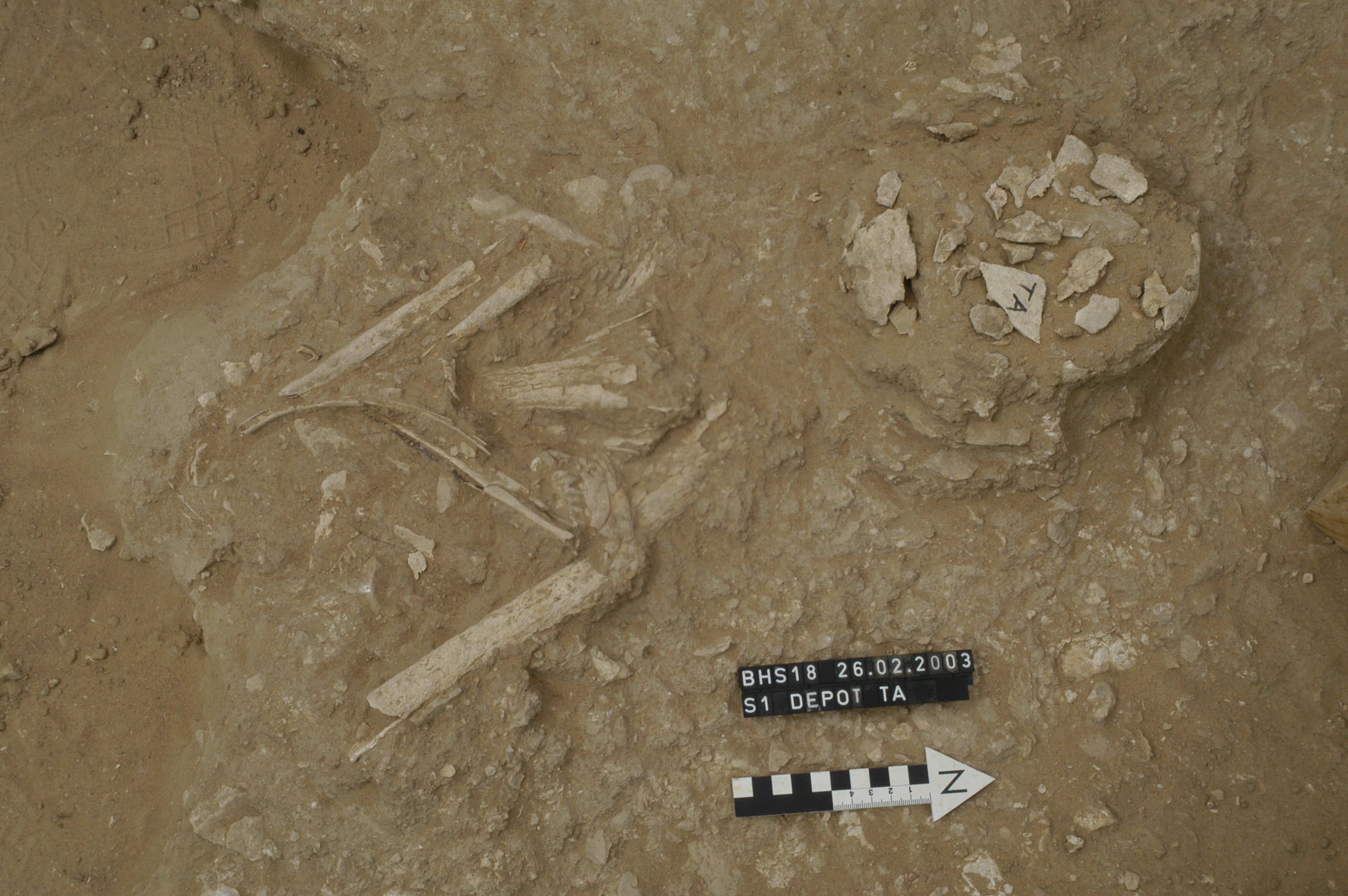
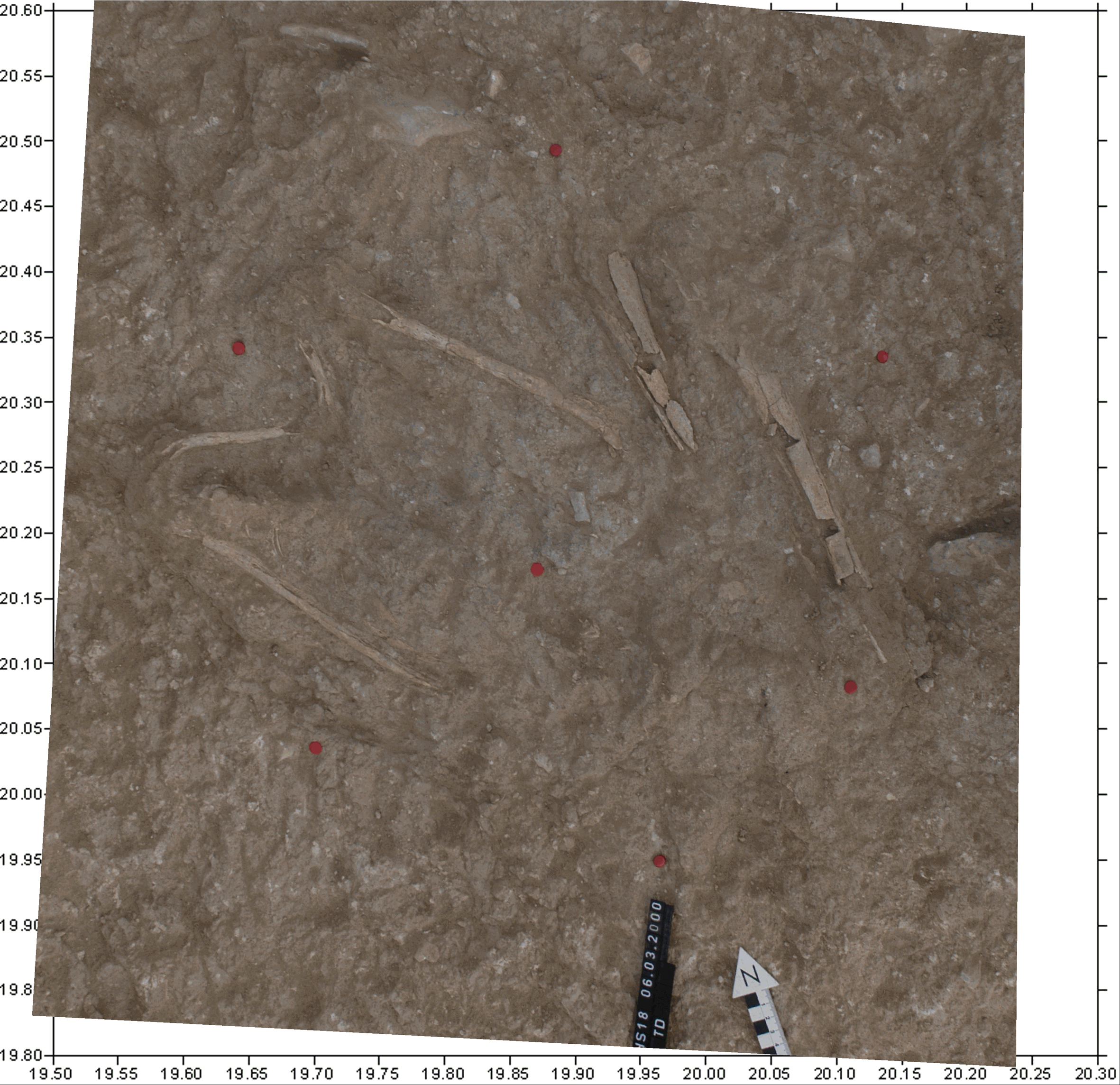
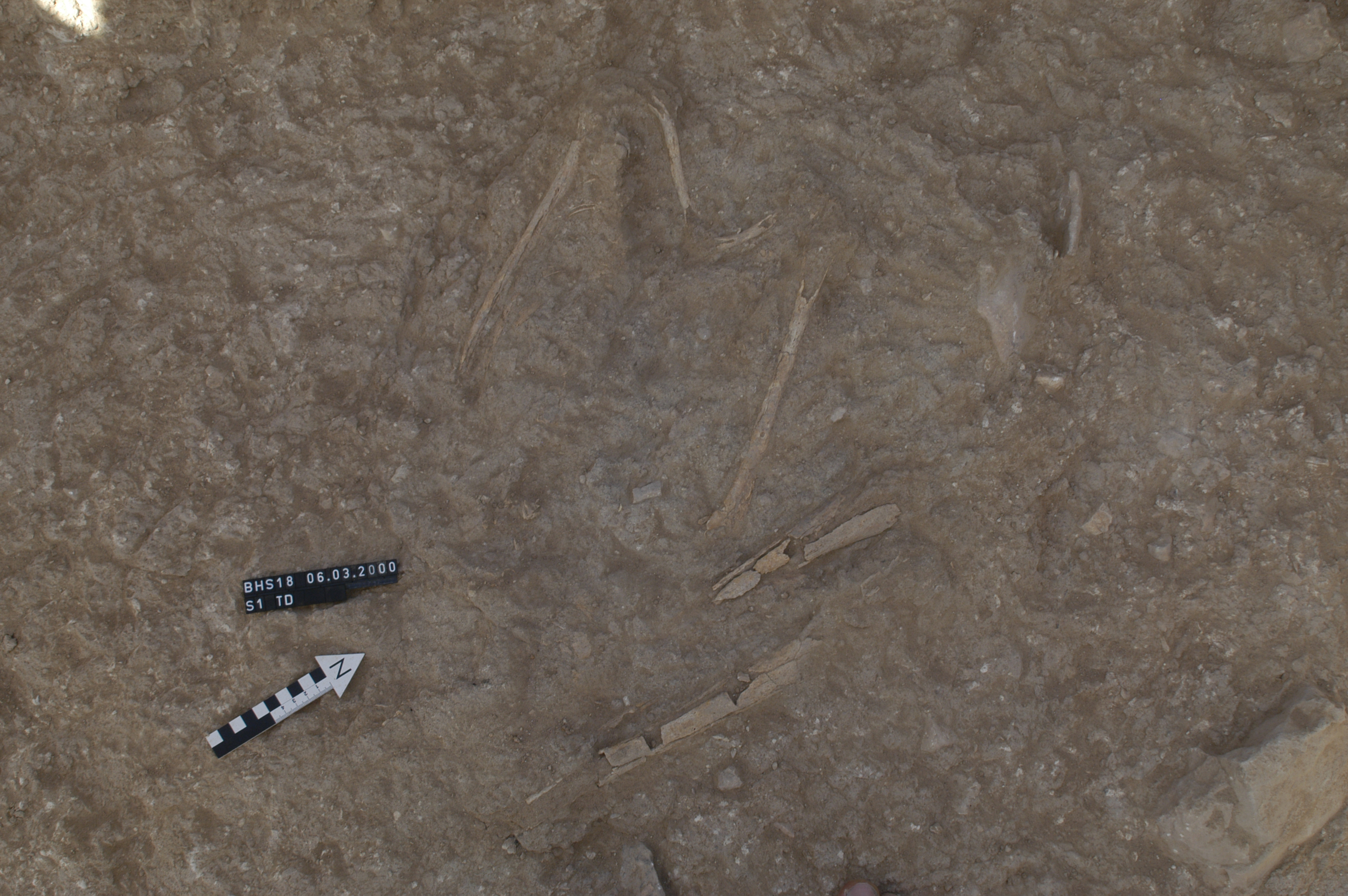
Inidvidual TC:
Secondary burial TC in situ
Full Resolution
Full Resolution
Inidvidual TC:
Secondary burial TC. Orthorectified.
Full Resolution
Full Resolution
Inidvidual TC:
Detail in situ, fingers, mandible
Full Resolution
Full Resolution
Inidvidual TD:
TD in situ. Orthorectified picture.
Full Resolution
Full Resolution
Inidvidual TD:
Individual TD in situ on the upcoming rock. pit
Full Resolution
Full Resolution

Inidvidual TG:
In situ together with TP, To u.a.
Full Resolution
Full Resolution
Inidvidual TG:
Clavicle TG
Full Resolution
Full Resolution

Inidvidual TG:
TI and TK with ophiolite
Full Resolution
Full Resolution

Inidvidual TG:
Muscle marks TG
Full Resolution
Full Resolution
Inidvidual TG:
Shell held in the hand
Full Resolution
Full Resolution

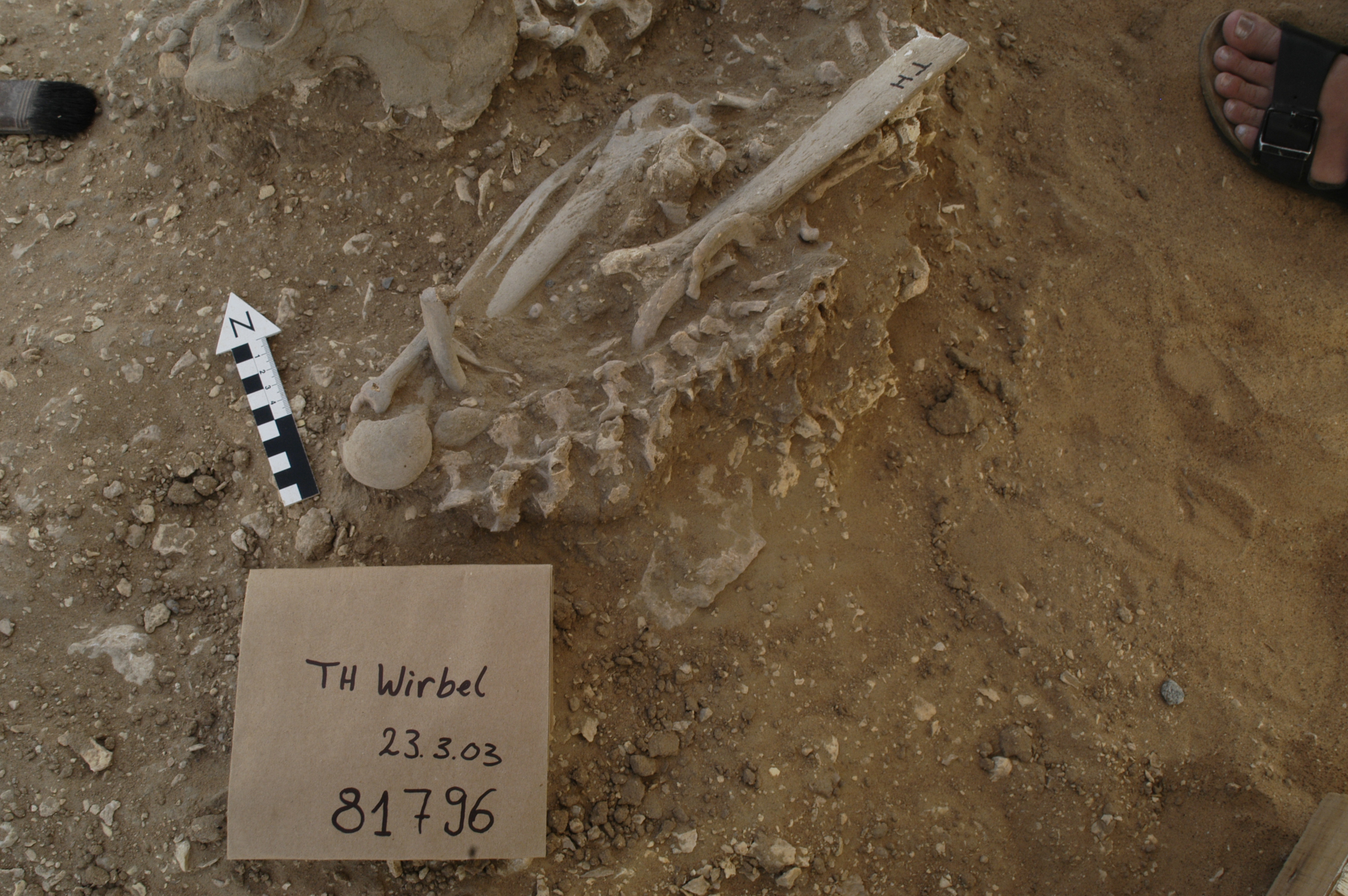
Inidvidual TH:
In situ with TI, TG, TJ
Full Resolution
Full Resolution
Inidvidual TH:
Spinal column in situ
Full Resolution
Full Resolution
Inidvidual TI:
In situ, together with ES, TH
Full Resolution
Full Resolution
Inidvidual TI:
TI and TK with ophiolite
Full Resolution
Full Resolution

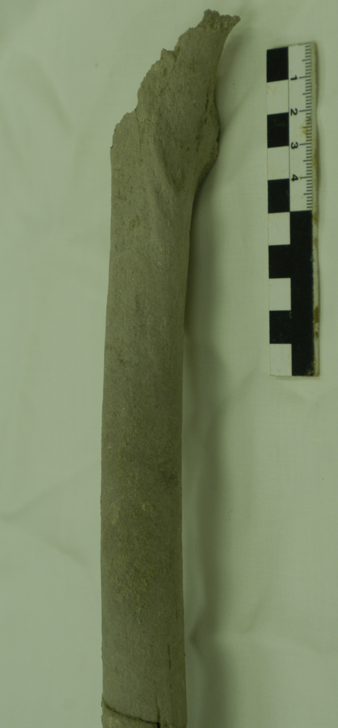
Inidvidual TJ:
In situ TJ and TM
Full Resolution
Full Resolution
Inidvidual TJ:
Occupational marker of stress, femur individual TJ/TM
Full Resolution
Full Resolution
Inidvidual TM:
TJ and TM in situ
Full Resolution
Full Resolution

Inidvidual TM:
Occupational marker of stress, femur individual TJ/TM
Full Resolution
Full Resolution
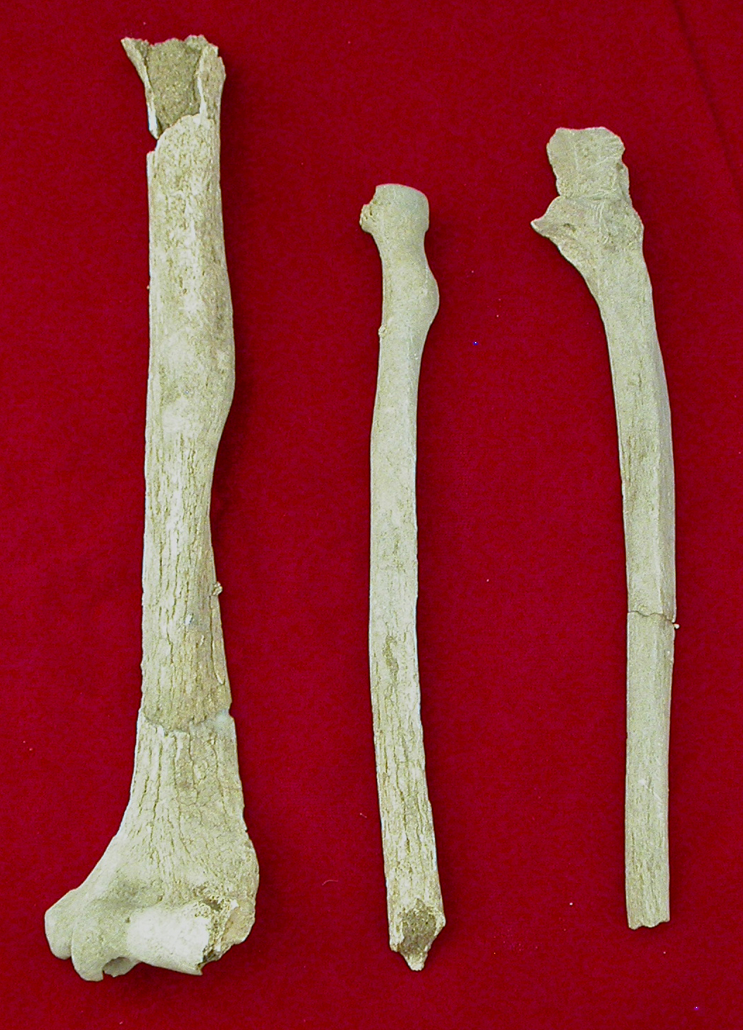
Inidvidual TN:
Arm bones with muscle imprints
Full Resolution
Full Resolution
Inidvidual TN:
Strongly developed muscle marks at the humerus and radius of individual TN
Full Resolution
Full Resolution
TO mandible
Full Resolution
Full Resolution
Inidvidual TP:
Steg S1-AX, individuals TO, TP, TG, ES in situ. Orthorectified picture.
Full Resolution
Full Resolution

Inidvidual TP:
Secondary burials of TX, TU and TP. The stone was laying underneath individual TW
Full Resolution
Full Resolution
TP mandible
Full Resolution
Full Resolution
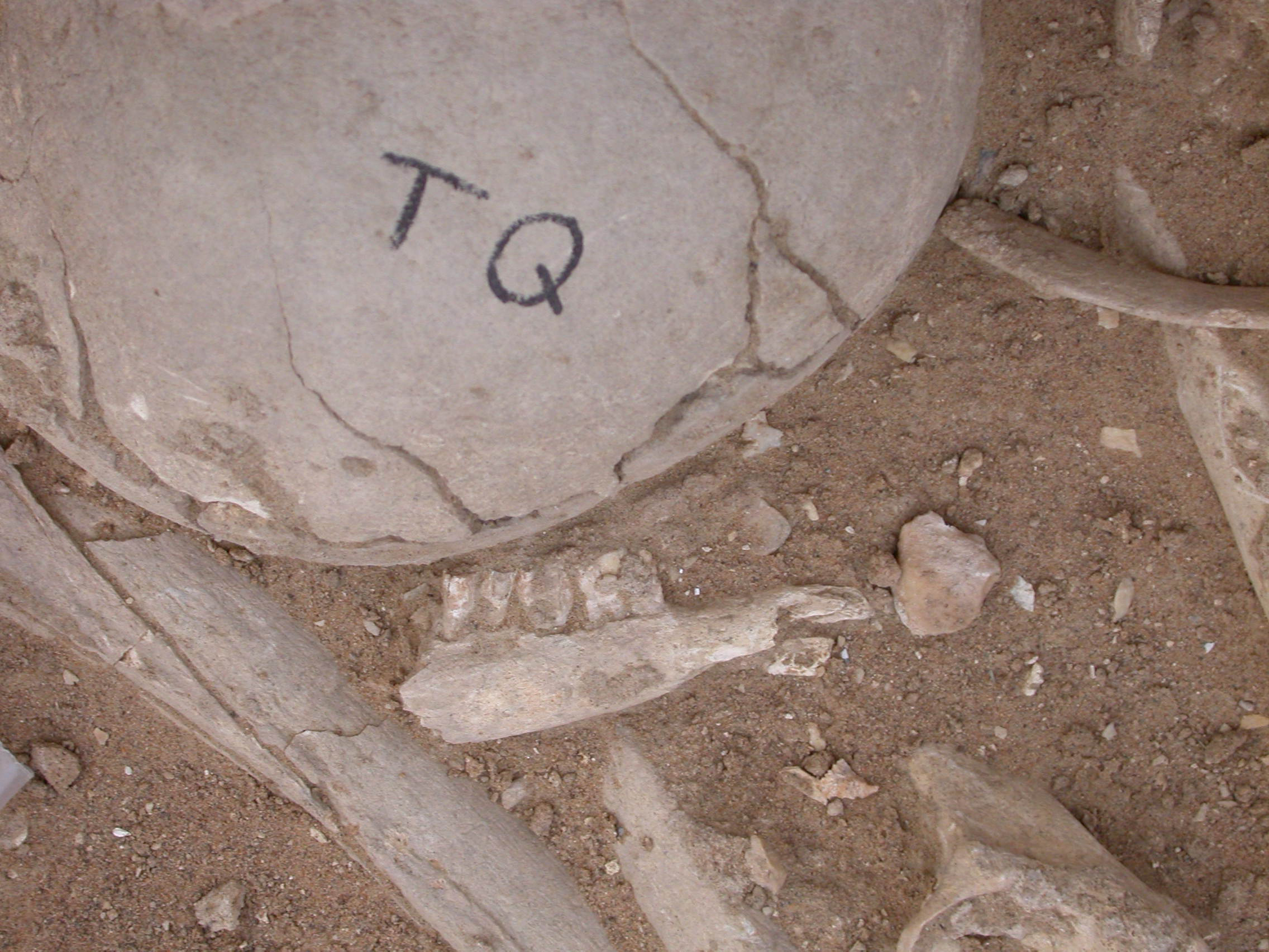
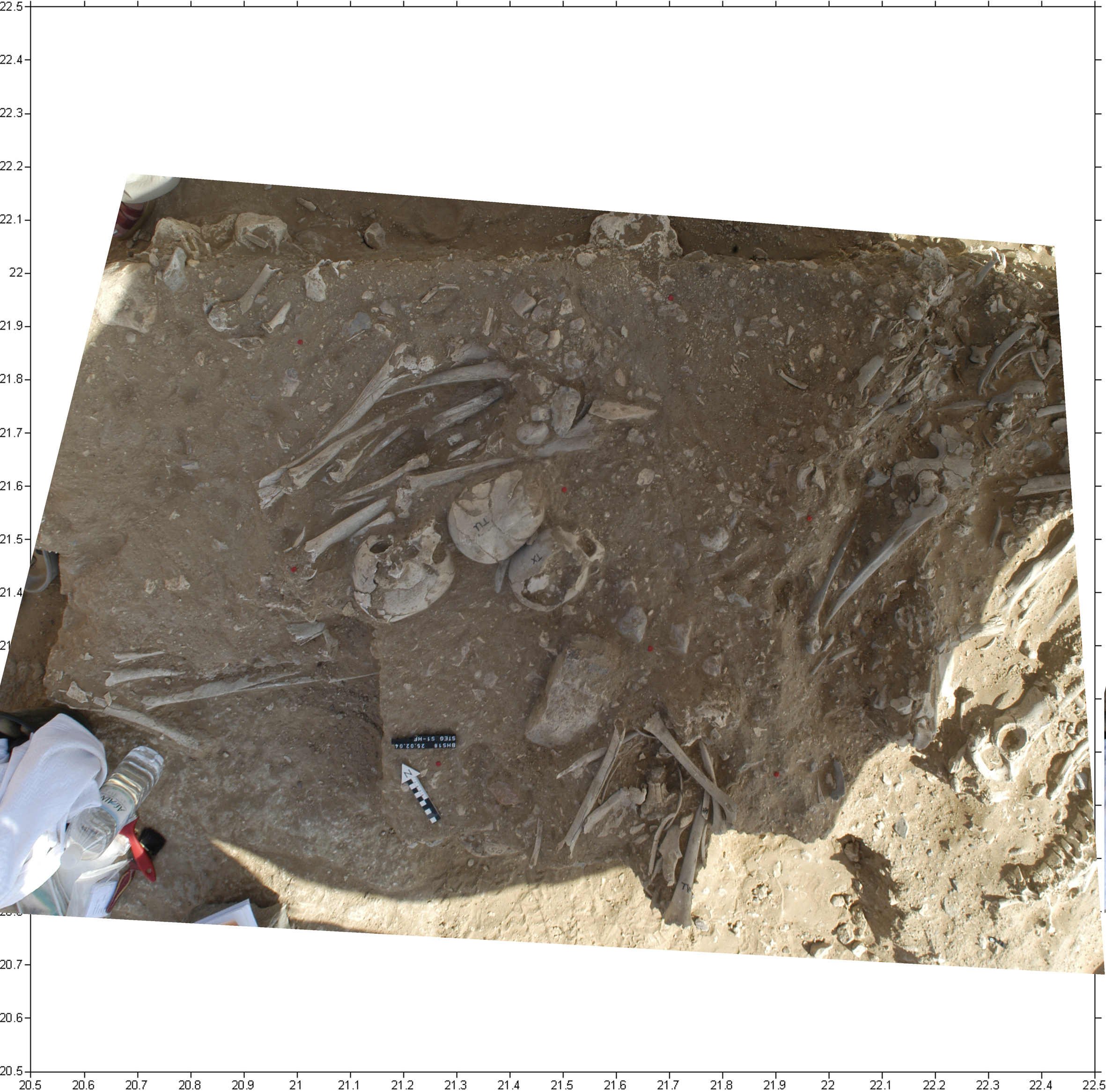
Inidvidual TQ:
Skull in situ with jaw of sheep or goat
Full Resolution
Full Resolution
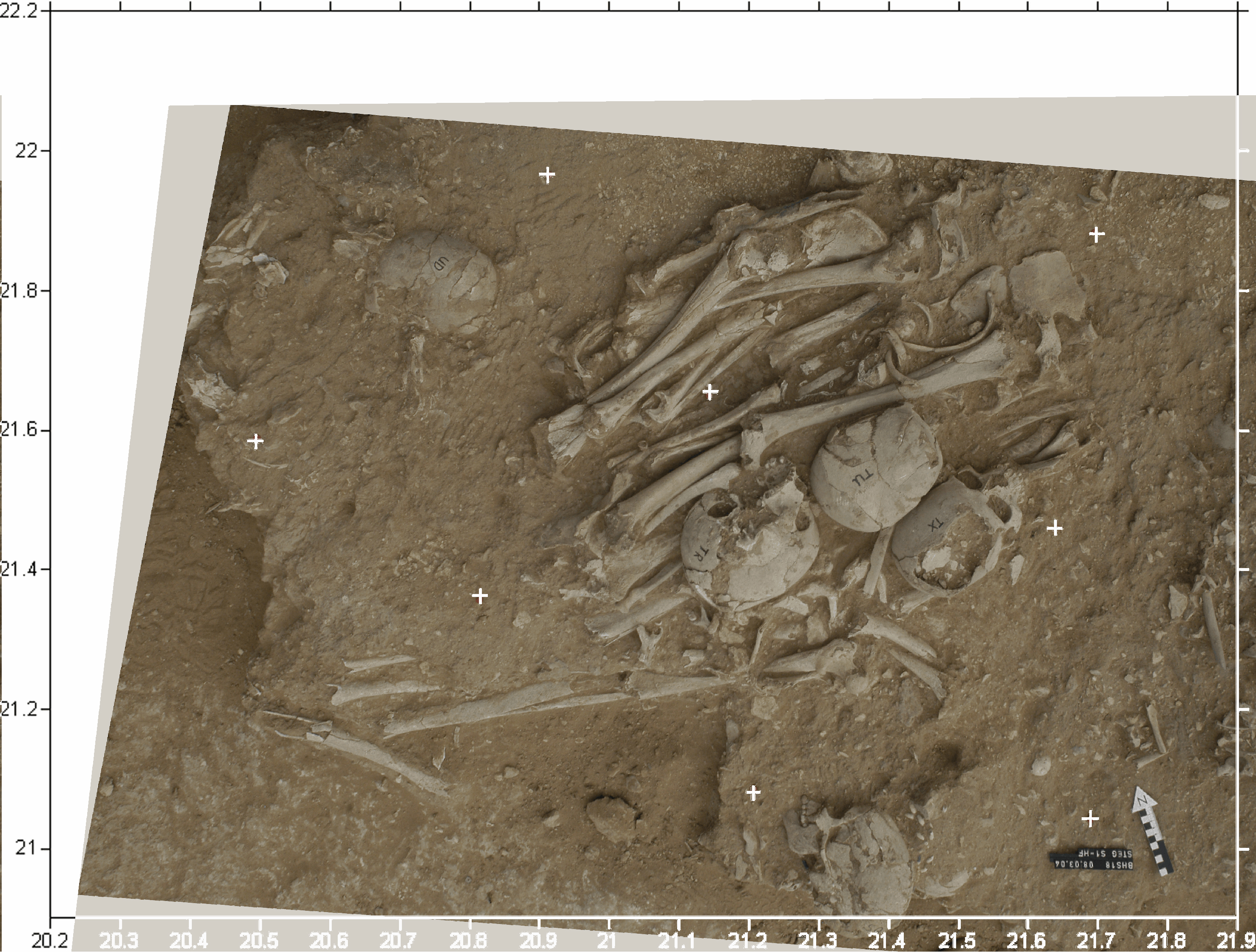
Inidvidual TQ:
Skull UD. Depot TX, TR, TU, TQ in situ
Full Resolution
Full Resolution

Inidvidual TQ:
Depot TR, TQ, TU, TX and skull of burial SQ
Full Resolution
Full Resolution

Inidvidual TQ:
Depot TR, TQ, TU, TX and UD and UF in situ.
Full Resolution
Full Resolution

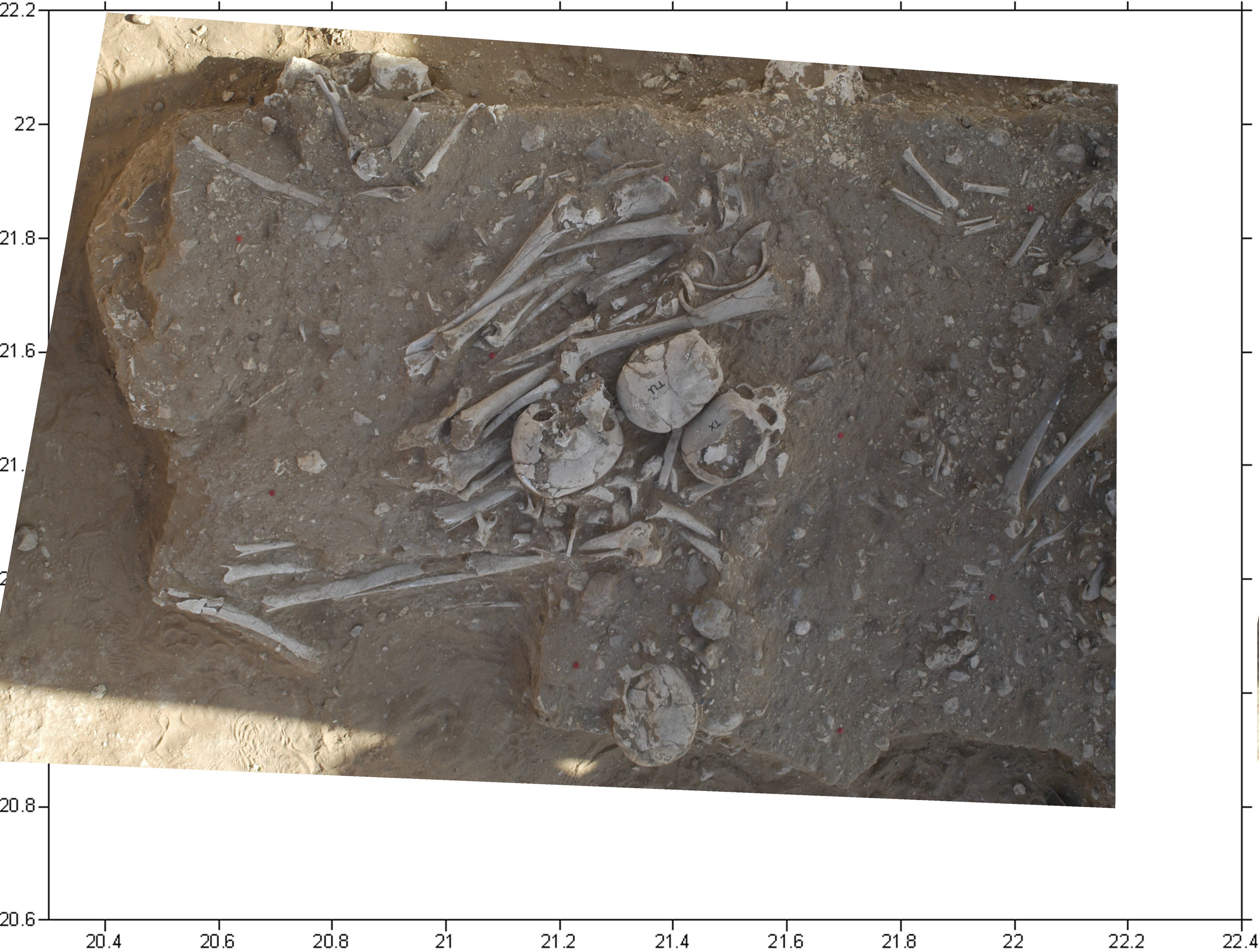
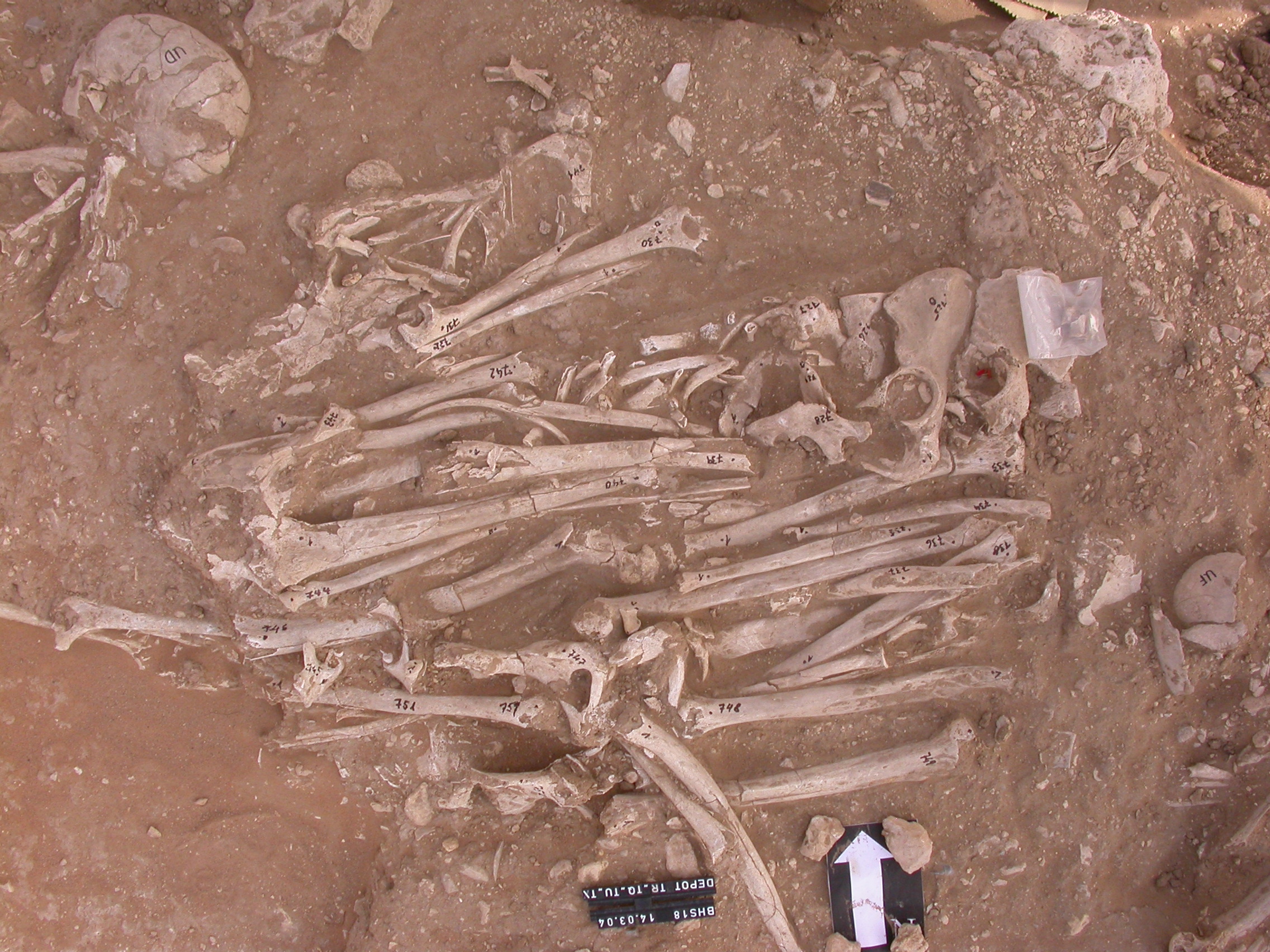
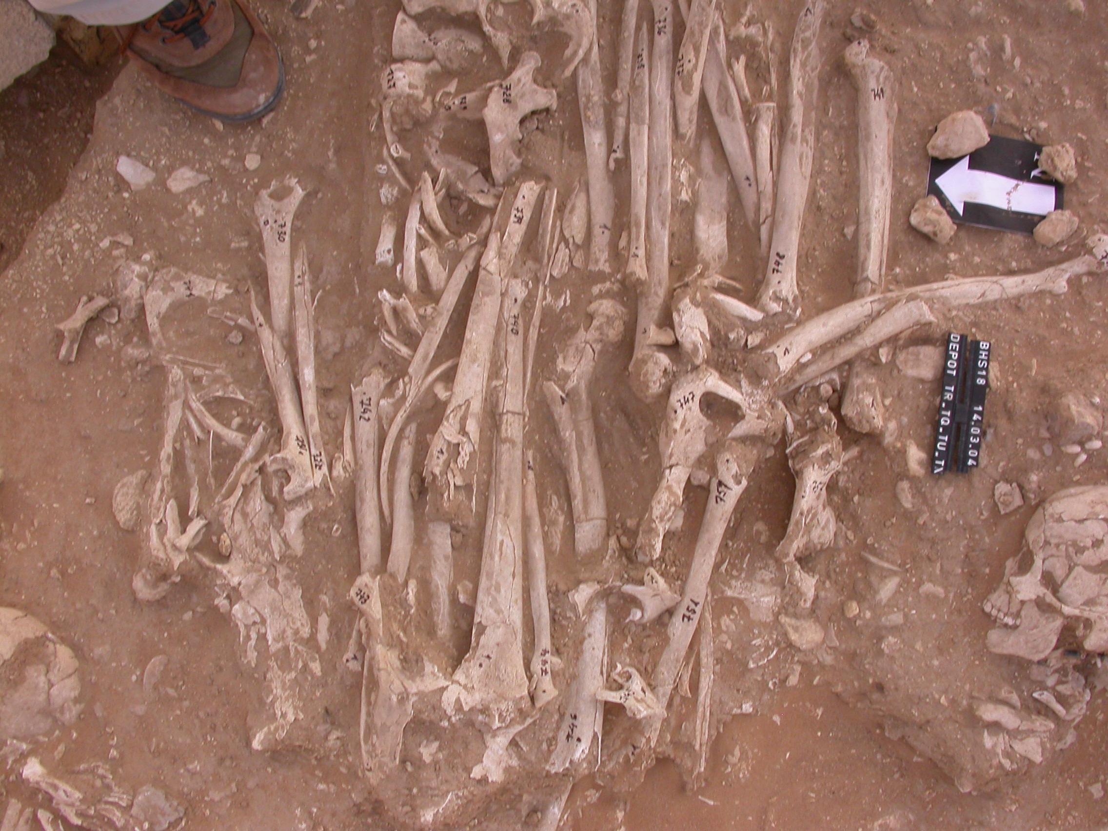
Inidvidual TQ:
TR, TU, TX, TQ depot in situ
Full Resolution
Full Resolution

Inidvidual TR:
Orthorectified picure of S1-AX with individuals TU, TX and TR
Full Resolution
Full Resolution

Inidvidual TR:
TU, TX, SQ and TR, orthorectified picture of Steg S1-Ax
Full Resolution
Full Resolution

Inidvidual TR:
TU, UD, TR, TX. Orthorectified picture.
Full Resolution
Full Resolution

Inidvidual TR:
TU, TX, TR orthorectified
Full Resolution
Full Resolution

Inidvidual TR:
Skull UD. Depot TX, TR, TU, TQ in situ
Full Resolution
Full Resolution

Inidvidual TR:
Depot TR, TQ, TU, TX and skull of burial SQ
Full Resolution
Full Resolution

Inidvidual TR:
Depot TR, TQ, TU, TX and UD and UF in situ.
Full Resolution
Full Resolution

Inidvidual TR:
TR, TU, TX, TQ depot in situ
Full Resolution
Full Resolution
Inidvidual TT:
Skull TT with adornments, bottom side after recovery
Full Resolution
Full Resolution
Inidvidual TU:
Orthorectified picure of S1-AX with individuals TU, TX and TR
Full Resolution
Full Resolution
Inidvidual TU:
TU, TX, SQ and TR, orthorectified picture of Steg S1-Ax
Full Resolution
Full Resolution
Inidvidual TU:
TU, UD, TR, TX. Orthorectified picture.
Full Resolution
Full Resolution

Inidvidual TU:
TU, TX, TR orthorectified
Full Resolution
Full Resolution

Inidvidual TU:
Skull UD. Depot TX, TR, TU, TQ in situ
Full Resolution
Full Resolution

Inidvidual TU:
Depot TR, TQ, TU, TX and skull of burial SQ
Full Resolution
Full Resolution
Inidvidual TU:
Depot TR, TQ, TU, TX and UD and UF in situ.
Full Resolution
Full Resolution
Inidvidual TU:
TR, TU, TX, TQ depot in situ
Full Resolution
Full Resolution

Inidvidual TU:
Secondary burials of TX, TU and TP. The stone was laying underneath individual TW
Full Resolution
Full Resolution
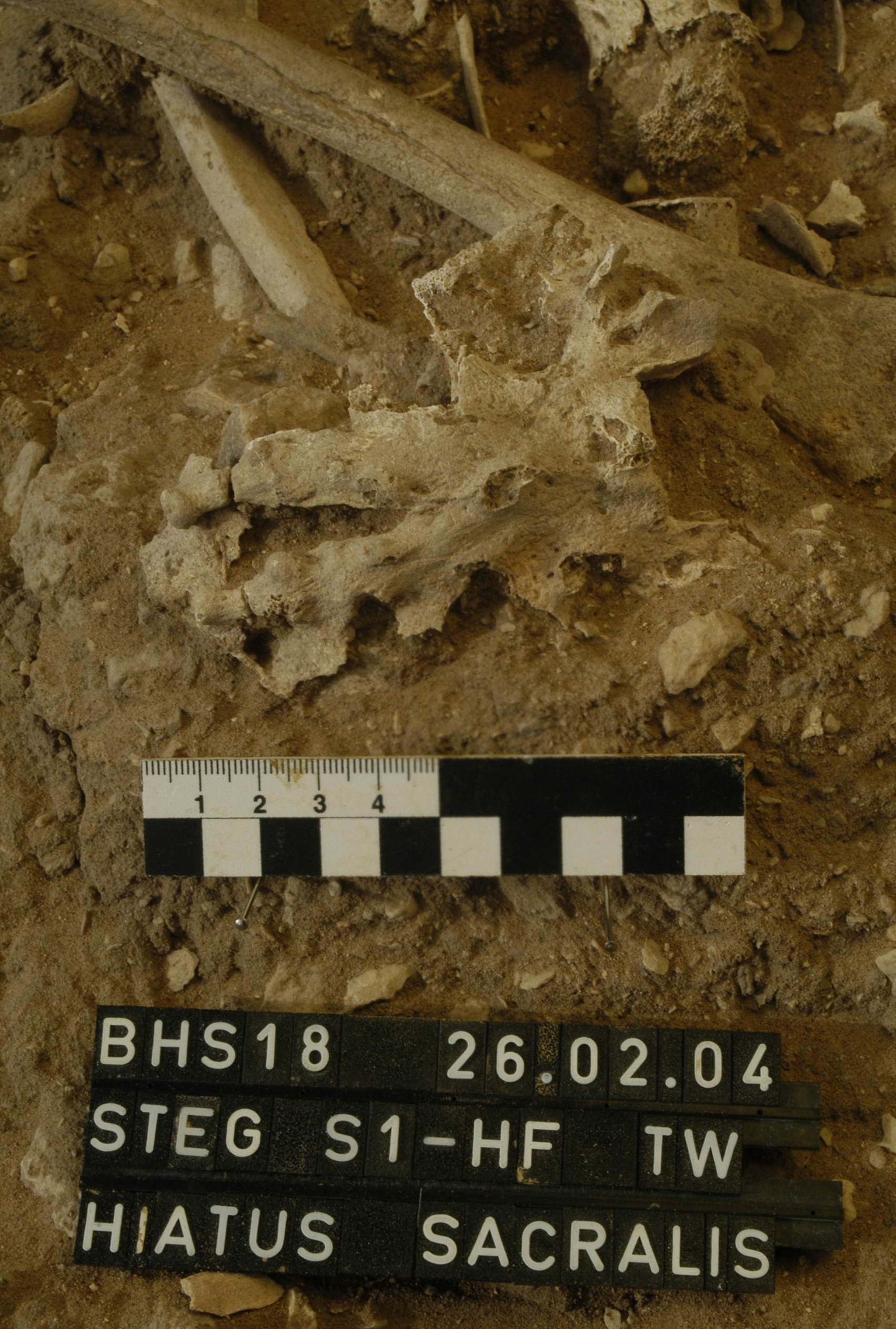
Inidvidual TW:
TW, Os sacrum with Hiatus sacralis. Spina bifida
Full Resolution
Full Resolution
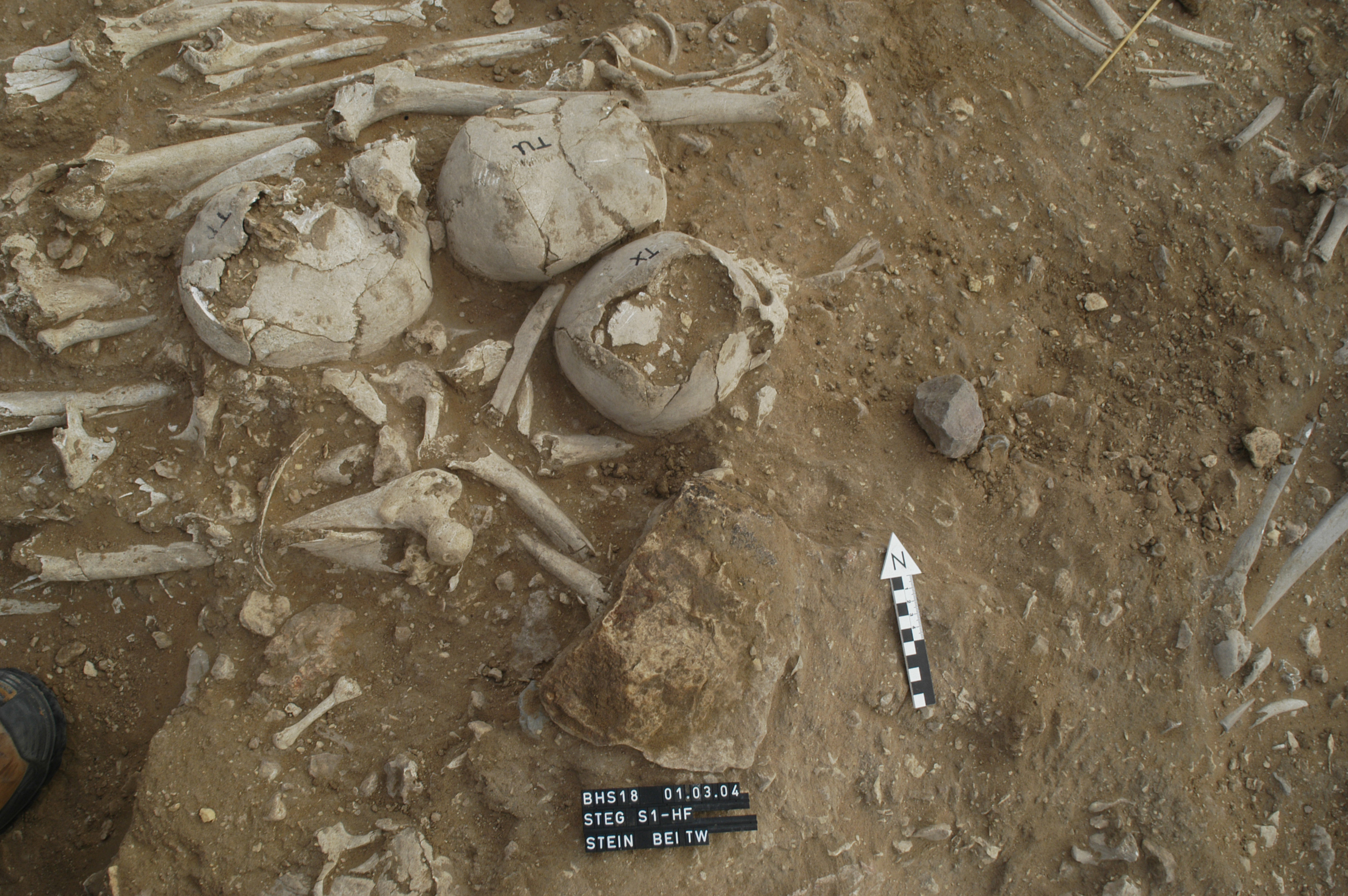
Inidvidual TW:
Secondary burials of TX, TU and TP. The stone was laying underneath individual TW
Full Resolution
Full Resolution
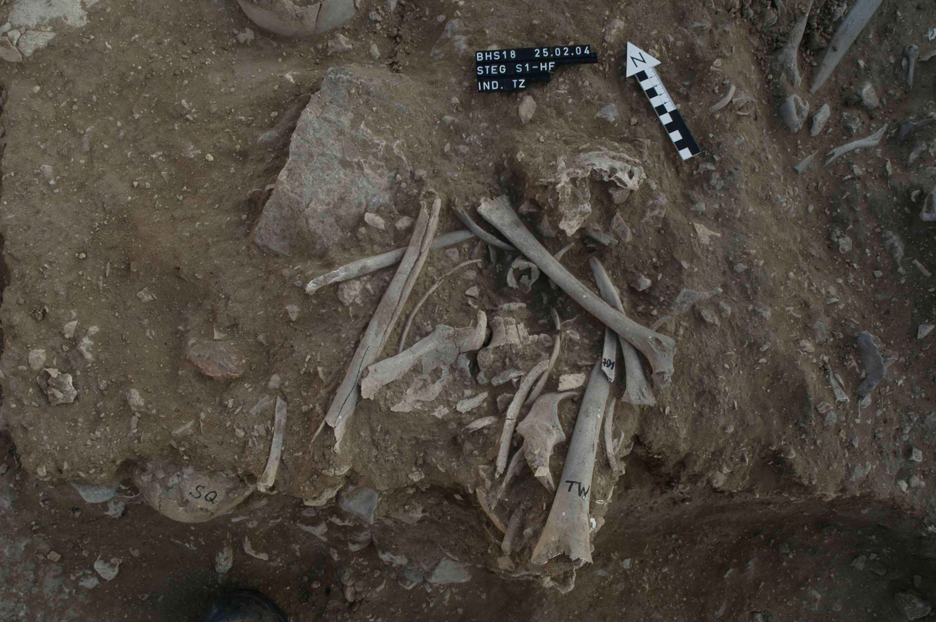
Inidvidual TW:
Bones individual TZ (infantile), mixed with TW. Steg S1-Ax
Full Resolution
Full Resolution

Inidvidual TX:
Orthorectified picure of S1-AX with individuals TU, TX and TR
Full Resolution
Full Resolution

Inidvidual TX:
TU, TX, SQ and TR, orthorectified picture of Steg S1-Ax
Full Resolution
Full Resolution

Inidvidual TX:
TU, UD, TR, TX. Orthorectified picture.
Full Resolution
Full Resolution
Inidvidual TX:
TU, TX, TR orthorectified
Full Resolution
Full Resolution

Inidvidual TX:
Skull UD. Depot TX, TR, TU, TQ in situ
Full Resolution
Full Resolution

Inidvidual TX:
Depot TR, TQ, TU, TX and skull of burial SQ
Full Resolution
Full Resolution

Inidvidual TX:
Depot TR, TQ, TU, TX and UD and UF in situ.
Full Resolution
Full Resolution

Inidvidual TX:
TR, TU, TX, TQ depot in situ
Full Resolution
Full Resolution

Inidvidual TX:
Secondary burials of TX, TU and TP. The stone was laying underneath individual TW
Full Resolution
Full Resolution
Inidvidual TX:
Skull with lesion at the left Os zygomaticum
Full Resolution
Full Resolution

Inidvidual TX:
Os zygomaticum, note the sharp-edged edge
Full Resolution
Full Resolution
Inidvidual TY:
TY, lesion at Femur.
Full Resolution
Full Resolution

Inidvidual TZ:
Bones individual TZ (infantile), mixed with TW. Steg S1-Ax
Full Resolution
Full Resolution

Inidvidual UD:
TU, UD, TR, TX. Orthorectified picture.
Full Resolution
Full Resolution

Inidvidual UD:
Skull UD. Depot TX, TR, TU, TQ in situ
Full Resolution
Full Resolution

Inidvidual UD:
Depot TR, TQ, TU, TX and UD and UF in situ.
Full Resolution
Full Resolution

Inidvidual UF:
Depot TR, TQ, TU, TX and UD and UF in situ.
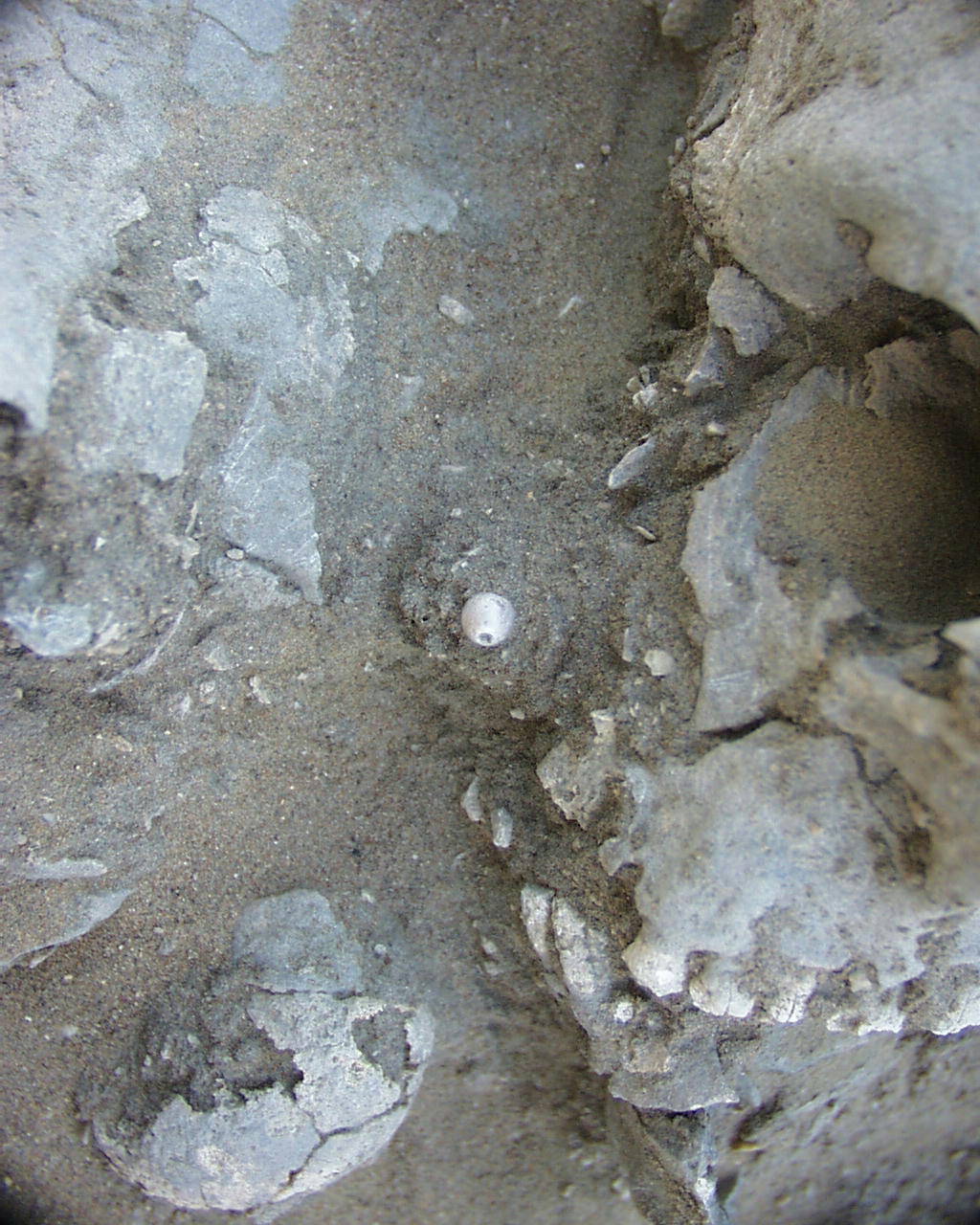
Full Resolution
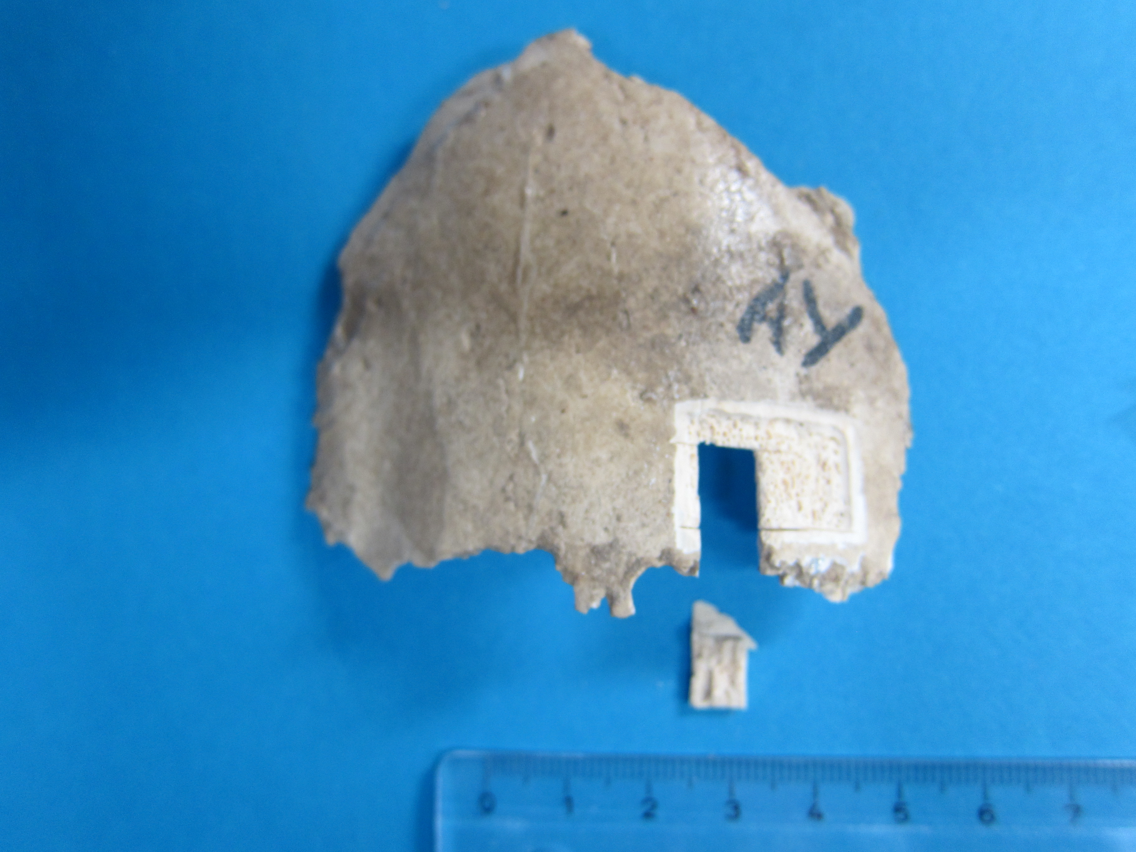
Full Resolution
Inidvidual UJ:
Clavicle with small bracelet wrapped around. This was a unique finding.
Full Resolution
Full Resolution

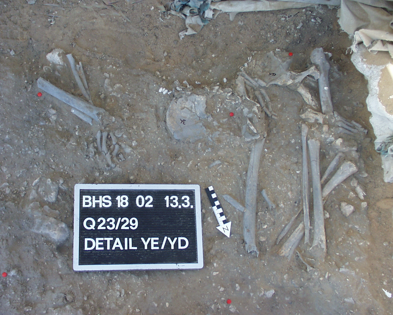
Inidvidual XD:
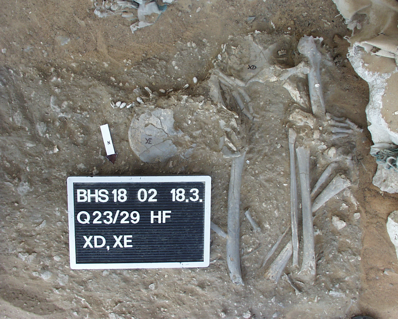
individuals XE and XD, (formerly known as YE and YD). Main area (Hauptfläche HF, Square 23/29)
Full Resolution
Full Resolution
Inidvidual XD:
Detail adornments individuals XD and XE. A disturbance of the burials can be seen, only the lower parts of the skeletons from the pelvis to the feet are preserved. Adornments at pelvis.
Full Resolution
Full Resolution
Inidvidual XE:
individuals XE and XD, (formerly known as YE and YD). Main area (Hauptfläche HF, Square 23/29)
Full Resolution
Full Resolution

Inidvidual XE:
Detail adornments individuals XD and XE. A disturbance of the burials can be seen, only the lower parts of the skeletons from the pelvis to the feet are preserved. Adornments at pelvis
Full Resolution
Full Resolution

Inidvidual YA:
FD, FY, GA, GB, GD, GE, GP, KL, YA in situ
Full Resolution
Full Resolution

Inidvidual YA:
FD, FY, GA, GB, GD, GE, GP, KL, YA in situ
Full Resolution
Full Resolution
Inidvidual YJ:
YJ fracture
Full Resolution
Full Resolution
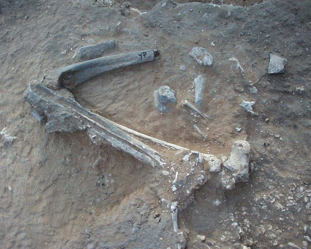
Inidvidual YP:
YP in situ
Full Resolution
Full Resolution

Inidvidual YR:
LL and YR in situ
Full Resolution
Full Resolution
Inidvidual YR:
YR in situ
Full Resolution
Full Resolution

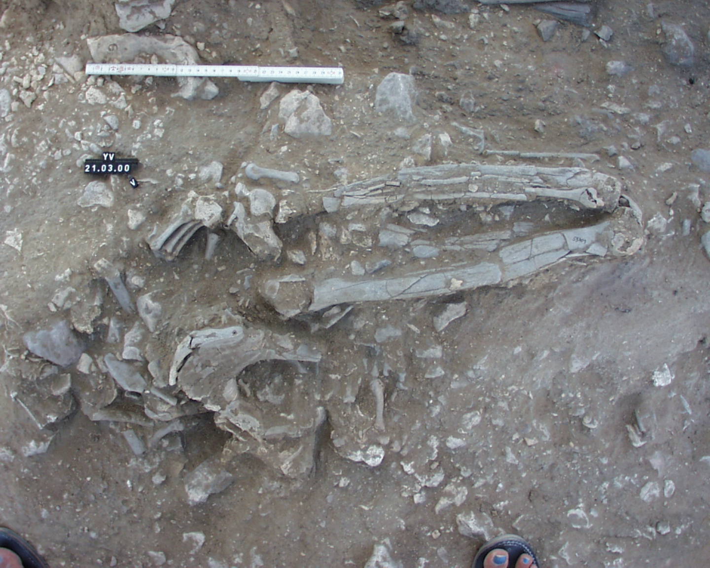
Inidvidual YT:
YT and EV in situ
Full Resolution
Full Resolution
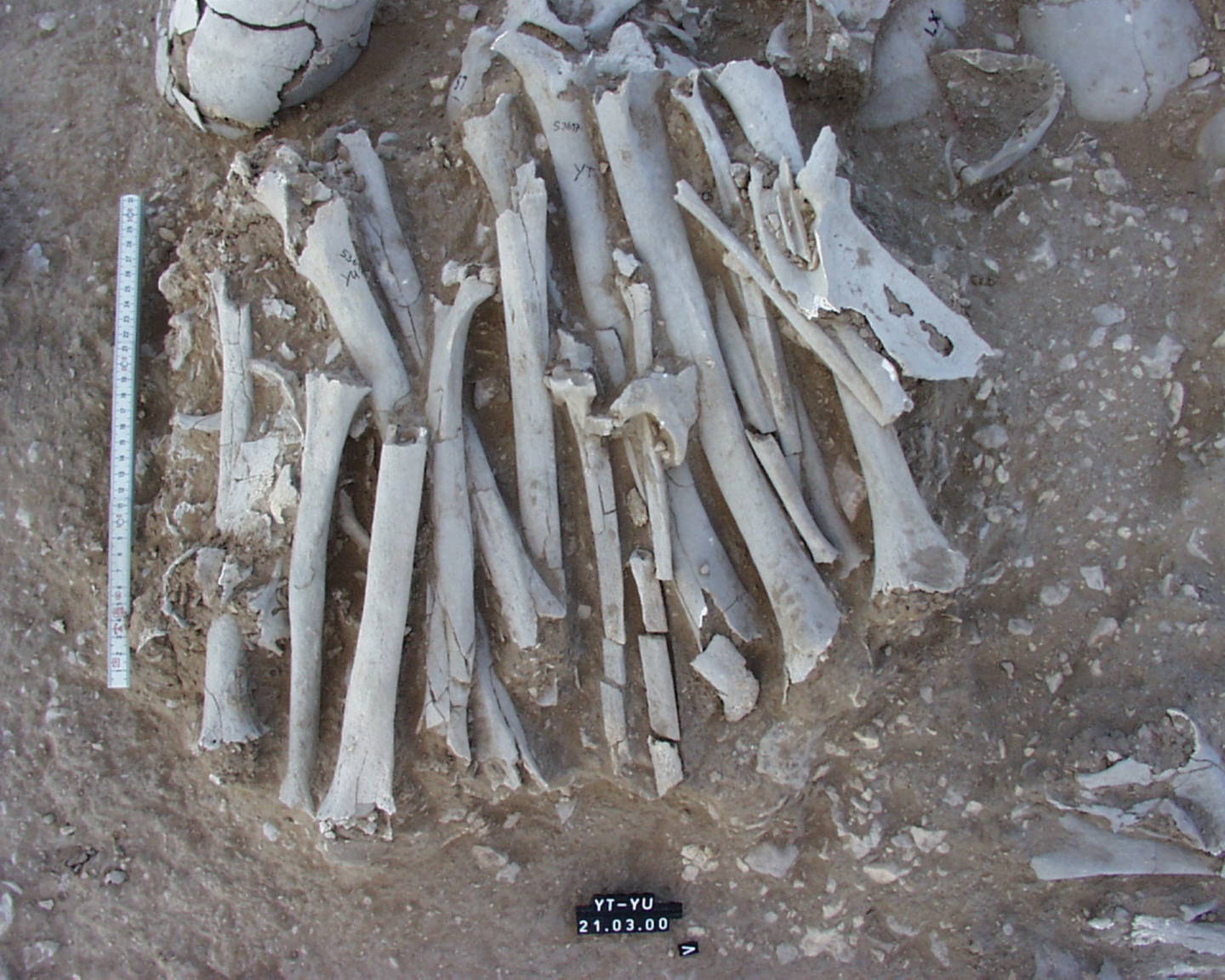
Inidvidual YT:
YT and YU in situ
Full Resolution
Full Resolution

Inidvidual YU:
YT and YU in situ
Full Resolution
Full Resolution
Inidvidual YV:
YV in situ
Full Resolution
Full Resolution
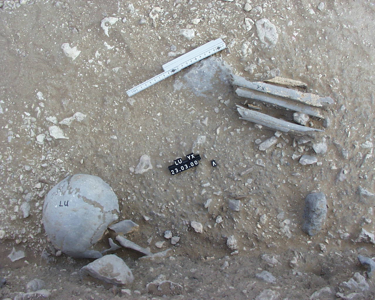
Inidvidual YX:
skull LU and longbone fragments YX in situ
Full Resolution
Full Resolution
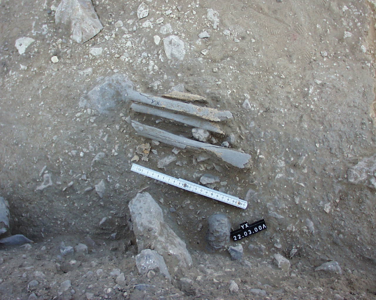
Inidvidual YX:
YX in situ
Full Resolution
Full Resolution